
EBV Infection Associated Neoplastic Lesions and Imaging Characteristics
-
摘要: Epstein-Barr病毒(EBV)属γ疱疹病毒,具有嗜淋巴细胞性,EBV是人类感冒病毒群中常见的成员,也是第一个被发现的人类肿瘤病毒。EBV感染主要通过呼吸道分泌物及唾液感染口,B淋巴细胞和上皮细胞是EBV宿主细胞。EBV首次感染常发生于幼年及儿童期。EBV相关性病变常见的包括传染性单核细胞增多症、嗜血细胞综合征,慢性慢性活动性EBV感染、EBV感染相关性自身免疫性疾病。EBV感染还可以引起感染细胞的增殖性病变,如各种良性淋巴增殖性病变,及伯基特淋巴瘤(BL)、霍奇金淋巴瘤(HL)、T/自然杀伤(NK)细胞淋巴瘤等恶性淋巴瘤,还可发生鼻咽癌、胃癌等上皮源性恶性肿瘤。本文对儿童常见的EBV感染相关性肿瘤性病变及影像学表现予以综述。
-
关键词:
- 影像学 /
- 儿童 /
- Epstein-Barr病毒 /
- EBV相关淋巴增殖性病变
Abstract: Epstein-Barr virus (EBV), also known as a gamma herpesviru, has the characteristics of lymphophilic tissue. EBV is one of the most common human viruses of the human cold virus group and the first human tumor virus found. Infection with EBV occurs by the oral transfer of saliva and respiratory secretions. Human B-lymphocytes and epithelial cells are EBV-susceptible host cells. First EBV infection often occurs in infancy and in childhood. EBV-associated diseases include infectious mononucleosis, EB virus associated hemophagocytic lymphoproliferative disorder (EBV-HLH), chronic active EBV Infection, EBV infection-associated autoimmune disease. EBV infection can also cause the proliferative diseases of the infected cells, such as various non-malignant lymphoproliferative diseases, lymphomatoid granulomatosis, EBV associated with many more malignant diseases including Burkitt′s lymphoma, Hodgkin′s lymphoma, Diffuse Large B-Cell Lymphoma, NK/T cell lymphoma, nasopharyngeal carcinoma, a subset of gastric carcinomas, and many more. This paper mainly focuses on EBV associated lymphoproliferative diseases and imaging characteristics in children. -
-
![]()
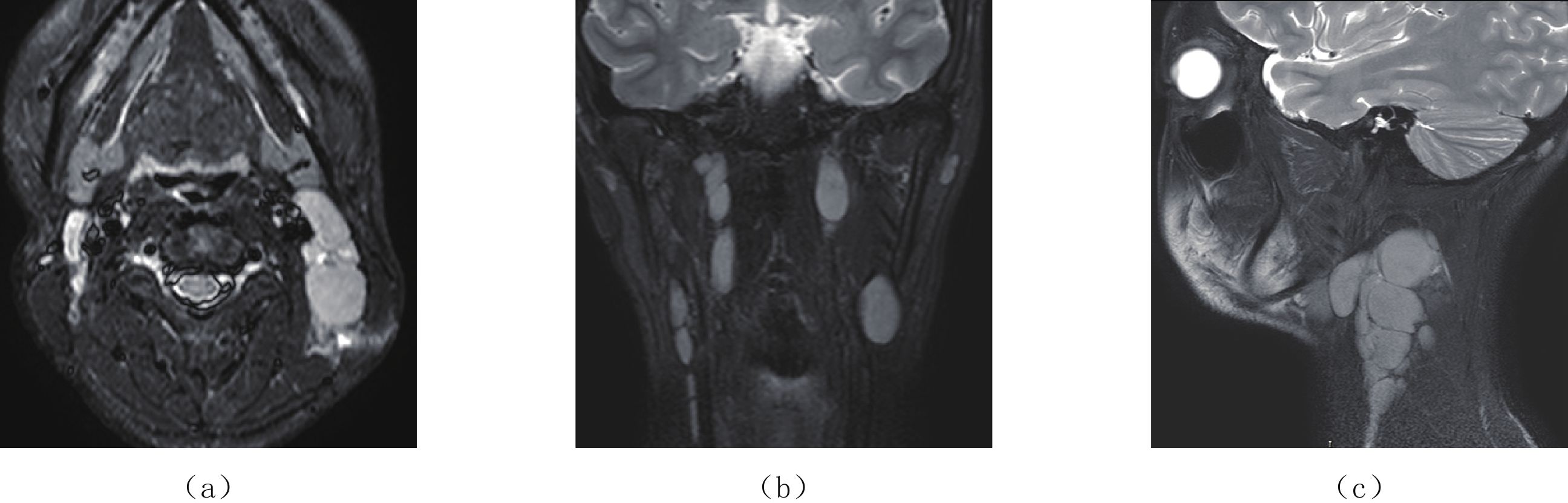
图 1 16岁,男孩,EBV相关性经典型霍奇金淋巴瘤
(a)~(c)分别为颈部MR T2压脂序列的轴面、冠状面及矢状面图,显示颈部肌间,咽旁间隙多发增大淋巴结,边缘清晰锐利,压脂均匀高信号。
Figure 1. A 16-year-old boy was diagnosed as EBV associated stereotypical Hodgkin lymphoma
![]()
图 2 14岁,男孩,EBV相关伯基特淋巴瘤
腹部CT平扫。(a)和(b)CT轴面,(c)和(d)冠状面。胰头周围的十二指肠降段及水平段肠壁明显增厚,僵直,小肠的肠腔粗细不均匀,累及十二指肠大乳头,壶腹部胆管梗阻,胆总管明显扩张。
Figure 2. A 14-year-old boy was diagnosed as EBV associated Burkitt lymphoma
![]()
图 3 7岁,男孩,EBV相关性伯基特淋巴瘤
腹部CT平扫。(a)~(c)轴面,(d)冠状面。腹腔内巨大肿物,软组织密度,外观结节状,与肠系膜,小肠,腹膜分界不清,腹腔内肠系膜肿胀,肠管肿胀,腹腔少许积液;心膈角区可见增大淋巴结。
Figure 3. A 7-year-old boy was diagnosed as EBV associated Burkitt lymphoma
![]()
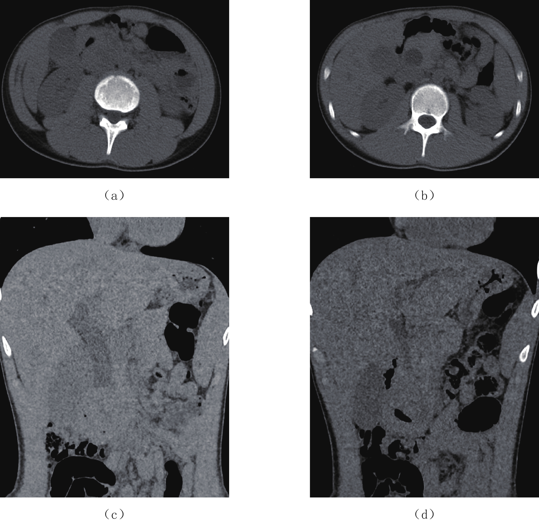
图 4 11.5岁,男孩,EBV相关性B淋巴母细胞型淋巴瘤
腹部增强CT。(a)和(b)腹部增强CT轴面及冠状面。表现为双肾外形增大,增强后双肾可见广泛分布占位性病变,强化程度低于正常肾实质。(c)~(e)腹部MR轴面T2序列、冠状面T2压脂序列及弥散序列,表现为双肾外形大,肾实质散在等T2信号灶,T2压脂等信号,病灶明显弥散受限。同时脊柱骨信号不均匀,提示存在骨髓浸润。
Figure 4. A 11.5-year-old boy was diagnosed as EBV associated B lymphoblastic lymphoma
![]()
图 5 4岁,男孩,EB病毒相关弥漫大B淋巴瘤,累及中枢神经系统
(a)头颅CT平扫;(b)~(g)分别为头颅MR的T1WI、T2WI、T2FLAIR、DWI、ADC及T1增强序列。头颅CT平扫左顶叶见稍高密度团块影,病灶周围可见水肿带;MR见左顶叶病变区脑回肿胀,病灶呈环状短T1信号,T1短信号环FLAIR高信号,弥散受限,并强化;环状信号中心区呈稍长T1信号,T2呈不均匀等/高信号,T2FLAIR呈低信号,无弥散受限,无强化。提示为恶性病灶中心区存在的坏死区。
Figure 5. A 4-year-old boy was diagnosed as EBV-associated diffuse large B lymphoma involving central nervous system
![]()
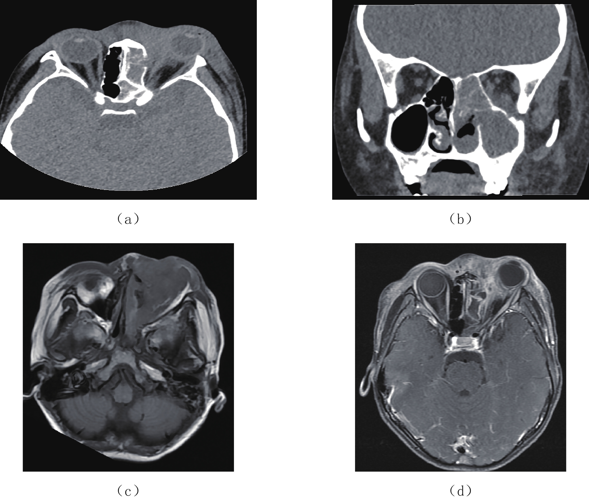
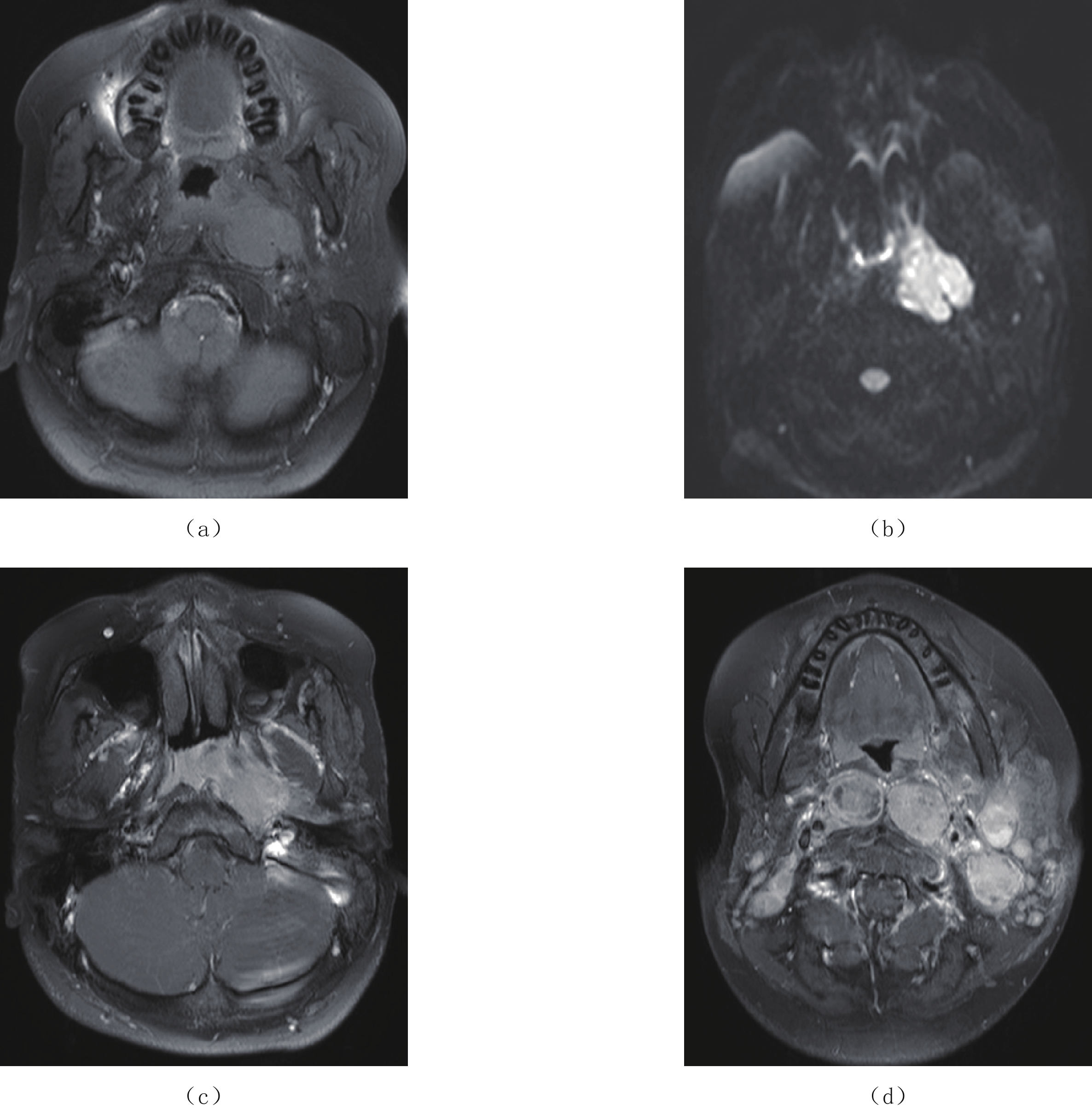
图 6 11岁,男孩,鼻型结外NK/T细胞淋巴瘤
(a)和(b)副鼻窦CT平扫轴面及冠状面;(c)和(d)鼻窦MR T1WI及T1WI增强序列。鼻窦CT可见颌面及眶周软组织弥漫肿,边界不清,左侧眶周病变重;左侧鼻腔,左侧鼻窦粘膜软组织增厚;左侧眶壁,左侧上颌窦,鼻中隔,左侧鼻甲骨破坏。病变向左侧眶内延伸,左眼球外凸。MR显示病变呈等T1稍长T2信号,明显强化。
Figure 6. A 11-year-old boy was diagnosed as Nasal extranodal NK/T cell lymphoma
![]()
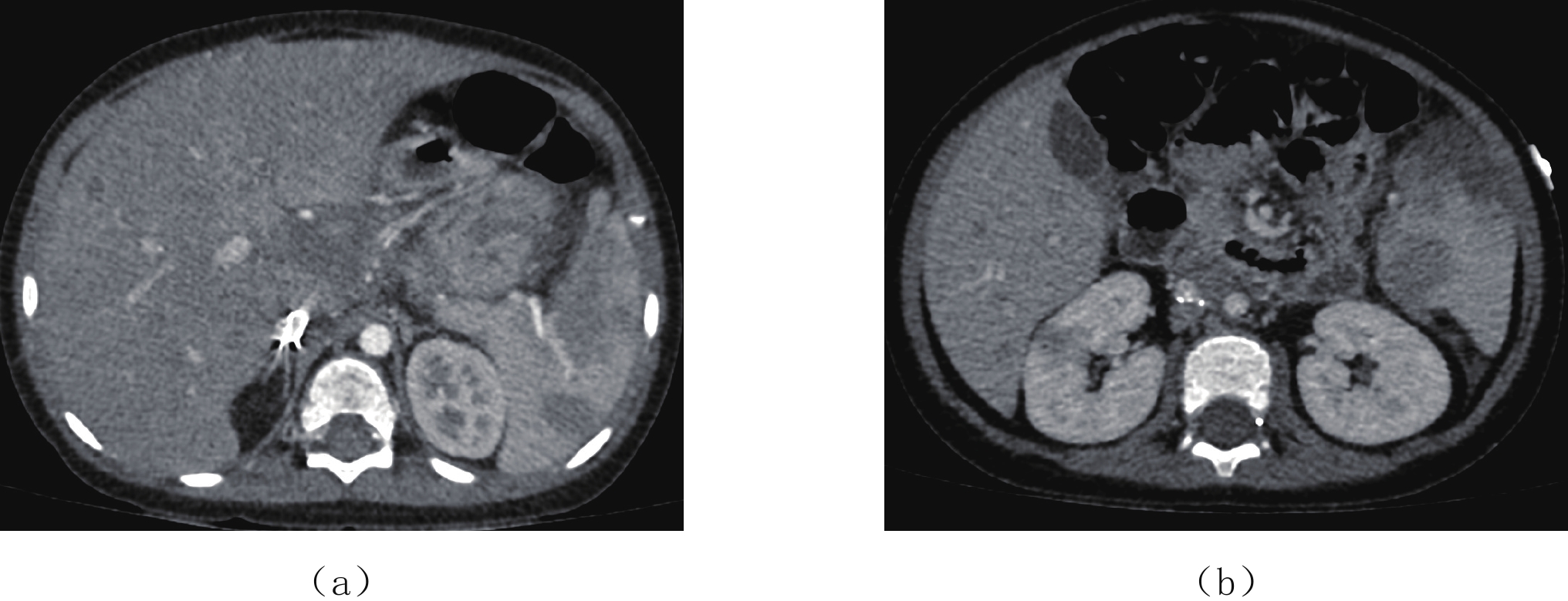
图 7 2岁,男孩,EBV相关性T细胞性淋巴瘤
腹部CT增强。(a)和(b)为CT轴面,可见肝脏实质内,肝门区,脾脏,肾脏多发瘤灶,轻度强化,强化程度较周围实质脏器程度减低。
Figure 7. A 2-year-old boy was diagnosed as EBV associated T-cell lymphoma
![]()
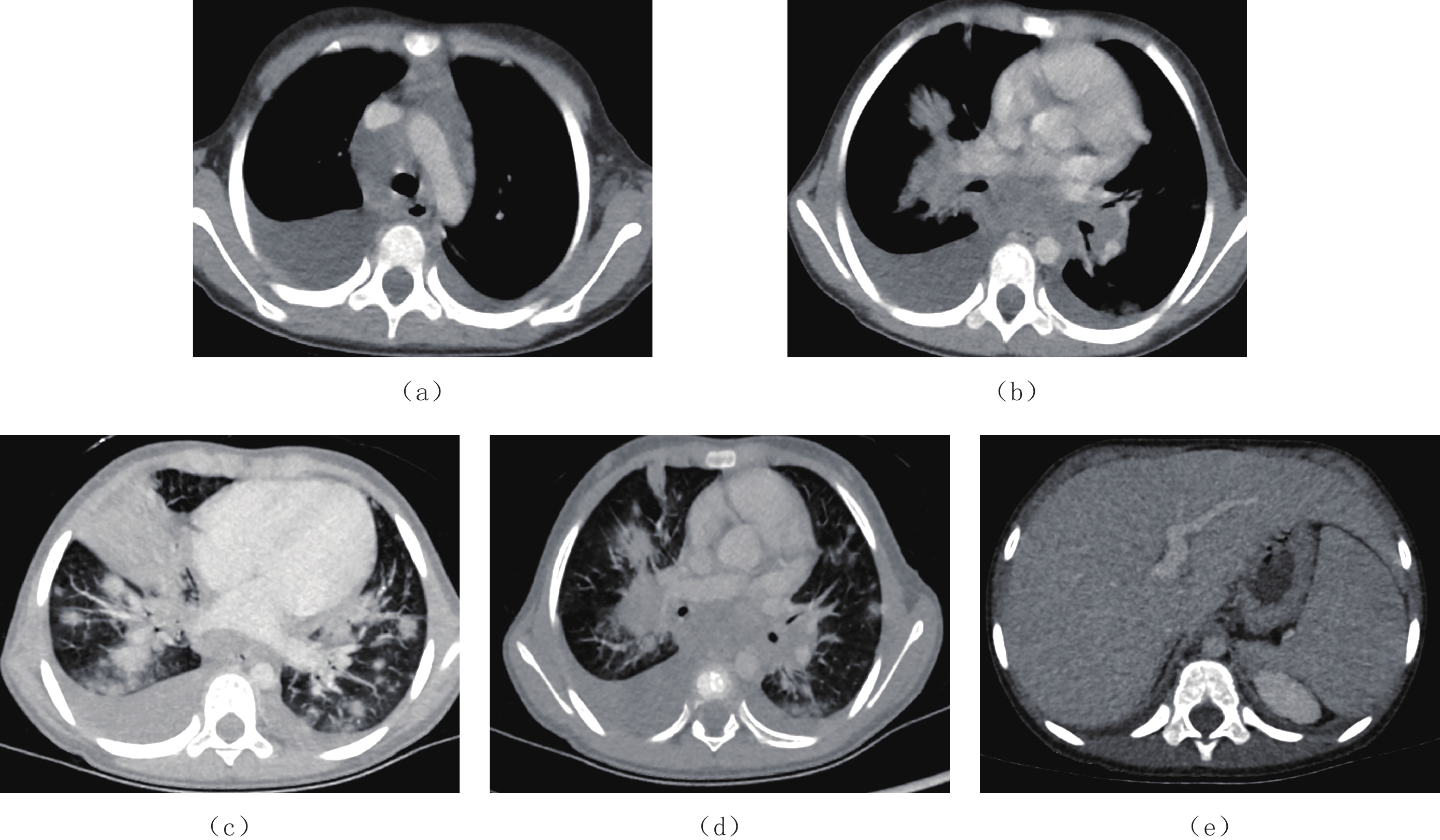
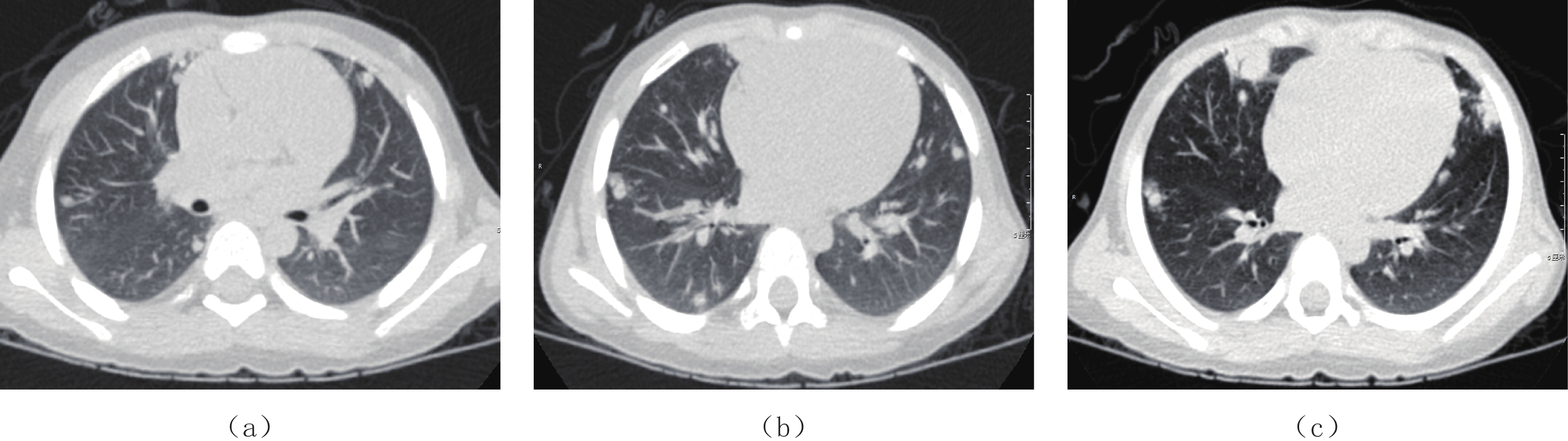
图 8 女孩,8岁,EBV相关T细胞淋巴组织增殖性疾病
胸部CT增强。两肺散在结节影及团片影,部分病灶内可见含气支气管穿行。纵隔、气管隆突、肺门淋巴结肿大;双侧胸腔积液;腹部层面肝脾大。
Figure 8. A 8-year-old girl was diagnosed as EBV-associated T cell lymphoproliferative disease
![]()
图 9 4岁,男孩,EBV相关淋巴瘤样肉芽肿
胸部CT平扫可见双肺散在小结节影,边缘略呈分叶状,双肺野外带为著,右肺舌叶可见团片影,内见含气支气管穿行征象。肺门淋巴结略增大。
Figure 9. A 4-year-old boy was diagnosed as EBV-associated lymphoma granuloma
-
[1] 艾军红, 谢正德, 申昆玲. EB病毒及儿童EB病毒相关疾病[J]. 中华实用儿科临床杂志, 2016,31(22): 1683−1686. doi: 10.3760/cma.j.issn.2095-428X.2016.22.002 AI J H, XIE Z D, SHEN K L. Epstein-Barr virus and Epstein-Barr virus associated diseases in children[J]. Journal of Applied Clinical Pediatrics, 2016, 31(22): 1683−1686. (in Chinese). doi: 10.3760/cma.j.issn.2095-428X.2016.22.002
[2] 贾雁琳, 郝彦琴. EB病毒感染机制及临床研究进展[J]. 中华临床医师杂志(电子版), 2019,13(8): 624−626. JIA Y L, HAO Y Q. Epstein-Barr virus infection: Mechanism and clinical research[J]. Chinese Journal of Clinicians (Electronic Edition), 2019, 13(8): 624−626. (in Chinese).
[3] 刘茂艳, 詹学. 儿童EB病毒原发性感染相关疾病研究进展[J]. 临床医学进展, 2022,12(3): 2211−2217. doi: 10.12677/ACM.2022.123318 LIU M Y, ZHAN X. Related research progress of Epstein-Barr virus associated diseases with primary infection in children[J]. Advances in Clinical Medicine, 2022, 12(3): 2211−2217. (in Chinese). doi: 10.12677/ACM.2022.123318
[4] Gulley M L, TANG W H. Laboratory assays for Epstein-Barr virus-related disease[J]. Journal of Molecular Diagnostics, 2018, 10(4): 279−292.
[5] 谢正德, 申昆玲. EB病毒感染实验室诊断及临床应用专家共识[J]. 中华实验和临床病毒学杂志, 2018,32(1): 1−8. doi: 10.3760/cma.j.issn.1003-9279.2018.01.000 XIE Z D, SHEN K L. Expert consensus on laboratory diagnosis and clinical application of EB virus infection[J]. Chinese Journal of Experimental and Clinical Virology, 2018, 32(1): 1−8. (in Chinese). doi: 10.3760/cma.j.issn.1003-9279.2018.01.000
[6] GULLEY M L, GLASER S L, CRAIG F E. et al. Guidelines for interpreting EBER in situ hybridization and LMP1 immunohistochemical tests for detecting Epstein-Barr virus in Hodgkin lymphoma[J]. American Journal of Clinical Pathology, 2002, 117: 259−267. doi: 10.1309/MMAU-0QYH-7BHA-W8C2
[7] AYEE R, OFORI M E O, WRIGHT E, et al. Epstein-Barr virus associated lymphomas and epithelia cancers in humans[J]. Journal Cancer, 2020, 11(7): 1737−1750. doi: 10.7150/jca.37282
[8] 王滕滕, 袁田, 张翼鷟. EB病毒与其相关淋巴瘤的研究进展[J]. 中国实验血液学杂志, 2014,22(6): 1775−1779. WANG T T, YUAN T, ZHANG Y Z. Progress of EB virus and its associated lymphoma[J]. Chinese Journal of Experimental Hematology, 2014, 22(6): 1775−1779. (in Chinese).
[9] VOCKERODT M, CADER F Z, SHANNON-LOWE C, et al. Epstein-Barr virus and the origin of Hodgkin lymphoma[J]. Chinese Journal of Cancer, 2014, 33(12): 591−597.
[10] WANG S, MEDEIROS L J, XU-MONETTE Z Y, et al. Epstein-Barr virus-positive nodular lymphocyte predominant Hodgkin lymphoma[J]. Annals of Diagnostic Pathology, 2014, 18: 203−209. doi: 10.1016/j.anndiagpath.2014.03.007
[11] SWERDLOW S H. WHO classification of tumours of haematopoietic and lymphoid tissues[J]. WHO Classification of Tumours, 2008, 22008: 439.
[12] LEVINE P H, ABLASHI D V, BERARD C W, et al. Elevated antibody titers to Epstein-Barr virus in Hodgkin's disease[J]. Cancer, 1971, 27: 416−421. doi: 10.1002/1097-0142(197102)27:2<416::AID-CNCR2820270227>3.0.CO;2-W
[13] WU T C, MANN R B, CHARACHE P, et al. Detection of EBV gene expression in reed-sternberg cells of Hodgkin's disease[J]. International Journal of Cancer, 1990, 46: 801−804. doi: 10.1002/ijc.2910460509
[14] SWERDLOW S H, CAMPO E, PILERI S A, et al. The 2016 revision of the World Health Organization (WHO) classification of lymphoid neoplasms[J]. Blood, 2016, 27(20): 2375−2390.
[15] EPSTEIN M A. Virus particles in cultured lymphoblasts from Burkitt's lymphoma[J]. Lancet, 1964, 1: 702−703.
[16] ROCHFORD R, MOORMANN A M. Burkitt's lymphoma[J]. Epstein Barr Virus Volume 1: Springer, 2015: 267−285.
[17] SHANNON-LOWE C, RICKINSON A B, BELL A I. Epstein-Barr virus-associated lymphomas[J]. Philosophical Transactions of the Royal Society of London, Series B, Biological Sciences, 2017, 372: 20160271. doi: 10.1098/rstb.2016.0271
[18] LEE H Y, KIM H S, PARK J W, et al. Atypical imaging features of Epstein-Barr virus-positive primary central nervous system lymphomas in patients without AIDS[J]. American Journal of Neuroradiology, 2013, 34(8): 1562−1567. doi: 10.3174/ajnr.A3429
[19] ZHANG N, ZUO Y, JIANG L, et al. Epstein-Barr virus and neurological diseases[J]. Frontiers in Molecular Biosciences, 2022, 8: 816098. doi: 10.3389/fmolb.2021.816098
[20] TSE E, KWONG Y L. NK/T-cell lymphomas[J]. Best Practice & Research. Clinical Haematology, 2019, 32(3): 253−261.
[21] TSE E, KWONG Y L. Recent advances in the diagnosis and treatment of natural killer cell malignancies[J]. Cancers (Basel), 2022, 14(3): 597. doi: 10.3390/cancers14030597
[22] SALEM A E, ZAKI Y H, EL-HUSSIENY G, et al. An overview of selected rare B-cell lymphoproliferative disorders: Imaging, histopathologic, and clinical features[J]. Cancers (Basel), 2021, 13(22): 5853. doi: 10.3390/cancers13225853
[23] SIRAJUDDIN A, RAPARIA K, LEWIS V A, et al. Primary pulmonary lymphoid lesions: Radiologic and pathologic findings[J]. Radiographics, 2016, 36(1): 53−70. doi: 10.1148/rg.2016140339
[24] MIMI C Y, YUAN J M. Epidemiology of nasopharyngeal carcinoma[J]. Seminars in Cancer Biology, 2002, 12(6): 421−429. doi: 10.1016/S1044579X02000858
[25] LEE H M, OKUDA K S, GONZÁLEZ F E, et al. Current perspectives on nasopharyngeal carcinoma[J]. Advances in Experimental Medicine and Biology, 2019, 1164: 11−34.
[26] KING A, WONG L, LAW B, et al. MR imaging criteria for the detection of nasopharyngeal carcinoma: Discrimination of early-stage primary tumors from benign hyperplasia[J]. American Journal of Neuroradiology, 2017, 39(3): 515−523.
[27] PICKHARDT P J, SIEGEL M J, HAYASHI R J, et al. Posttransplantation lymphoproliferative disorder in children: Clinical, histopathologic, and imaging features[J]. Radiology, 2000, 217(1): 16−25. doi: 10.1148/radiology.217.1.r00oc3816
[28] 林芳, 沈茜, 徐虹, 等. 儿童肾移植术后淋巴组织增生性疾病两例报告暨文献复习[J]. 中华肾脏病杂志, 2021,37(3): 183−190. doi: 10.3760/cma.j.cn441217-20200613-00029 LIN F, SHEN Q, XU H, et al. Post-transplantation lymphoproliferative disorders of kidney in children: Two cases report and literature review[J]. Chinese Journal of Nephrology, 2021, 37(3): 183−190. (in Chinese). doi: 10.3760/cma.j.cn441217-20200613-00029
[29] BAKER A, ESTEBAN F R, JUAN T C, et al. Current practices on diagnosis, prevention and treatment of post-transplant lymphoproliferative disorder in pediatric patients after solid organ transplantation: Results of ERN transplantchild healthcare working group survey[J]. Children (Basel), 2021, 8(8): 661. DOI: 10.3390/children8080661.
[30] KARIMZADEH M, TABIBZADEH A, MOGHOOFEI M, et al. As evidence-based tumorigenic role of Epstein-Barr virus miR-BART1-3p in neurological tumors[J]. Asian Pacific Journal of Cancer Prevention, 2021, 22(1): 257−266. doi: 10.31557/APJCP.2021.22.1.257
[31] ZAVALA-VEGA S, CASTRO-ESCARPULLI G, HERNÁNDEZ-SANTOS H, et al. An overview of the infection of CMV, HSV 1/2 and EBV in Mexican patients with glioblastoma multiforme[J]. Pathology, Research and Practice, 2017, 213(3): 271−276. DOI: 10.1016/j.prp.2016.12.006.
[32] MAGG T, SCHOBER T, WALZ C, et al. Epstein-Barr virus+smooth muscle tumors as manifestation of primary immunodeficiency disorders[J]. Frontiers in Immunology, 2018, 9: 368. DOI: 10.3389/fimmu.2018.00368.
-
期刊类型引用(3)
1. 卢振如,王婷. 成人肠道套细胞淋巴瘤合并回盲型肠套叠一例. 中国医学科学院学报. 2024(03): 458-461 .  百度学术
百度学术
2. 王婷,田松林,郑珂. MR小肠造影对肠道T细胞淋巴瘤与B细胞淋巴瘤的鉴别效能分析. 罕少疾病杂志. 2024(10): 95-97 . 百度学术
3. 陈衍池. 超声诊断肠梗阻的价值分析. 中国医疗器械信息. 2023(14): 16-18+176 . 百度学术
其他类型引用(0)
 下载:
下载:


计量
- 文章访问数: 490
- HTML全文浏览量: 269
- PDF下载量: 20
- 被引次数: 3


