
Methods Research on Correction for Intracranial Metal Artifacts Based on Prior Image Utilization
-
摘要:
目的:为了有效抑制颅内金属伪影对术后复查的影响,提出一种基于先验图像的金属伪影校正算法。方法:首先对颅内金属伪影的CT图像进行自适应滤波、阈值分割和K均值聚类获得只含金属物质的图像和先验图像;以先验图像为参照,在金属物质的投影轨迹中,对原始图像的投影数据进行插值校正得到修复后的图像,并将其与只含金属物质图像融合,获得最终校正图像。为了验证该算法抑制CT金属伪影的有效性,利用仿真单金属伪影和颅内金属伪影的CT图像进行伪影校正。结果:与线性插值算法和ADN算法相比,该算法校正后的图像结构相似性和峰值信噪比最大,而均方根误差最小。由两位高年资医生对该算法去除CT金属伪影的主观评价得分高于线性插值算法和ADN算法的主观评价得分。结论:该算法在有效保留图像边缘信息的同时,可有效地抑制CT金属伪影。
Abstract:Objective: A metal artifact correction algorithm based on prior image is proposed to suppress the effect of intracranial metal artifacts on postoperative review. Method: The computed tomography (CT) image compensating for the intracranial metal artifact is obtained by adaptive filtering, threshold segmentation, and K mean clustering. In projecting the trajectory of metallic matter, the interpolation correction of the original image’s projection data is obtained using the prior image as a reference, and the image is fused with the metal-containing material only image to produce the final corrected image. To verify the efficacy of this algorithm in suppressing CT metal artifacts, artifact correction was performed using CT images of simulated isolated metal artifacts and intracranial metal artifacts. Results: Compared with linear interpolation and the ADN algorithms, this algorithm achieves the highest structure similarity and peak signal-to-noise ratio and lowest root mean square error in corrected images. Two senior doctors subjectively scored this algorithm higher than the linear interpolation and ADN algorithms for removing CT metal artifacts. Conclusion: This algorithm effectively suppresses metal artifacts while preserving image edge information.
-
Keywords:
- CT metal artifact simulation /
- prior image /
- metal artifact /
- adaptive filter
-
-
![]()
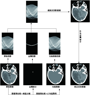
图 1 本文提出金属伪影校正的流程图
Figure 1. Flow chart of the proposed metal artifact correction method
表 1 比较LI-MAR和FP-MAR校正后,单金属图像上SSIM值、PSNR值和RMSE值
Table 1 Comparison between SSIM, PSNR, and RMSE values for isolated metal artifact images after LI-MAR and FP-MAR correction
评价指标 LI-MAR ADN FP-MAR SSIM 0.721 0.992 0.994 PSNR 33.200 44.640 45.200 RMSE 0.193 0.022 0.024  下载: 导出CSV
表 2 比较LI-MAR、ADN和FP-MAR校正后,单金属图像的主观评分
Table 2 Comparison of subjective scores on isolated metal images after LI-MAR and FP-MAR corrections
评价指标 常规CT图像 LI-MAR FP-MAR ADN 评价者1 3.20 3.9 4.5 4.2 评价者2 3.10 3.7 4.3 4.4 评价者得分取均值 3.15 3.8 4.4 4.3 T检验 P < 0.05
下载: 导出CSV
-
[1] DING Y H, GHOZY S, DAI D, et al. Rabbit elastase aneurysm model mimics the recurrence rate of human intracranial aneurysms following platinum coil embolization[J]. American Journal of Neuroradiology. 2022, 43(5): 741-747.
[2] 刘远来, 孙异春, 何永超, 等. Solitaire支架与lvis支架辅助弹簧圈栓塞对颅内宽颈动脉瘤的疗效对比[J]. 南方医科大学学报, 2020, 40(3): 423-426. DOI: 10.12122/j.issn.1673-4254.2020.03.23. LIU Y L, SUN Y C, HE Y C, et al. Treatment efficacy of Solitaire stent- and Lvis stent-assisted coil embolization for intracranial wide-neck carotid aneurysm[J]. Journal of Southern Medical University, 2020, 40(3): 423-426. DOI:10.12122/j.issn.1673-4254.2020.03.23. (in Chinese).
[3] DAI W, LING H, SUN Y, et al. The comparison of neuronavigation combined with CT three-dimensional angiography vs. CT angiography in the guidance of clipping treatment in distal intracranial aneurysm surgery: A retrospective clinical study[J]. Annals of Translational Medicine, 2022, 10(10): 572-578.
[4] KANANI A, YAZDI M, OWRANGI A M, et al. Metal artifact reduction in cervix brachytherapy with titanium applicators using dual-energy CT through virtual monoenergetic images and an iterative algorithm: A phantom study[J]. Brachytherapy, 2022, 21(6): 933-942.
[5] 陈宗桂, 陆思璇, 管海辰, 等. IMAR技术在去除腰椎内固定金属伪影中的应用价值[J]. 中国CT和MRI杂志, 2023, 21(11): 167-169. DOI: 10.3969/j.issn.1672-5131.2023.11.051. CHEN Z G, LU S X, GUAN H C, et al. Value of IMAR Iterative algorithm in removing metal artifacts of lumbarInternal fixation[J]. Chinese Journal of CT and MRI, 2023, 21(11): 167-169. DOI:10.3969/j.issn.1672-5131.2023.11.051. (in Chinese).
[6] SCHWARZ G M, HUBER S, WASSIPAUL C, et al. Influence of scan parameters of single and dual-energy CT protocols in combination with metal artifact suppression algorithms for THA: An EX Vivo study[J]. Journal of Bone and Joint Surgery-american Volume, 2023, 105(8): 620-629.
[7] 于佳弘, 张昆鹏, 靳爽, 等. 弦图插值结合UNIT网络图像转换的CT金属伪影校正[J]. 南方医科大学学报, 2023, 43(7): 1214-1223. DOI: 10.12122/j.issn.1673-4254.2023.07.18. YU J H, ZHANG K P, JIN S, et al. CT metal artifact correction using chord graph interpolation combined with UNIT network image conversion[J]. Journal of Southern Medical University, 2023, 43(7): 1214-1223. DOI: 10.12122/j.issn.1673-4254.2023.07.18. (in Chinese).
[8] ZHANG Z, YANG M, XU L, et al. An innovative metal artifact reduction algorithm based on Res-U-Net GANs[J]. Current Medical Imaging, 2023, 19(13): 1549-1560.
[9] YOON H, LEE K Y, BECHWATI I. CLIMAR: Classified linear interpolation based metal artifact reduction for severe metal artifact reduction in X-ray CT imaging[J]. Physics in Medicine&Biology, 2021, 66(7): 1-13.
[10] 齐宏亮, 凌庆, 李翰威, 等. 基于先验图像的CT图像金属伪影校正算法[J]. 北京生物医学工程, 2023, 42(4): 384-389, 413. DOI: 10.3969/j.issn.1002-3208.2023.04.008. QI H L, LING Q, LI H W, et al. Metal artifact correction algorithm for CT images based on prior images[J]. Befjing Blomedical Engineering, 2023, 42(4): 384-389, 413. DOI: 10.3969/j.issn.1002-3208.2023.04.008. (in Chinese).
[11] SCHMIDT A M A, GRUNZ J P, PETRITSCH B, et al. Combination of iterative metal artifact reduction and virtual monoenergetic reconstruction using split-filter dual-energy CT in patients with dental artifact on head and neck CT[J]. American Journal of Roentgenology, 2022, 218(4): 716-727.
[12] TRABZONLU T A, TERRAZAS M, MOZAFFARY A, et al. Application of iterative metal artifact reduction algorithm to CT urography for patients with hip prostheses[J]. American Journal of Roentgenology, 2020, 214(1): 137-143.
[13] JACOBS F, SUNDERMANN E, de Sutter B, et al. A fast algorithm to calculate the exact radiological path through a pixel or voxel space[J]. Journal of Computing and Information Technology, 1998, 6(1): 89-94.
[14] LIAO H F, LIN W A, ZHOU S K, et al. ADN: Artifact disentanglementnetwork for unsupervised metal artifact reduction[J]. IEEE TransMed Imaging, 2020, 39(3): 634-643. DOI: 10.1109/TMI.2019.2933425.
[15] 王军. 基于投影数据修正的CT金属伪影消除方法研究[D]. 南京: 东南大学, 2013. DOI: 10.7666/d.Y2438396. WANG J. Research on CT metal artifact elimination method based on projection data correction[D]. Nanjing: Southeast University, 2013. DOI:10.7666/d.Y2438396.(in Chinese).
[16] SARA U, AKTER M, UDDIN M S. Image quality assessment through FSIM, SSIM, MSE and PSNR-A comparative study[J]. Journal of Computer and Communications, 2019, 7(3): 8-18. DOI: 10.4236/jcc.2019.73002.
[17] MATTHIEU B, LOTHAR S. Metal artifact reduction in CT using tissue-class modeling and adaptive prefiltering[J]. Medical Physics, 2006, 33(8): 2852-2859. DOI: 10.1118/1.2218062.
[18] 郑艳梅, 岳向江, 李翠, 等. 基于均方误差的变形图像的质量评价[J]. 测控技术, 2018, 37(8): 117-120. DOI: 10.19708/j.ckjs.2018.08.027. ZHENG Y M, YUE X J, LI C, et al. Quality evaluation of deformed images based on mean square error[J]. Measurement and Control Technology, 2018, 37(8): 117-120. DOI: 10.19708/j.ckjs.2018.08.027. (in Chinese).
[19] ANHAUS J A, KILLERMANN P, SEDLMAIR M, et al. Nonlinearly scaled prior image-controlled frequency split for high-frequency metal artifact reduction in computed tomography[J]. Medical Physics, 2022, 49(9): 5870-5885.
[20] GONDIM T P, MEYER J B, BAUMANN C, et al. Total hip prosthesis CT with single-energy projection based metallic artifact reduction: Impact on the visualization of specific periprosthetic soft tissue structures[J]. Skeletal Radiology, 2014, 43(9): 1237-1246. DOI: 10.1007/s00256-014-1923-5.


计量
- 文章访问数: 207
- HTML全文浏览量: 51
- PDF下载量: 35


