
Advance in Silicon Photomultiplier for All-Digital Positron Emission Tomography
-
摘要:
近年来,硅光电倍增器(SiPM)凭借其出色的性能表现,已经成为正电子发射断层成像(PET)中的首选光电转换器件。SiPM具有单光子分辨能力和低于100 ps的时间分辨率,使得精确测量光子到达时间成为可能,催生了飞行时间PET、光子计数计算机断层扫描、正电子素寿命显像等新兴应用领域,这些应用又对SiPM的性能提出了更高的挑战。因此,如何将SiPM性能推进至其物理极限已成为新一代SiPM的研究的关键方向。在传统的SiPM架构中,信号经过多次处理和模数转换,带来噪声增加和时间性能恶化的问题,从而限制了SiPM的性能潜力。随着半导体制造工艺的快速发展,SiPM可在标准CMOS工艺节点上制造,标志着可以将数字逻辑集成在SiPM器件内,这是SiPM领域的一次重大突破,使我们能在单一SiPM内实现更精确的时间、能量、位置信息获取,为推进SiPM达到性能极限提供了一条可能的途径。本文综述SiPM的发展历史、工作原理和性能参数,分析传统SiPM的局限性,梳理数字SiPM研究的关键问题,介绍当前几种数字化SiPM架构,最后对数字SiPM的关键技术进行总结和展望。
Abstract:In recent years, silicon photomultipliers (SiPMs) have emerged as preferred photoelectric conversion devices in positron emission tomography (PET) due to their outstanding performance. SiPMs possess single-photon resolution capability and time resolution below 100 ps, enabling precise photon arrival time measurements. These advances paved the way for emerging applications such as time-of-flight PET (TOF-PET), photon counting CT, and positron emission lifetime imaging, presenting new challenges to SiPM performance, the advancing of which to their physical limits has become a key focus area in next-generation SiPM research. In traditional SiPM architectures, signal processing and analog-to-digital conversion introduce noise and degrade time performance, thereby limiting the full SiPM potential. With the recent and rapid development of semiconductor manufacturing processes, SiPMs could be manufactured on standard CMOS process nodes, which marks a significant breakthrough in the SiPM field, allowing for the integration of digital logic within SiPM devices. This advancement opens the possibility of achieving more precise time, energy, and position information within a single SiPM, thereby providing potential possibilities to push SiPMs to their performance limits. In this study, we reviewed the development history, working principles, and performance parameters of SiPMs. We analyzed the limitations of traditional SiPMs, outlined key aspects of digital SiPM research, and introduced various current digital SiPM architectures. Finally, we summarized and anticipated key technologies in digital SiPMs.
-
卵巢恶性肿瘤中上皮恶性肿瘤最常见,约占卵巢恶性肿瘤的85%~90%,主要包括浆液性囊腺癌、黏液性囊腺癌、透明细胞癌及子宫内膜样癌等[1]。由于恶性程度较高、发展快,该类肿瘤预后往往不佳[2]。同时因为缺乏特异症状和诊断方法,一般难以早期诊断,约 60%~70% 临床确诊时已属晚期[2-3]。
目前 CT和血清肿瘤标志物CA125、HE4在其术前诊断中作用越来越突出[1],我们通过对156例卵巢上皮肿瘤进行研究,探讨CT联合血清CA125及人附睾蛋白4(human epididymis protein 4,HE4)对卵巢上皮恶性肿瘤的诊断价值。
1. 资料和方法
1.1 研究对象
对2017年1月至2021年5月间于淄博市博山区医院、淄博市第一医院及上海中医药大学附属龙华医院金山分院诊治的156例卵巢上皮肿瘤进行研究。其中良性72例(浆液性囊腺瘤46例,浆液性乳头状囊腺瘤3例,浆液性腺纤维瘤和囊腺纤维瘤1例,黏液性囊腺瘤19例,良性子宫内膜样肿瘤2例,勃勒纳瘤1例),恶性84例,年龄26~71岁,平均年龄(51±13.5)岁。
入选标准:卵巢原发上皮肿瘤首次确诊,既往无卵巢肿瘤病史及卵巢手术史;未并发其他肿瘤,无肿瘤病史或放、化疗等治疗史;术后经病理学确诊。按国际妇产科联盟(international federation of gynecology and obstetrics,FIGO)对卵巢上皮恶性肿瘤的分期[4],84例恶性肿瘤中Ⅰ期15例,Ⅱ期21例,Ⅲ 期37例,Ⅳ 期11例。其中浆液性囊腺癌61例、黏液性囊腺癌16例、子宫内膜样腺癌5例、透明细胞癌2例。156例病例为连续收集,入组路线图见图1。
![]() 图 1 156例卵巢上皮肿瘤病例入组路线图Figure 1. Enrollment diagram of 156 cases of ovarian epithelial tumors
图 1 156例卵巢上皮肿瘤病例入组路线图Figure 1. Enrollment diagram of 156 cases of ovarian epithelial tumors1.2 血清CA125及HE4检测
全部病例清晨采集空腹静脉血约5 mL,自然抗凝后以3000 r/min离10 min,取上清液采用酶联免疫吸附法测定血清中HE4和CA125水平,检测试剂盒购自北京中杉金桥生物有限公司,具体检测步骤严格按照试剂盒说明书进行操作。
血清CA125和HE4参考值分别设定为0~35 U/mL和0~72 pmol/L,血清CA125>35 U/mL为阳性,≤35 U/mL为阴性;血清HE4高于72 pmol/L为阳性,≤72 pmol/L为阴性。
1.3 CT检查及诊断
采用GE Light Speed 64排CT及Siemens Definition 64排CT进行检查。层厚5 mm,间隔5 mm,扫描范围从膈上至耻骨联合水平,每例均行平扫及动脉、静脉期增强扫描。采用高压注射器经肘静脉注射非离子对比剂碘海醇(300 mgI/mL)80~100 mL,注射流率3 mL/s,注射对比剂后30 s和55 s后分别行动脉期、静脉期扫描。
由两位高年资CT医师共同进行诊断,参照周康荣等[4]诊断卵巢上皮性恶性肿瘤标准,根据病变的部位、数目、形态、大小、密度、边界、强化特点、与周围结构的关系及腹、盆腔积液等改变进行诊断及分期。
1.4 统计学处理
根据最终病理诊断结果,对CT、血清CA125、HE4及联合诊断的结果进行统计学分析。采用SPSS 20.0软件,计数资料采用(%)表示,组间比较采用χ
${}^2 $ 检验,P<0.05具有统计学意义。2. 结果
血清CA125、HE4水平在卵巢上皮良、恶性肿瘤组中阳性率的比较详见表1,结果显示恶性组血清CA125及HE4阳性率均显著高于良性组。
表 1 血清CA125、HE4在卵巢上皮良、恶性肿瘤组中阳性率的比较Table 1. Comparison among positive rates of serum CA125 and HE4 in benign and malignant ovarian epithelial tumors分组 CA125 HE4 恶性肿瘤组(n=84) 85.71%(72/84) 80.95%(68/84) 良性肿瘤组(n=72) 38.89%(28/72) 22.22%(16/72) χ${} ^2$ 36.94 53.81 P 0.000 0.000 CT、血清CA125、HE4单独及联合应用对卵巢上皮恶性肿瘤的诊断结果比较显示CA125诊断的灵敏度高于HE4,HE4诊断特异度高于CA125,CA125联合HE4诊断的准确率高于CT;CT+CA125+HE4诊断准确率高于单独CT或肿瘤标志物诊断(表2),部分病例CT图像见图2~图5。
表 2 CT、血清CA125、HE4单独及联合应用对卵巢上皮恶性肿瘤的诊断结果比较Table 2. Comparison among the diagnostic results of CT, serum CA125, HE4 alone and combination application in epithelial ovarian cancer诊断项目 灵敏度/% 特异度/% 阳性预测值/% 阴性预测值/% 准确率/% CA125 85.71a 61.11 72.00 78.57 74.36 HE4 80.95 77.78b 80.95b 77.78 79.49b CA125+HE4 90.48c 83.33c 86.36c 88.24c 87.18c CT 83.33 80.56 83.33 80.56 82.05 CT+CA125+HE4 95.24d 88.89d 90.91d 94.18d 92.31d χ2 9.86 18.59 12.99 9.99 21.30 P 0.043 0.001 0.011 0.041 0.000 注:a-高于HE4;b-高于CA125;c-高于CA125、HE4及CT;d-高于CA125、HE4、CA125+HE4及CT。 ![]() 图 2 46岁女性,右侧卵巢黏液性囊腺癌Figure 2. A 46-year-old female, mucinous cystadenocarcinoma in the right ovary
图 2 46岁女性,右侧卵巢黏液性囊腺癌Figure 2. A 46-year-old female, mucinous cystadenocarcinoma in the right ovary![]() 图 3 70岁女性,右侧卵巢浆液性囊腺癌Figure 3. A 70-year-old female, serous cystadenocarcinoma in the right ovary
图 3 70岁女性,右侧卵巢浆液性囊腺癌Figure 3. A 70-year-old female, serous cystadenocarcinoma in the right ovary![]() 图 4 43岁女性,左侧卵巢透明细胞癌Figure 4. A 43-year-old female,clear cell carcinoma in the left ovarian
图 4 43岁女性,左侧卵巢透明细胞癌Figure 4. A 43-year-old female,clear cell carcinoma in the left ovarian![]() 图 5 55岁女性,右侧卵巢子宫内膜样腺癌Figure 5. A 55-year-old female, endometrioid adenocarcinoma in the right ovarian
图 5 55岁女性,右侧卵巢子宫内膜样腺癌Figure 5. A 55-year-old female, endometrioid adenocarcinoma in the right ovarian3. 讨论
卵巢恶性肿瘤致死率居女性生殖系统恶性肿瘤之首[4]。该类肿瘤主要包括上皮细胞来源恶性肿瘤、性索间质来源恶性肿瘤、生殖细胞来源恶性肿瘤及转移性肿瘤,其中绝大多数为上皮细胞来源,以浆液性囊腺癌最为多见,其他尚有黏液性囊腺癌、透明细胞癌及内膜样腺癌、未分化癌等[5-6]。由于恶性肿瘤5年生存率由发病早期的90% 下降至晚期的25%~30%,因而及时确诊并积极治疗是改善预后、延长生存的关键[6-7]。
CA125是一种高分子质量的糖蛋白,作为目前妇科应用最广泛的肿瘤标志物,主要存在于间皮细胞组织、苗勒管上皮、间皮细胞及苗勒管衍生物发生的肿瘤中,如卵巢上皮癌、输卵管癌、子宫内膜癌、宫颈腺癌及间皮细胞癌等[8-9]。约85.0% 的晚期卵巢癌血清CA125升高,手术后和化疗奏效时水平下降,肿瘤复发会再度升高,因此广泛应用于卵巢上皮恶性肿瘤的临床诊断、疗效观察与监测[10-11]。其不足之处在于特异度不高,正常排卵期、子宫内膜异位症、子宫肌瘤、盆腔炎、卵巢过度刺激综合征、以及非卵巢癌的恶性肿瘤如肺癌、胃癌等状况下也会升高,导致假阳性;同时CA125在早期卵巢癌表达率较低,对浆液性癌以外恶性肿瘤如黏液性癌等的检出率也较低[8,11]。
本研究中CA125诊断的灵敏度较高,达到85.70%,但特异度、阳性预测值、阴性预测值及诊断准确率均较低,也说明单独应用CA125并不适合卵巢癌的筛查及早期诊断。
HE4是一种人附睾分泌蛋白,在卵巢癌组织中表达水平明显升高,但在正常卵巢组织中一般不表达,在癌旁组织和良性肿瘤中有低水平的表达,因而是鉴别卵巢癌的新型肿瘤标志物,具有简单易测、创伤性小、受干扰因素少的优势[12-13]。
HE4在鉴别卵巢肿瘤的良恶性时准确率较高,尤其是对Ⅰ期卵巢癌的敏感度明显高于CA125,不足之处在于HE4对绝经前后肿瘤的诊断效能亦不同,绝经、年龄越大往往HE4水平有所升高因而对绝经前恶性肿瘤的诊断能力更高;此外多项研究证实HE4在卵巢透明细胞癌和黏液性癌中表达率较低[10-12],如联合CA125则能提高敏感性及特异度[14]。
本研究显示HE4诊断的灵敏度低于CA125,但特异度、阳性预测值、阴性预测值及诊断准确率均高于CA125,HE4联合CA125则具有较高的诊断效能,灵敏度、特异度及诊断准确率高于两者单独诊断。
CT技术的发展有利于显示恶性肿瘤病变本身及继发改变的细节,从而及时诊断和准确分期[15-16]。总结本组资料并复习相关文献,我们认为卵巢上皮性恶性肿瘤的CT一般具有下述特征[4,17-18]:早期主要表现为囊性或囊实性,病情发展呈囊实性或部分实性;体积较大,一般直径大于4 cm;呈多房囊腔,肿瘤囊壁及囊腔内分隔厚薄不均匀,最大可超过3 cm;增强瘤体实性部分较明显强化,囊内可见明显强化的壁结节;后期往往伴有腹、盆腔积液及周边结构侵犯、淋巴结及远处转移,有时可见到较明显的肿瘤血管及两侧卵巢同时发病。
本组CT诊断准确率为82.12%,对大部分Ⅱ期肿瘤和全部 Ⅲ、Ⅳ 期肿瘤均得以正确诊断及分期。漏诊者均为单发体积较小的Ⅰ、Ⅱ期囊性肿瘤,因为体积较小、实性成分少且强化不明显而误认为良性囊腺瘤;3例良性肿瘤因为体积较大且实性成分强化较明显而误诊为恶性。因此CT不能单独根据病灶大小、强化程度等对病灶性质进行判断。此外卵巢转移瘤和原发性肿瘤有时具有相似的CT表现,而肿瘤标志物CA125及HE4则可以一定程度上弥补CT的不足。
本组资料证实,CT联合血清CA125及HE4对卵巢上皮恶性肿瘤诊断的灵敏度、特异度及准确率分别为95.21%、88.92% 及92.32%,明显高于单独的CT或肿瘤标志物检测。
综上所述,CT联合血清CA125及HE4对卵巢上皮恶性肿瘤的诊断具有重要价值,有利于早期诊断及准确分期,从而为临床治疗提供可靠的依据,是术前鉴别卵巢上皮肿瘤良恶性的有效组合,值得临床推广应用。
-
![]()
图 2 新型SiPM结构的横截面示意图
Figure 2. Schematic diagrams of the new SiPM structure cross-sections
![]()
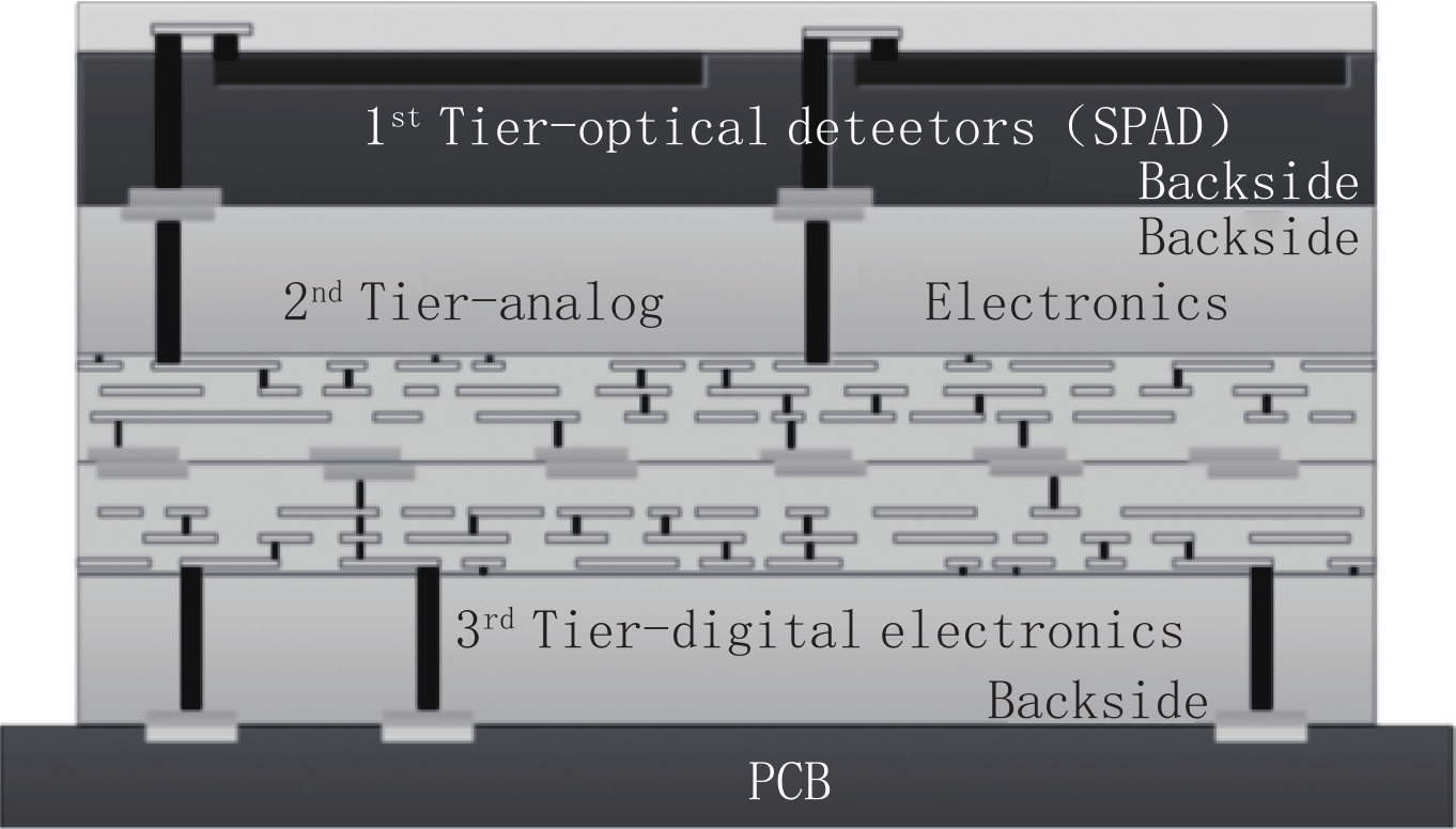
图 6 三维封装数字SiPM横截面示意图
Figure 6. Schematic 3D package digital SiPM cross-section diagram.
-
[1] 苗旺, 李翰山. 强辐射背景下光电成像探测性能计算方法[J]. 电光与控制, 2018, 25(12): 11−15. DOI: 10.3969/j.issn.1671-637X.2018.12.003. MIAO W, LI H S. Calculation method of photoelectric imaging detection performance in strong radiation background[J]. Electronics Optics & Control, 2018, 25(12): 11−15. DOI: 10.3969/j.issn.1671-637X.2018.12.003. (in Chinese).
[2] 向雨琰, 李松, 马跃. 光电倍增管输出电子流脉冲堆叠对光子计数法测距的影响[J]. 物理学报, 2022, 71(21): 163−175. XIANG Y Y, LI S, MA Y. Effect of pile-up of electron flow pulse from photomultiplier tube on ranging by photon counting[J]. Acta Physica Sinica, 2022, 71(21): 163−175. (in Chinese).
[3] 张雨, 张健, 张国光. 采用硅光电倍增管和塑闪阵列的便携式快中子成像系统的可行性研究[J]. 核技术, 2021, 44(3): 46−53. ZHANG Y, ZHANG J, ZHANG G G. Feasibility study of portable fast neutron imaging system using silicon photomultiplier and plastic scintillator array[J]. Nuclear Techniques, 2021, 44(3): 46−53. (in Chinese).
[4] 陈星, 周畅, 刘可为, 等. 基于宽禁带半导体氧化物微纳材料的紫外探测器研究进展[J]. 中国光学, 2022, 15(5): 912−928. CHENG X, ZHOU C, LIU K W, et al. Review of ultraviolet photodetectors based on micro/nano-structured wide bandgap semiconductor oxide[J]. Chinese Journal of Optics, 2022, 15(5): 912−928. (in Chinese).
[5] 苗泉龙, 代雷, 李佰成, 等. 基于SiPM和TCMPC的时间分辨拉曼散射测量技术研究[J]. 光谱学与光谱分析, 2018, 38(5): 1444−1450. MIAO Q L, DAI L, LI B C, et al. Time-resolved raman scattering measurement based on SiPM and TCMPC method[J]. Spectroscopy and Spectral Analysis, 2018, 38(5): 1444−1450. (in Chinese).
[6] 陈建光, 倪旭翔, 袁波, 等. SiPM激光雷达阳光下探测概率性能分析[J]. 光电工程, 2021, 48(10): 29−38. CHENG J G, NI X X, YUAN B, et al. Analysis of detection probability perf ormance of SiPM LiDAR under sunlight[J]. Opto-Electronic Engineering, 2021, 48(10): 29−38. (in Chinese).
[7] 刘鸿彬, 李铭, 王凤香, 等. 一种少光子高精度多波束激光雷达系统及验证[J]. 红外与毫米波学报, 2019, 38(4): 535−541. DOI: 10.11972/j.issn.1001-9014.2019.04.021. LIU H B, LI M, WANG F X, et al. A high accuracy multi-beam lidar system and its verification on several photons[J]. Journal of Infrared and Millimeter Waves, 2019, 38(4): 535−541. DOI: 10.11972/j.issn.1001-9014.2019.04.021. (in Chinese).
[8] TN3000系列SiPM数据手册[EB/OL]. [2023-11-12]. https://www.joinbon.com/Uploads/file/20191126/20191126171114_23598.pdf. TN3000系列SiPM数据手册[EB/OL]. [2023-11-12]. https://www.joinbon.com/Uploads/file/20191126/20191126171114_23598.pdf.
[9] C-series SiPM Sensors[EB/OL]. [2023-11-02]. https://www.onsemi.com/pdf/datasheet/microc-series-d.pdf.
[10] AFBR-S4N33C013 NUV-HD single silicon photo multiplier[EB/OL]. [2023-11-02]. https://docs.broadcom.com/doc/AFBR-S4N33C013-DS.
[11] LECOQ P, MOREL C, PRIOR J O, et al. Roadmap toward the 10ps time-of-flight PET challenge[J]. Physics in Medicine & Biology, 2020, 65(21): 21RM01.
[12] LECOQ P. Pushing the limits in time-of-flight PET imaging[J]. IEEE Transactions on Radiation and Plasma Medical Sciences, 2017, 1(6): 473−485. DOI: 10.1109/TRPMS.2017.2756674.
[13] ASCENZO N D’, ANTONECCHIA E, BRENSING A, et al. A novel high photon detection efficiency silicon photomultiplier with shallow junction in 0.35 μm CMOS[J]. IEEE Electron Device Letters, 2019, 40(9): 1471−1474. DOI: 10.1109/LED.2019.2929499.
[14] LIANG X, D’ASCENZO N, BROCKHERDE W, et al. Silicon photomultipliers with area Up to 9 mm2 in a 0.35 μm CMOS process[J]. IEEE Journal of the Electron Devices Society, 2019, 7: 239−251. DOI: 10.1109/JEDS.2019.2893802.
[15] D’ASCENZO N, SAVELIEV V, XIE Q. China silicon photomultiplier technology[C]//2016 IEEE Photonics Conference (IPC). Waikoloa, HI, USA: IEEE, 2016.
[16] OHL R S. Light-sensitive electric device: United States, 2402662[P]. 1951-06-12.
[17] McKAY K G. Avalanche breakdown in silicon[J]. Physical Review, 1954, 94(4): 877−884. DOI: 10.1103/PhysRev.94.877.
[18] MCINTYRE R J. Theory of microplasma instability in silicon[J]. Journal of Applied Physics, 1961, 32(6): 983−995. DOI: 10.1063/1.1736199.
[19] HAITZ R H. Model for the electrical behavior of a microplasma[J]. Journal of Applied Physics, 1964, 35(5): 1370−1376. DOI: 10.1063/1.1713636.
[20] RENKER D. Geiger-mode avalanche photodiodes, history, properties and problems[J]. Nuclear Instruments and Methods in Physics Research Section A: Accelerators, Spectrometers, Detectors and Associated Equipment, 2006, 567(1): 48-56.
[21] VASILE S, WILSON R J, SHERA S, et al. High gain avalanche photodiode arrays for DIRC applications[J]. IEEE Transactions on Nuclear Science, 1999, 46(4): 848−852. DOI: 10.1109/23.790690.
[22] GHIONI M, COVA S, ZAPPA F, et al. Compact active quenching circuit for fast photon counting with avalanche photodiodes[J]. Review of Scientific Instruments, 1996, 67(10): 3440−3448. DOI: 10.1063/1.1147156.
[23] ANTICH P P, TSYGANOV E N, MALAKHOV N A, et al. Avalanche photo diode with local negative feedback sensitive to UV, blue and green light[J]. Nuclear Instruments and Methods in Physics Research Section A: Accelerators, Spectrometers, Detectors and Associated Equipment, 1997, 389(3): 491-498.
[24] GOLOVIN V. Avalanche photodetector: Russia, 2142175[P].
[25] SAVELIEV V, GOLOVIN V. Silicon avalanche photodiodes on the base of metal-resistor-semiconductor (MRS) structures[J]. Nuclear Instruments and Methods in Physics Research Section A: Accelerators, Spectrometers, Detectors and Associated Equipment, 2000, 442(1-3): 223-229.
[26] NINKOVIĆ J, ANDRIČEK L, LIEMANN G, et al. SiMPl-novel high QE photosensor[J]. Nuclear Instruments and Methods in Physics Research Section A: Accelerators, Spectrometers, Detectors and Associated Equipment, 2009, 610(1): 142-144.
[27] 韩德俊, 张国青, 胡小波, 等. 一种新结构硅光电倍增器(SiPM)及其应用研究[C]//第九届全国光电技术学术交流会论文集. 2010: 23-26. [28] SUN F, DUAN N, LO G Q. Novel silicon photomultiplier with vertical bulk-Si quenching resistors[J]. IEEE Electron Device Letters, 2013, 34(5): 653−655. DOI: 10.1109/LED.2013.2250900.
[29] RENSCHLER M, PAINTER W, BISCONTI F, et al. Characterization of hamamatsu 64-channel TSV SiPMs[J]. Nuclear Instruments and Methods in Physics Research Section A: Accelerators, Spectrometers, Detectors and Associated Equipment, 2018, 888: 257-267.
[30] ACERBI F, PATERNOSTER G, GOLA A, et al. High-density silicon photomultipliers: Performance and linearity evaluation for high efficiency and dynamic-range applications[J]. IEEE Journal of Quantum Electronics, 2018, 54(2): 1−7.
[31] D’ASCENZO N, XIE Q. Possible layout solutions for the improvement of the dark rate of geiger mode avalanche structures in the globalfoundries bcdlite 0.18 μm CMOS technology[J]. Journal of Instrumentation, 2018, 13(4): T04007−T04007. DOI: 10.1088/1748-0221/13/04/T04007.
[32] GUNDACKER S, HEERING A. The silicon photomultiplier: Fundamentals and applications of a modern solid-state photon detector[J]. Physics in Medicine & Biology, 2020, 65(17): 17TR01. DOI: 10.1088/1361-6560/ab7b2d.
[33] SAVELIEV V. Silicon photomultiplier-new Era of photon detection[M]//YOUNG K. Advances in Optical and Photonic Devices. InTech, 2010. DOI: 10.5772/7150.
[34] SEITZ P, THEUWISSEN A J. Single-photon imaging: 160[M]. Berlin, Heidelberg: Springer Berlin Heidelberg, 2011. DOI: 10.1007/978-3-642-18443-7.
[35] ACERBI F, GUNDACKER S. Understanding and simulating SiPMs[J]. Nuclear Instruments and Methods in Physics Research Section A: Accelerators, Spectrometers, Detectors and Associated Equipment, 2019, 926: 16-35. DOI: 10.1016/j.nima.2018.11.118.
[36] RENKER D, LORENZ E. Advances in solid state photon detectors[J]. Journal of Instrumentation, 2009, 4(4): P04004−P04004. DOI: 10.1088/1748-0221/4/04/P04004.
[37] CALÒ P P, CICIRIELLO F, PETRIGNANI S, et al. SiPM readout electronics[J]. Nuclear Instruments and Methods in Physics Research Section A: Accelerators, Spectrometers, Detectors and Associated Equipment, 2019, 926: 57-68.
[38] PARK H, YI M, LEE J S. Silicon photomultiplier signal readout and multiplexing techniques for positron emission tomography: A review[J]. Biomedical Engineering Letters, 2022, 12(3): 263−283. DOI: 10.1007/s13534-022-00234-y.
[39] NAKAMURA K Z, BAN S, ICHIKAWA A K, et al. Front-end electronics for the SiPM-readout gaseous TPC for neutrinoless double-beta decay search[J]. IEEE Transactions on Nuclear Science, 2020, 67(7): 1772−1776. DOI: 10.1109/TNS.2020.2988716.
[40] BISOGNI M G, DEL GUERRA A, BELCARI N. Medical applications of silicon photomultipliers[J]. Nuclear Instruments and Methods in Physics Research Section A: Accelerators, Spectrometers, Detectors and Associated Equipment, 2019, 926: 118-128.
[41] SCHAART D R, CHARBON E, FRACH T, et al. Advances in digital SiPMs and their application in biomedical imaging[J]. Nuclear Instruments and Methods in Physics Research Section A: Accelerators, Spectrometers, Detectors and Associated Equipment, 2016, 809: 31-52.
[42] FRACH T, PRESCHER G, DEGENHARDT C, et al. The digital silicon photomultiplier-principle of operation and intrinsic detector performance[C]//2009 IEEE Nuclear Science Symposium Conference Record (NSS/MIC). Orlando, 2009: 1959-1965.
[43] FRACH T, PRESCHER G, DEGENHARDT C, The digital silicon photomultiplier-system architecture and performance evaluation[C]//IEEE Nuclear Science Symposuim & Medical Imaging Conference. Knoxville, USA, 2010: 1722-1727. DOI: 10.1109/NSSMIC.2010.5874069.
[44] DEGENHARDT C, ZWAANS B, FRACH T, et al. Arrays of digital silicon photomultipliers-intrinsic performance and application to scintillator readout[C]//IEEE Nuclear Science Symposuim & Medical Imaging Conference. Knoxville, 2010: 1954-1956. DOI: 10.1109/NSSMIC.2010.5874115.
[45] BRUNNER S E, SCHAART D R. BGO as a hybrid scintillator/cherenkov radiator for cost-effective time-of-flight PET[J]. Physics in Medicine and Biology, 2017, 62(11): 4421−4439. DOI: 10.1088/1361-6560/aa6a49.
[46] FRACH T. Optimization of the digital silicon photomultiplier for cherenkov light detection[J]. Journal of Instrumentation, 2012, 7(1): C01112−C01112. DOI: 10.1088/1748-0221/7/01/C01112.
[47] WEISSLER B, GEBHARDT P, DUEPPENBECKER P M, et al. A digital preclinical PET/MRI insert and initial results[J]. IEEE Transactions on Medical Imaging, 2015, 34(11): 2258−2270. DOI: 10.1109/TMI.2015.2427993.
[48] SCHUG D, WEHNER J, DUEPPENBECKER P M, et al. PET performance and MRI compatibility evaluation of a digital, ToF-Capable PET/MRI insert equipped with clinical scintillators[J]. Physics in Medicine and Biology, 2015, 60(18): 7045−7067. DOI: 10.1088/0031-9155/60/18/7045.
[49] VEERAPPAN C, RICHARDSON J, WALKER R, et al. A 160×128 single-photon image sensor with on-pixel 55 ps 10 b time-to-digital converter[C]//2011 IEEE International Solid-State Circuits Conference. San Francisco, CA, USA: IEEE, 2011: 312-314.
[50] MEIJLINK J R, VEERAPPAN C, SEIFERT S, et al. First measurement of scintillation photon arrival statistics using a high-granularity solid-state photosensor enabling time-stamping of up to 20, 480 single photons[C]//2011 IEEE Nuclear Science Symposium Conference Record. Valencia, Spain: IEEE, 2011: 2254-2257. DOI: 10.1109/NSSMIC.2011.6152491.
[51] NOLET F, DUBOIS F, ROY N, et al. Digital SiPM channel integrated in CMOS 65 nm with 17.5 ps FWHM single photon timing resolution[J]. Nuclear Instruments and Methods in Physics Research Section A: Accelerators, Spectrometers, Detectors and Associated Equipment, 2018, 912: 29-32. DOI: 10.1016/j.nima.2017.10.022.
[52] NOLET F, LEMAIRE W, DUBOIS F, et al. A 256 pixelated SPAD readout ASIC with in-pixel TDC and embedded digital signal processing for uniformity and skew correction[J]. Nuclear Instruments and Methods in Physics Research Section A: Accelerators, Spectrometers, Detectors and Associated Equipment, 2020, 949: 162891.
[53] BERUBE B L, RHEAUME V P, PARENT S, et al. Implementation study of single photon avalanche diodes (SPAD) in 0.8 μm HV CMOS technology[J]. IEEE Transactions on Nuclear Science, 2015, 62(3): 710−718. DOI: 10.1109/TNS.2015.2424852.
[54] TABACCHINI V, WESTERWOUDT V, BORGHI G, et al. Probabilities of triggering and validation in a digital silicon photomultiplier[J]. Journal of Instrumentation, 2014, 9(6): P06016−P06016. DOI: 10.1088/1748-0221/9/06/P06016.
[55] BRUNNER S E, GRUBER L, HIRTL A, et al. A comprehensive characterization of the time resolution of the Philips Digital Photon Counter[J]. Journal of Instrumentation, 2016, 11(11): P11004−P11004. DOI: 10.1088/1748-0221/11/11/P11004.
[56] MANDAI S, VENIALGO E, CHARBON E. Timing optimization utilizing order statistics and multichannel digital silicon photomultipliers[J]. Optics Letters, 2014, 39(3): 552. DOI: 10.1364/OL.39.000552.
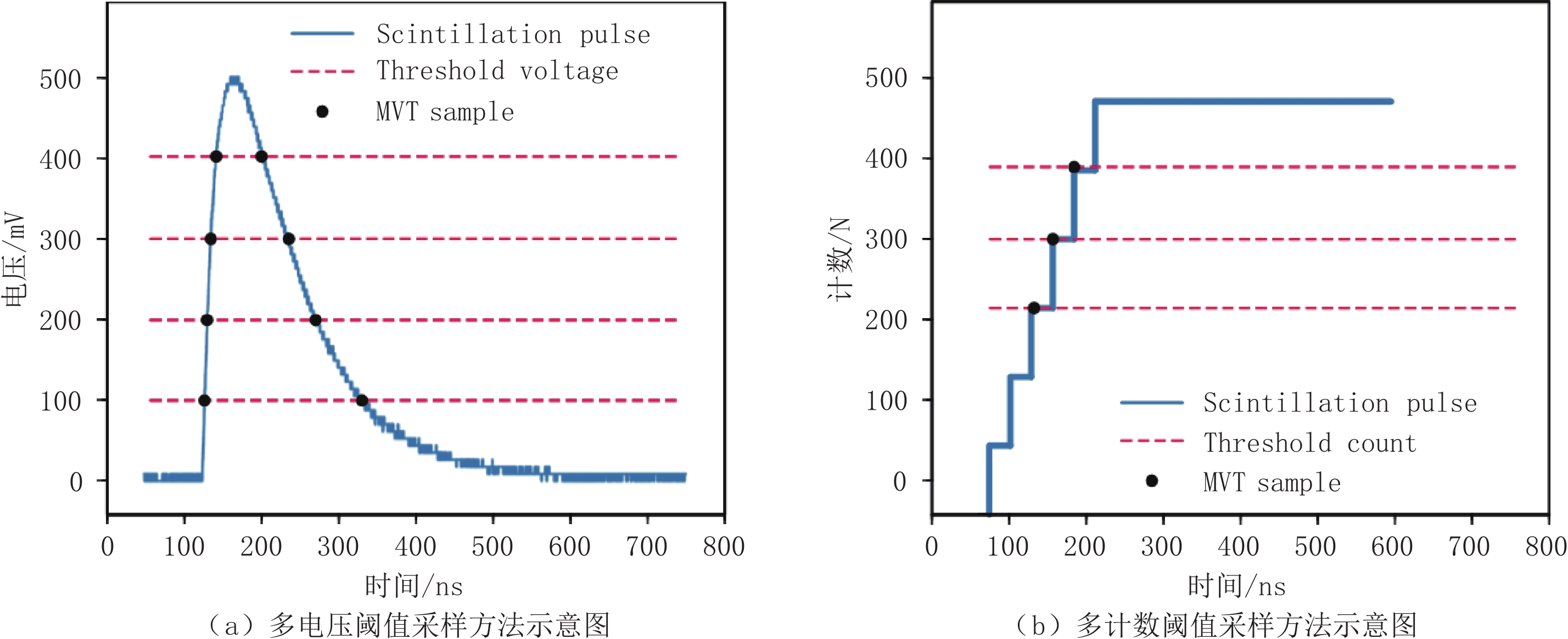
[57] XIE Q, KAO C M, HSIAU Z, et al. A new approach for pulse processing in positron emission tomography[J]. IEEE Transactions on Nuclear Science, 2005, 52(4): 988−995. DOI: 10.1109/TNS.2005.852966.
[58] KIM H, KAO C M, XIE Q, et al. A multi-threshold sampling method for TOF-PET signal processing[J]. Nuclear Instruments and Methods in Physics Research Section A: Accelerators, Spectrometers, Detectors and Associated Equipment, 2009, 602(2): 618-621.
-
期刊类型引用(6)
1. 李新经,袁珊. CA199和HE4及VEGF在卵巢交界性和恶性上皮性肿瘤中的表达及诊断价值. 医药论坛杂志. 2025(02): 211-215 .  百度学术
百度学术
2. 王丽雯,李静芳,李晓红,余鑫. 血清miR-200c表达水平与复发性卵巢癌的相关性及临床意义. 中国性科学. 2024(03): 58-61 . 百度学术
3. 岳红 ,汤进 ,陈钢 . 血清ALP、HE4联合SII指数与卵巢癌肿瘤良恶性程度相关性及预后影响因素分析. 临床和实验医学杂志. 2024(22): 2412-2416 . 百度学术
4. 张建泉,符纪宁,蔡淑华,沈智勇. 卵巢癌MRI表现与Ki-67表达及预后的关系. 中国CT和MRI杂志. 2023(06): 130-132 . 百度学术
5. 王丽雯,李静芳,李晓红,余鑫,杨伟,高艳章. 上皮性卵巢癌患者血清miR-200a、miR-200b水平变化及意义. 山东医药. 2023(26): 58-61 . 百度学术
6. 谢敏霞,林琳. HE4与CA125联合检测在妇科肿瘤诊断中的作用评价. 深圳中西医结合杂志. 2023(15): 64-67 . 百度学术
其他类型引用(0)

 下载:
下载:


计量
- 文章访问数: 523
- HTML全文浏览量: 71
- PDF下载量: 109
- 被引次数: 6


