
Analysis of the Value of GSI Scanning Combined with “Double-low” Technology in Head and Neck CTA in Middle-aged and Elderly Individuals
-
摘要:
目的:探讨头颈部CTA中GSI扫描联合“双低”技术在中老年人群体的应用价值。材料和方法:回顾性收集2023年2月至12月行头颈CTA检查患者的影像资料150例,根据不同的扫描方式分为A、B和C 3组,各50例。A组和B组均为常规螺旋扫描,管电压分别为120 kV和100 kV,均为管电流自动调制300~500 mA,对比剂用量及流速均为固定55 mL和4.5 mL/s,盐水以与对比剂相同用量及流速进行注射。C组能谱扫描,平扫组管电压为80 kV,增强组管电压为80~140 kV瞬时切换,管电流均为恒定的320 mA,采用个性化对比剂注射方案。对3组影像资料进行主观评价、客观评价及比较对比剂用量、平均注射速率。结果:B组头颈部各血管平均CT值大于A组和C组,差异具有统计学意义;主动脉弓层面,B组SNR及CNR参数优于A组和C组,差异具有统计学意义;其余血管层面C组SNR、CNR、SD参数均优于A组和B组,除颈总动脉层面,差异具有统计学意义;C组DLP相较于A组和B组分别降低53.9%和27.6%,对比剂用量降低27.2%。C组对比剂平均注射速率为(3.9±0.5)mL/s低于A组和B组固定流速4.5 mL/s。结论:在中老年人群体中,以GSI扫描模式联合“双低”技术行头颈CTA可有效降低辐射剂量、减少对比剂用量及改善图像质量。
Abstract:Objective: This study aimed to investigate the value of gemstone spectral imaging (GSI) scanning combined with "double-low" technology in head and neck computed tomography angiography (CTA) in middle-aged and elderly individuals. Materials and Methods: One hundred fifty patients who underwent head and neck CTA from February to December 2023 were retrospectively collected and divided into three groups, comprising 50 patients each, according to different scanning modalities; A, B, and C. Groups A and B underwent conventional spiral scans, with tube voltages of 120 kV and 100 kV, respectively, and tube currents of 300~500 mA were automatically modulated, and the dose and flow rate of the contrast agent were fixed at 55 mL, 4.5 mL/s, saline with the same dose and flow rate for flushing and group C energy spectrum scanning, the tube voltage of the flat scanning group was 80 kV, the tube voltage of the enhancement group was 80~140 kV instantaneous switching, and the tube current was a constant 320 mA in all cases, with a personalized contrast agent injection scheme. The subjective evaluation, objective evaluation, and comparison of contrast use dose were performed on the imaging data of all groups. Results: The mean CT values of head and neck vessels in group B were greater than those in groups A and C, with statistically significant differences. At the aortic arch level, the SNR and CNR parameters in group B were better than those in groups A and C, with statistically significant differences. At the level of the remaining blood vessels, the SNR, CNR, and SD parameters of group C were better than those in groups A and B, excluding the common carotid artery level, with statistically significant differences. The radiation dose in group C was 53.9% and 27.6% lower than that in groups A and B, respectively, and the contrast dose was 27.2% lower. Conclusion: In middle-aged and elderly individuals, head and neck CTA with GSI scanning combined with “double-low” technique can effectively reduce radiation and contrast doses and improve image quality.
-
根据中国心血管健康与疾病报告指出2003年至2020年脑血管疾病死亡率整体呈增长趋势[1]。其中脑卒中已经成为我国成人致死致残的首位原因,且脑卒中年轻化趋势明显,40~64岁人群标化后占比为71.80%[2],因此对于中老年人群体脑血管疾病的早期诊断十分关键。
头颈CTA是目前诊断头颈部血管病变、观察血管解剖和血管病变以外疾病血供来源的重要影像方法[3]。但其存在CT电离辐射具有引起DNA突变和损伤,导致癌症的潜在可能性[4],所以辐射剂量的降低尤为重要。除此之外,碘对比剂的使用也是热门话题,大剂量地使用高浓度对比剂会增加对比剂肾病(contrast inducted nephropathy,CIN)的发生概率[5]。
王丽敏等[6]发现我国老年人多重慢性病的患病率为13%~98%,这表明中老年人群体普遍存在身体机能下降等问题,因此更易诱发CIN。而能谱CT扫描具有检查速度快、成像清晰与辐射剂量低等优势[7],可有效减少辐射损伤。马光明等[8]及徐军等[9]都已证实GSI扫描联合“双低”技术在头颈部CTA中应用的可行性和优越性。
在本领域中对于中老年群体行头颈CTA扫描方案进行特定优化及改善的研究较少且临床中为确保影像图像质量,“双低”技术的实际应用较少,但对于中老年人群体的影像检查具有推广的必要性。本研究旨在中老年人群体中,以GSI扫描联合“双低”技术行头颈CTA检查与常规头颈CTA扫描进行系统比较并探讨其具体的应用价值及阐述推广的必要性。
1. 资料与方法
1.1 一般资料
搜集2023年2月至2023年12月于中国科学技术大学附属第一医院行头颈CTA检查的患者,按照扫描方式的不同分为3组,分别为A组120 kV螺旋扫描、B组100 kV螺旋扫描与C组80~140 kV瞬时切换能谱扫描,每组50例。
纳入标准为:①明确行头颈CTA患者;②年龄≥45岁;③BMI指数 < 27 kg/m2。排除标准为:①碘过敏史者;②甲状腺功能亢进者;③具有心衰、肝肾功能不全者;④无法配合行头颈CTA检查者。本研究已得到医院伦理委员会审理通过。
1.2 设备与方法
所有患者检查均在GE revolution CT行头颈CTA双期扫描检查,扫描范围从气管隆突至颅顶,平扫组与增强组扫描范围一致。
A组平扫参数:管电压120 kV,管电流自动调制300~500 mA,螺距为0.992∶1,扫描层厚为0.625 mm,机架旋转速度为0.5 s/r,噪声指数为11.0。增强扫描参数与平扫相同;B组仅将管电压改为100 kV,其余参数与A组一致;C组采用低剂量扫描,平扫参数:管电压为80 kV,管电流恒定320 mA,其余与A组平扫参数相同。增强扫描参数:管电压改为80~140 kV瞬时切换,管电流恒定320 mA。
3组对比剂均采用浓度为350 mgI/mL碘海醇。A组和B组对比剂用量和盐水用量均为固定55 mL,流速为4.5 mL/s;C组采用个性化低对比剂注射方案,每位患者对比剂具体用量为0.6 mL/kg,对比剂流速为对比剂用量/10 s,盐水用量与对比剂一致,流速为1.2倍对比剂流速。3组患者均优先从右手肘正中静脉接针,注入对比剂,采用团注追踪触发法,增强组的监测层面与扫描起始层面气管隆突出保持一致,并圈画圆形感兴趣区(region of interest ROI),并将其置于降主动脉中心,触发阈值设置为100 HU。
1.3 图像后处理
扫描结束后,3组图像进行重建及减影。A组和B组平扫及增强组均重建层厚为0.625 mm,自适应统计迭代算法(adaptive statistical iterative reconstruction,ASIR)为50%图像。C组平扫组重建层厚为0.625 mm,ASIR为50%图像,增强组重建层厚为0.625 mm,ASIR为60%,65 keV的单能量图像[10]。
最后将图像传入AW4.7工作站进行后处理,得到多平面重组、最大密度投影、曲面重建及容积再现三维图像。
1.4 图像质量的主观与客观评价
1.4.1 主观评价
采用双盲法,由两名工作经验为5年以上的影像诊断医师采用4分制[11]对各分支的图像质量进行主观评分。1分:颅内及颈部动脉显示不清,无法诊断;2分:颅内及颈部动脉轮廓显影模糊,与背景图像分界不清,低于诊断标准;3分:颅内及颈部动脉轮廓显影清晰,诊断可靠度较高;4分:颅内及颈部动脉轮廓显影锐利,细节显示清晰。1~2分为图像不能满足诊断需要,3~4分为图像质量较好,可满足诊断需求。
对上腔静脉对比剂滞留情况采用3分制进行评分,评分标准:3分,上腔静脉血管显示良好,无对比剂滞留伪影;2分,上腔静脉显示较好,有少许对比剂滞留伪影,但不影响诊断准确性;1分,上腔静脉有对比剂滞留伪影,影响诊断准确性[12]。
1.4.2 客观评价
在主动脉弓、左颈总动脉、左颈内动脉及左大脑中动脉的血管中圈画圆形ROI,面积覆盖2/3血管,避开钙化斑块及血管壁,记录血管CT值以及图像噪声值(standard deviation,SD)。并在相同的血管层面,选择相对应的肌肉,放置大小相同的ROI测取背景信号。其肌肉的选取,肩部层面为胸大肌,颈部层面为胸锁乳突肌,头部层面为颞肌。每一层面血管CT值、SD值及背景信号各测量3次,取平均值。计算得出信噪比(signal-to-noise ratio,SNR)以及对比信噪比(contrast-to-noise ratio,CNR),计算公式分别为:
$$ {\mathrm{SNR}}=血管平均{\mathrm{CT}}值/血管平均{\mathrm{SD}}值 \text{,} $$ (1) $$ \mathrm{CNR}=\; \frac{\left(血管平均\mathrm{CT}值-肌肉平均\mathrm{CT}值\right)}{血管平均\mathrm{SD}值}。 $$ (2) 在计算机的剂量报告中,获取患者的剂量长度乘积(dose length product,DLP),以及CT容积剂量指数(CT dose index volume,CTDIvol),并计算有效剂量(effective dose,ED),计算公式为
$$ {\mathrm{ED}} = {\mathrm{DLP}}{\text{ }}\times{\text{ }}{{C}} \text{,} $$ (3) 其中C为换算因子。在头颈部,C=
0.0031 (mSv/mGy·cm)[13]。1.5 统计学分析
使用SPSS 26.0软件对3组患者的一般患者资料、辐射剂量参数和图像质量参数进行统计学分析。计量资料以(均数±标准差)表示,计数资料以频数和比例表示。对于3组图像质量主观评分使用克鲁斯卡尔-沃利斯(Kruskal-Wallis,K-W)检验分析,对于两位诊断医师对图像质量评分的一致则使用kappa检验进行评估,当k < 0.2,差;0.2≤k < 0.4,一般;0.4≤k < 0.6,中等;0.6≤k < 0.8,较强;0.8≤k < 1,优异。
将3组数据进行正态性检验,若满足正态分布采用单因素方差分析(one-way analysis of variance,One-way ANOVA),组间两两比较采用最小显著性差异法(least-significant difference,LSD)。若不满足正态分布,使用K-W检验,并使用邦费罗尼校正(Bonferroni correction)进行组间比较。P<0.05为差异有统计学意义。
2. 结果
2.1 患者一般资料及主观评价
患者性别、年龄及BMI指数无统计学差异(表1)。A组主观评分为(3.08±0.274),B组为(3.10±0.303),C组为(3.12±0.328),无统计学差异,均满足临床诊断。两名影像诊断医师对3组图像质量评分的一致性较强,Kappa值为0.811。
表 1 患者的一般资料Table 1. Patients’ general information项目 组别 统计检验 A组(n=50) B组(n=50) C组(n=50) $F/\chi^2 $ P 性别 34∶16 32∶18 27∶23 $\chi^2 $=2.207 0.332 年龄 61.02±10.25 65.46±9.45 62.90±9.29 F=2.655 0.074 BMI 23.80±1.84 23.28±1.39 23.53±1.63 F=1.285 0.280 上腔静脉对比剂滞留评分A组(2.04±0.75)、B组(2.04±0.67)、C组主观评分为(2.6±0.57),差异具有统计学意义,且Kappa值为0.788,两名影像诊断医师对上腔静脉对比剂滞留情况的评分一致性较强。
2.2 客观评价
实验中,C组辐射剂量参数DLP(497.04±26.84)、CTDIvol(5.65±0.00)及ED(1.54±0.9)均低于其他两组(表2)。3组图像中头颈各血管平均CT值,B组高于A组和C组,差异具有统计学意义;在大脑中动脉、颈内动脉颈及颈总动脉层面,C组SNR、CNR及SD值均优于A组和B组,除颈总动脉层面,差异具有统计学意义。而在主动脉弓层面,C组图像质量参数较差,差异具有统计学意义(表3)。
表 2 DLP、CTDIvol及ED的比较Table 2. Comparison of DLP, CTDIvol, and ED项目 组别 统计检验 A组 B组 C组 F P DLP/(mGy·cm) 1078.40 ±70.47686.51±40.10 497.04±26.84 1808.23 0.000 CTDIvol/mGy 13.08±0.43 8.43±0.16 5.65±0.00 9977.39 0.000 ED/mSv 3.34±0.22 2.13±0.12 1.54±0.90 1801.38 0.000 表 3 客观评价Table 3. Objective evaluation项目 组别 统计检验 A组 B组 C组 F P 血管平均CT值/HU 左侧大脑中动脉 409.79±52.93 480.63±55.47 403.15±55.98 30.697 0.000 左侧颈内动脉 415.92±56.54 490.26±56.59 419.81±53.47 28.370 0.000 左侧颈总动脉 425.56±57.97 497.53±57.60 427.66±60.15 24.439 0.000 主动脉弓动脉 408.15±46.07 483.11±52.22 429.49±54.44 28.640 0.000 SD/HU 左侧大脑中动脉 14.23±1.57 15.90±1.49 12.67±1.79 45.599 0.000 左侧颈内动脉 14.34±2.07 15.80±1.82 12.37±1.53 44.313 0.000 左侧颈总动脉 12.68±1.94 14.51±1.34 12.42±1.54 24.489 0.000 主动脉弓动脉 24.64±4.96 27.29±4.36 25.10±4.01 8.231 0.000 SNR 左侧大脑中动脉 29.11±4.78 30.45±4.32 32.24±5.24 5.349 0.006 左侧颈内动脉 29.51±5.34 31.41±5.10 34.57±5.62 11.384 0.000 左侧颈总动脉 34.11±5.84 34.52±4.90 34.76±5.47 0.180 0.835 主动脉弓动脉 17.31±4.37 18.16±3.58 15.58±2.93 6.393 0.002 CNR 左侧大脑中动脉 24.64±4.52 26.54±3.99 27.11±4.68 4.304 0.015 左侧颈内动脉 25.29±5.07 27.53±4.74 29.17±5.38 7.399 0.001 左侧颈总动脉 29.16±5.55 30.20±4.66 30.38±5.03 0.523 0.594 主动脉弓动脉 14.97±3.97 16.07±3.36 13.72±2.88 5.805 0.004 主观评分 3.06±0.37 3.12±0.44 3.14±0.41 0.528 0.591 2.3 对比剂的使用
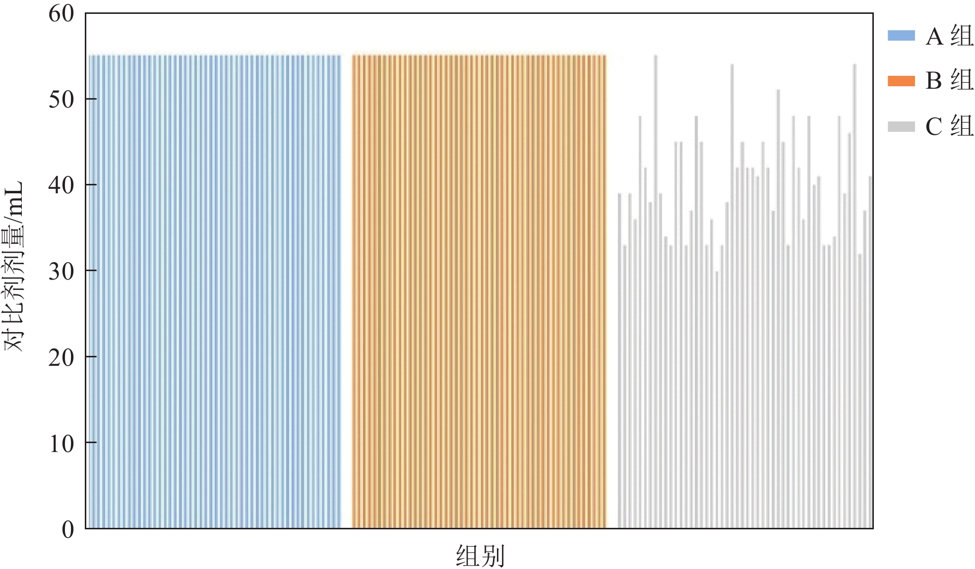
与A组和B两组采用的固定对比剂注射方案相比,C组采用基于体重的低用量个性化对比剂注射方案,其使用的对比剂剂量较A组和B组减少27.2%(图1)。
3. 讨论
头颈CTA由于无创性、检查迅速和图像直观等优点[14],其重要价值得到了普遍认可。尤其是在老龄化进程日益加快的如今,脑血管疾病又好发于中老年人群体,头颈CTA的临床应用更是广泛。但头颈CTA存在的电离辐射和CIN风险不可忽视。
能谱CT的宝石探测器具有更快的初始响应速度以及更好地稳定性,减少了余晖效应[15],提高了X线利用效率,降低了辐射剂量。降低管电压和管电流也是减少辐射剂量的方法之一[16]。以往的研究中,通常采取低管电压扫描来降低扫描辐射剂量。
在本研究中由于平扫组仅用于减影,对于图像质量并无影响。因此将C组的平扫管电压调整为80 kV,管电流调整为恒定320 mA,从而降低辐射剂量。C组辐射剂量参数DLP较A组降低53.9%、较B组降低27.6%。但过低的管电压和管电流会导致X线的穿透力降低,从而增加图像噪声,降低图像质量。因此本研究的管电流没有一味的进行降低。
在进行具体实验前,选取不同管电流进行扫描,均衡比较不同管电流扫描条件下影像图像的辐射剂量参数及图像参数后,最终选定管电流为320 mA。C组在主动脉弓层面SNR、CNR及SD参数略差于A组和B组。这是因为主动脉弓层面骨性结构更多且C组的低管电流扫描导致X射线量减少,从而增加图像噪声。而能谱CT联合ASIR技术可以有效抑制硬化伪影和降低图像噪声,从而改善图像质量[17]。Johnson等[18]发现单能量60 keV左右时能提高血管和周围组织的信噪比,因此本研究选择65 keV单能量图像。
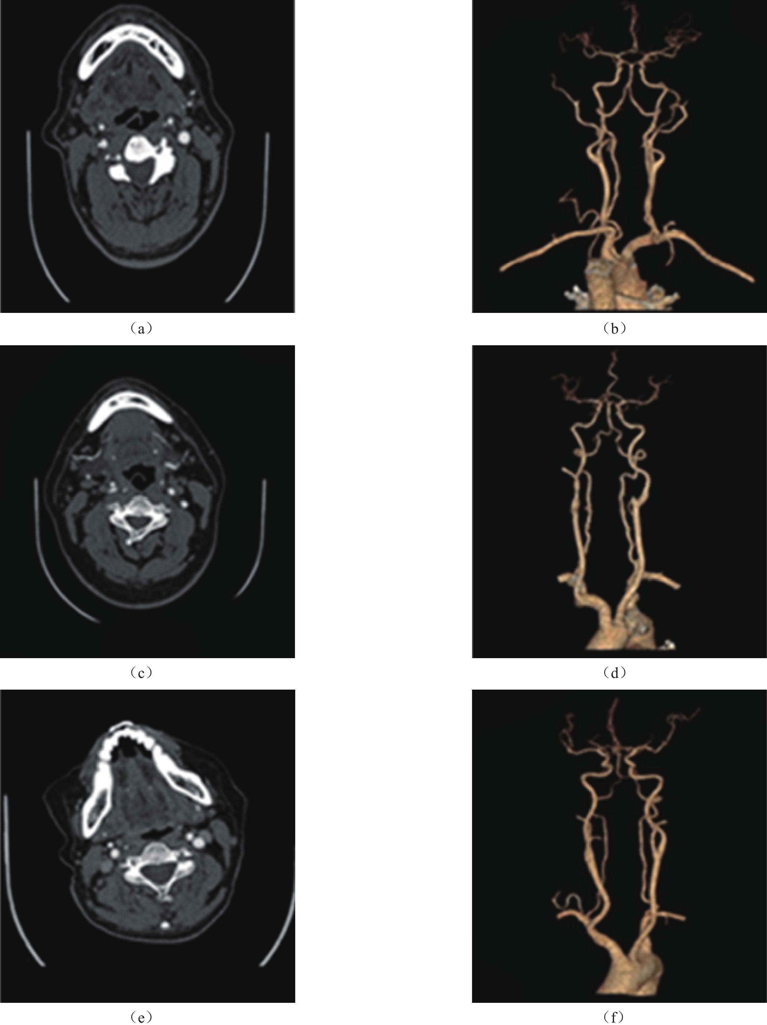
由表2所示,C组在除主动脉弓血管层面的平均SD值(12.67±1.79)、(12.37±1.53)、(12.42±1.54)、SNR(32.24±5.24)、(34.57±5.62)、(34.76±5.47)和CNR(27.11±4.68)、(29.17±5.38)、(30.38±5.03)参数均优于A组和B组,且除颈总动脉血管层面,差异具有统计学意义。在图2中,也能直接观察到C组经能谱扫描后得到的CTA图像,后重建的血管VR图,图像质量更好。
![]() 图 2 3组血管轴位图及 VR 图注:(a)颈内动脉轴位图像、(b)VR 图像。患者年龄 51 岁,采用管电压 120 kV,血管 CT 值为 401.90 HU、SD 值为 12.4、SNR 为 29.99、CNR 为 25.03,对比剂用量为 55 mL、主观评分为 3 分。(c)颈内动脉轴位图像、(d)VR 图像。患者年龄 73 岁,采用管电压 100 kV,血管 CT 值为 460.40 HU、SD 为 17.6、SNR 为 26.16、CNR 为 22.08,对比剂用量为 55 mL、主观评分为 3分。(e)颈内动脉轴位图像、(f)VR 图像。患者年龄 51 岁,采用 80/140 kVp,血管 CT 值为 433.50 HU、SD 为 11.7、SNR 为 37.05、CNR 为 31.05,对比剂用量为 33 mL、主观评分为 4 分。其中 100 kV 组 CT 值最高,80/140 kVp 组图像质量参数均优于其他两组,差异具有统计学意义,且 100 kV 及 120 kV 组 VR 图像均有钙化未去除。Figure 2. Axial and VR maps of three groups of vessels
图 2 3组血管轴位图及 VR 图注:(a)颈内动脉轴位图像、(b)VR 图像。患者年龄 51 岁,采用管电压 120 kV,血管 CT 值为 401.90 HU、SD 值为 12.4、SNR 为 29.99、CNR 为 25.03,对比剂用量为 55 mL、主观评分为 3 分。(c)颈内动脉轴位图像、(d)VR 图像。患者年龄 73 岁,采用管电压 100 kV,血管 CT 值为 460.40 HU、SD 为 17.6、SNR 为 26.16、CNR 为 22.08,对比剂用量为 55 mL、主观评分为 3分。(e)颈内动脉轴位图像、(f)VR 图像。患者年龄 51 岁,采用 80/140 kVp,血管 CT 值为 433.50 HU、SD 为 11.7、SNR 为 37.05、CNR 为 31.05,对比剂用量为 33 mL、主观评分为 4 分。其中 100 kV 组 CT 值最高,80/140 kVp 组图像质量参数均优于其他两组,差异具有统计学意义,且 100 kV 及 120 kV 组 VR 图像均有钙化未去除。Figure 2. Axial and VR maps of three groups of vessels对于上腔静脉对比剂滞留评分,C组主观评分为(2.6±0.57),高于A组(2.04±0.75)、B组(2.04±0.67),差异具有统计学意义。这是由于A组和B组的对比剂用量更大,流速更快,对比剂更易在上腔静脉积聚,从而更易形成硬束伪影[19]。而在C组中采用低剂量、低流速的个性化对比剂注射方案,并且将盐水速率设置为1.2倍对比剂注射速率进行盐水冲刷从而进一步降低上腔静脉对比剂伪影的产生,A组和B组在4.5 mL/s的流速上进一步提高流速会增加中老年人对比剂外渗风险,因此并未使用高速率的盐水冲刷技术。当C组出现严重的上腔静脉对比剂伪影时,能谱扫描模式仍可重建其它单能量图像,改善硬束伪影。
碘对比剂是CT增强检查中常用药物,然而大剂量使用对比剂增加不良反应和CIN的风险,发生概率与对比剂用量成正比。本研究C组采用个性化低对比剂注射方案,与A组和B组相比对比剂用量降低27.2%。C组在减少对比剂用量与注射时间恒定为10 s的条件下,C组流速为(3.84±0.447)mL/s与A组和B组固定流速4.5 mL/s相比,实现了对比剂的低流速注入,保护了患者血管,降低对比剂外渗风险。
随着年龄的增长,机体的生理状态随之发生改变。一方面细胞的老化和衰老,导致细胞修复能力的降低。而电离辐射对于DNA和染色体的初始伤害可能会引起细胞损伤,甚至死亡。中老年人群体更难修复辐射损伤,从而可能致使细胞向恶性肿瘤转化,并最终导致癌症[20-21]。另一方面在衰老的过程中,肾血浆流量减少和肾单位数降低[22-23]等因素均会引起肾小球滤过率降低,根据Glassock等[24]研究表明35岁之后,GFR每10年下降约5%~10%,对比剂在体内滞留时间更长,从而增加CIN风险。Song等[25]研究表明老年患者发生由对比剂引起的急性肾损伤几率更大,而65岁患者总发病率高达13.6%。
在临床检查中,对于婴幼儿群体的CT检查总会进行防护措施及扫描方案的调整,而对中老年人群体的CT扫描并没有过多改变。但由于中年老年群体独特的机体生理状态,CT增强检查的扫描方案应采取合理的优化。例如随着年龄的增长,人体脂质周转率的下降,中老年人群体的脂肪含量更高[26],而碘对比剂不被脂肪组织吸收。在肝脏的增强CT检查中,根据去脂体重计算对比剂用量,对比剂可进行合理减少且影像图像符合临床诊断要求[27]。影像精准化地实现不仅仅在于硬件设备的提升,更在于根据目标人群调整扫描参数,选择适合的扫描方案。
本研究存在一定的局限性。①本研究的影像图像没有与“金标准”DSA进行联合,使影像结果得到更为精准的评估;②本研究未纳入BMI>27 kg/m2的患者,对于肥胖体型患者应选择更大的管电流来确保图像质量。
综上所述,在中老年人群体中以GSI扫描联合“双低”技术行头颈CTA可以有效降低辐射剂量、对比剂用量及改善图像质量。临床上,对于BMI指数小于27 kg/m2的中老年人群体的患者行头颈CTA检查,值得推广应用。
-
![]()
图 2 3组血管轴位图及 VR 图
注:(a)颈内动脉轴位图像、(b)VR 图像。患者年龄 51 岁,采用管电压 120 kV,血管 CT 值为 401.90 HU、SD 值为 12.4、SNR 为 29.99、CNR 为 25.03,对比剂用量为 55 mL、主观评分为 3 分。(c)颈内动脉轴位图像、(d)VR 图像。患者年龄 73 岁,采用管电压 100 kV,血管 CT 值为 460.40 HU、SD 为 17.6、SNR 为 26.16、CNR 为 22.08,对比剂用量为 55 mL、主观评分为 3分。(e)颈内动脉轴位图像、(f)VR 图像。患者年龄 51 岁,采用 80/140 kVp,血管 CT 值为 433.50 HU、SD 为 11.7、SNR 为 37.05、CNR 为 31.05,对比剂用量为 33 mL、主观评分为 4 分。其中 100 kV 组 CT 值最高,80/140 kVp 组图像质量参数均优于其他两组,差异具有统计学意义,且 100 kV 及 120 kV 组 VR 图像均有钙化未去除。
Figure 2. Axial and VR maps of three groups of vessels
表 1 患者的一般资料
Table 1 Patients’ general information
项目 组别 统计检验 A组(n=50) B组(n=50) C组(n=50) $F/\chi^2 $ P 性别 34∶16 32∶18 27∶23 $\chi^2 $=2.207 0.332 年龄 61.02±10.25 65.46±9.45 62.90±9.29 F=2.655 0.074 BMI 23.80±1.84 23.28±1.39 23.53±1.63 F=1.285 0.280  下载: 导出CSV
表 2 DLP、CTDIvol及ED的比较
Table 2 Comparison of DLP, CTDIvol, and ED
项目 组别 统计检验 A组 B组 C组 F P DLP/(mGy·cm) 1078.40 ±70.47686.51±40.10 497.04±26.84 1808.23 0.000 CTDIvol/mGy 13.08±0.43 8.43±0.16 5.65±0.00 9977.39 0.000 ED/mSv 3.34±0.22 2.13±0.12 1.54±0.90 1801.38 0.000
下载: 导出CSV
表 3 客观评价
Table 3 Objective evaluation
项目 组别 统计检验 A组 B组 C组 F P 血管平均CT值/HU 左侧大脑中动脉 409.79±52.93 480.63±55.47 403.15±55.98 30.697 0.000 左侧颈内动脉 415.92±56.54 490.26±56.59 419.81±53.47 28.370 0.000 左侧颈总动脉 425.56±57.97 497.53±57.60 427.66±60.15 24.439 0.000 主动脉弓动脉 408.15±46.07 483.11±52.22 429.49±54.44 28.640 0.000 SD/HU 左侧大脑中动脉 14.23±1.57 15.90±1.49 12.67±1.79 45.599 0.000 左侧颈内动脉 14.34±2.07 15.80±1.82 12.37±1.53 44.313 0.000 左侧颈总动脉 12.68±1.94 14.51±1.34 12.42±1.54 24.489 0.000 主动脉弓动脉 24.64±4.96 27.29±4.36 25.10±4.01 8.231 0.000 SNR 左侧大脑中动脉 29.11±4.78 30.45±4.32 32.24±5.24 5.349 0.006 左侧颈内动脉 29.51±5.34 31.41±5.10 34.57±5.62 11.384 0.000 左侧颈总动脉 34.11±5.84 34.52±4.90 34.76±5.47 0.180 0.835 主动脉弓动脉 17.31±4.37 18.16±3.58 15.58±2.93 6.393 0.002 CNR 左侧大脑中动脉 24.64±4.52 26.54±3.99 27.11±4.68 4.304 0.015 左侧颈内动脉 25.29±5.07 27.53±4.74 29.17±5.38 7.399 0.001 左侧颈总动脉 29.16±5.55 30.20±4.66 30.38±5.03 0.523 0.594 主动脉弓动脉 14.97±3.97 16.07±3.36 13.72±2.88 5.805 0.004 主观评分 3.06±0.37 3.12±0.44 3.14±0.41 0.528 0.591
下载: 导出CSV
-
[1] 中国心血管健康与疾病报告编写组. 《中国心血管健康与疾病报告2022》概要[J]. 中国介入心脏病学杂志, 2023, 31(7): 485-508. DOI: 10.3969/j.issn.1004-8812.2023.07.002. [2] 中国心血管健康与疾病报告编写组. 《中国脑卒中防治报告2021》概要[J]. 中国脑血管病杂志, 2023, 20(11): 783-793. [3] 中华医学会放射学分会. 头颈部CT血管成像扫描方案与注射方案专家共识[J]. 中华放射学杂志, 2019, 53(2): 81-87. DOI: 10.3760/cma.j.issn.1005-1201.2019.02.001. Chinese Society of Radiology, Chinese Medical Association. Expert consensus of the head and neck CT angiography scanning and injection protocols[J]. Chinese Journal of Radiology, 2019, 53(2): 81-87. DOI: 10.3760/cma.j.issn.1005-1201.2019.02.001. (in Chinese).
[4] JI K, WANG Y, DU L, et al. Research progress on the biological effects of low-dose radiation in China[J]. Dose Response, 2019, 17(1): 1559325819833488. DOI: 10.1177/1559325819833488.
[5] 李飞. 低管电压低剂量低浓度造影剂在增强CT检查中预防造影剂肾病的临床研究[J]. 中国CT和MRI杂志, 2021, 19(3): 92-94. DOI: 10.3969/j.issn.1672-5131.2021.03.031. LI F. Low-dose low-concentration contrast agent combined with low tube voltage in renal CT enhancement examination[J]. Chinese Journal of CT and MRI, 2021, 19(3): 92-94. DOI: 10.3969/j.issn.1672-5131.2021.03.031. (in Chinese).
[6] 王丽敏, 陈志华, 张梅, 等. 中国老年人群慢性病患病状况和疾病负担研究[J]. 中华流行病学杂志, 2019, 40(3): 277-283. DOI: 10.3760/cma.j.issn.0254-6450.2019.03.005. WANG L M, CHEN Z H, ZAHNG M, et al. Study of the prevalence and disease burden of chronic disease in the elderly in China[J]. Chinese Journal of Epidemiology, 2019, 40(3): 277-283. DOI: 10.3760/cma.j.issn.0254-6450.2019.03.005. (in Chinese).
[7] 孙亮. 宝石能谱CT多参数成像检查在卵巢肿瘤良恶性鉴别诊断中的应用价值[J]. 中国民康医学, 2023, 35(10): 134-136. DOI: 10.3969/j.issn.1672-0369.2023.10.040. SUN L. Application value of gemstone spectral CT multi-parameter imaging in differential diagnosis of benign and malignant ovarian neoplasms[J]. Medical Journal of Chinese People’s Health, 2023, 35(10): 134-136. DOI: 10.3969/j.issn.1672-0369.2023.10.040. (in Chinese).
[8] 马光明, 陈静, 韩冬, 等. 能谱CT双低减影法在头颈CTA成像中的应用[J]. 临床放射学杂志, 2020, 39(5): 1009-1013. DOI: 10.13437/j.cnki.jcr.2020.05.037. MA G M, CHEN J, HAN D, et al. Subtraction artery angiography in head and neck with low radiation and contrast dose dual-energy spectral CT[J]. Journal of Clinical Radiology, 2020, 39(5): 1009-1013. DOI: 10.13437/j.cnki.jcr.2020.05.037. (in Chinese).
[9] 徐军, 胡孝梨, 罗昆, 等. “双低”扫描联合个性化对比剂注射方案在颅脑CTA中的应用价值[J]. 临床放射学杂志, 2022, 41(12): 2301-2305. DOI: 10.13437/j.cnki.jcr.2022.12.027. XU J, HU X L, LUO K, et al. The application value of“double low”scanning combined with personalized contrast injection protocol in cranial CTA[J]. Journal of Clinical Radiology, 2022, 41(12): 2301-2305. DOI: 10.13437/j.cnki.jcr.2022.12.027. (in Chinese).
[10] 王帅, 石磊, 郑敏文, 等. 双低剂量对比剂和最佳单能谱成像在头颈部CTA中的应用价值[J]. 中国医疗设备, 2016, 31(7): 9-11, 15. DOI: 10.3969/j.issn.1674-1633.2016.07.003. WANG S, SHI L, ZHEN M W, et al. Effectiveness of application of double low-dose contrast agent and optimal gemstone spectral imaging in head and neck CT angiography[J]. China Medical Equipment, 2016, 31(7): 9-11, 15. DOI: 10.3969/j.issn.1674-1633.2016.07.003. (in Chinese).
[11] 靖婧, 苗延巍. 优化低剂量一站式全脑CT灌注成像联合CTA扫描方案[J]. 中国医学影像技术, 2018, 34(3): 424-428. DOI: 10.13929/j.1003-3289.201706077. JING J, MIAO Y W. Optimization of scanning protocol in low-dose one-stop CT perfusion imaging combined with CTA of whole brain[J]. Chinese Journal of Medical Imaging Technology, 2018, 34(3): 424-428. DOI: 10.13929/j.1003-3289.201706077. (in Chinese).
[12] 张贺, 徐凯, 王冲, 等. 优化对比剂注射方案联合低剂量扫描模式在头颈部CTA中的应用[J]. 中国CT和MRI杂志, 2023, 21(2): 49-51. DOI: 10.3969/j.issn.1672-5131.2023.02.018. ZHANG H, XU K, WANG C, et al. Application of optimized contrast injection program combined with low dose scanning mode in head and neck CTA[J]. Chinese Journal of CT and MRI, 2023, 21(2): 49-51. DOI: 10.3969/j.issn.1672-5131.2023.02.018. (in Chinese).
[13] CHEN P A, CHEN C W, CHOU C C, et al. Quantitative and qualitative evaluation of low-dose craniocervical computed tomography angiography with a lower tube voltage protocol[J]. Acta Radiologica, 2019, 60(10): 1380-1389. DOI: 10.1177/0284185119825488.
[14] 冯晨, 方玉, 张德川, 等. 头颈CTA达峰时间相关因素的分析研究[J]. CT理论与应用研究, 2021, 30(6): 727-733. DOI: 10.15953/j.1004-4140.2021.30.06.08. FENG C, FANG Y, ZHANG D C, et al. The analysis of factors influencing the peak time of head and neck CTA[J]. CT Theory and Applications, 2021, 30(6): 727-733. DOI: 10.15953/j.1004-4140.2021.30.06.08. (in Chinese).
[15] 赵欣耘, 李曼. 颈动脉CTA过程中应用大螺距扫描配合能谱CT双低减影法的效果及对辐射剂量和对比剂用量的影响[J]. 影像科学与光化学, 2021, 39(3): 375-380. DOI: 10.7517/issn.1674-0475.201103. ZHAO X Y, LI M. Effect of large-pitch scanning combined with energy spectrum CT double-low subtraction in carotid CTA and the influence on the dose of radiation and contrast medium[J]. Imaging Science and Photochemistry, 2021, 39(3): 375-380. DOI: 10.7517/issn.1674-0475.201103. (in Chinese).
[16] LI L L, WANG H, SONG J, et al. A feasibility study of realizing low-dose abdominal CT using deep learning image reconstruction algorithm[J]. Journal of X-ray Science and Technology, 2021, 29(2): 361-372. DOI: 10.3233/XST-200826.
[17] 陆晓军, 罗春材, 齐叶青, 等. 深度学习图像重建算法在胸腹主动脉CTA图像质量中的临床应用研究[J]. 临床放射学杂志, 2022, 41(7): 1359-1364. DOI: 10.13437/j.cnki.jcr.2022.07.028. LU X J, LUN C C, QI Y Q, et al. Clinical application value of deep learning image reconstruction algorithm in improving thoracic and abdominal aorta image quality[J]. Journal of Clinical Radiology, 2022, 41(7): 1359-1364. DOI: 10.13437/j.cnki.jcr.2022.07.028. (in Chinese).
[18] JOHNSON T R, KRAUSS B, SEDLMAIR M, et al. Material differentiation by dual energy CT: Initial experience[J]. European Radiology, 2006, 17(6): 1510-1517. DOI: 10.1007/s00330-006-0517-6.
[19] 顾正山, 马燕, 普晓江, 等. 对比剂注射速率的不同对肺动脉CTA检查中上腔静脉伪影的影响[J]. 当代医药论丛, 2022, 20(5): 89-92. DOI: 10.3969/j.issn.2095-7629.2022.05.028. GU Z S, MA Y, PU X J, et al. Different rate of contrast medium injection on pulmonary artery CTA check upper vena cava artifacts[J]. Contemporary Medicine Symposium, 2022, 20(5): 89-92. DOI: 10.3969/j.issn.2095-7629.2022.05.028. (in Chinese).
[20] CARANTE M P, ALTIERI S, BORTOLUSSI S, et al. Modeling radiation-induced cell death: Role of different levels of DNA damage clustering[J]. Radiation and Environmental Biophysics, 2015, 54(3): 305-316. DOI: 10.1007/s00411-015-0601-x.
[21] BALLARINI F, ALTIERI S, BORTOLUSSI S, et al. The BIANCA model/code of radiation-induced cell death: Application to human cells exposed to different radiation types[J]. Radiation and Environmental Biophysics, 2014, 53(3): 525-533. DOI: 10.1007/s00411-014-0537-6.
[22] FANG Y, GONG A Y, HALLER S T, et al. The ageing kidney: Molecular mechanisms and clinical implications[J]. Ageing Research Reviews, 2020, 63: 101151. DOI: 10.1016/j.arr.2020.101151.
[23] ESPOSITO C, DAL C A. Functional changes in the aging kidney[J]. Journal of Nephrology, 2010, 23 (S15).
[24] DENIC A, GLASSOCK R J, RULE A D. Structural and functional changes with the aging kidney[J]. Advances in Chronic Kidney Disease, 2016, 23(1): 19-28. DOI: 10.1053/j.ackd.2015.08.004.
[25] SONG W, ZHANG T, PU J, et al. Incidence and risk of developing contrast-induced acute kidney injury following intravascular contrast administration in elderly patients[J]. Clinical Interventions in Aging, 2014, (9): 85-93. DOI:10.2147/CIA. S55157.
[26] ARNER P, BERNARD S, APPELSVED L L, et al. Adipose lipid turnover and long-term changes in body weight[J]. Nature Medicine, 2019, 25(9): 1385-1389. DOI: 10.1038/s41591-019-0565-5.
[27] 赵林伟, 李勇, 董国礼, 等. 基于去脂体质量确定肝脏双能量CT对比剂碘剂量研究[J]. 实用放射学杂志, 2022, (12): 2059-2063. DOI: 10.3969/j.issn.1002-1671.2022.12.036. ZHAO L W, LI Y, DONG G L, et al. Determination of optimal contrast media iodine dose based on lean body weight at hepatic dual-energy CT[J]. Journal of Practical Radiology, 2022, (12): 2059-2063. DOI: 10.3969/j.issn.1002-1671.2022.12.036. (in Chinese).


计量
- 文章访问数: 132
- HTML全文浏览量: 35
- PDF下载量: 23


