
Jejunal Diverticulum with Hepatic Portal Gas and Superior Mesenteric Vein Thrombosis: a Clinical Case Study
-
摘要:
总结多层螺旋CT(MSCT)诊断空肠憩室穿孔伴肝门静脉积气、肠系膜上静脉血栓及疗效评价,提高对本病的认识。回顾性分析1例空肠憩室穿孔伴肝门静脉积气、肠系膜上静脉血栓患者的临床资料、影像表现、治疗及预后,并总结分析相关文献。本例的临床表现主要为持续性腹痛、发热。MSCT可见空肠近段多发憩室,其中1个憩室壁破裂并与邻近肠系膜上静脉分支相通致肝门静脉积气,增强CT可见肠系膜上静脉内血栓。手术中可见空肠多发憩室,部分憩室充血水肿及渗出改变;另术中可见肠系膜上静脉内血栓。病理表现为空肠憩室内炎性细胞浸润、肠系膜上静脉内血栓。MSCT能明确诊断空肠憩室穿孔伴肝门静脉积气、肠系膜上静脉血栓,并确切显示憩室穿孔位置。空肠憩室穿孔伴肝门静脉积气、肠系上膜静脉血栓,提示患者病情危重,需积极进行手术治疗,挽救患者生命。
Abstract:This study aimed to summarize the diagnosis of jejunal diverticulum perforation with hepatic portal venous gas (HPVG) and superior mesenteric vein (SMV) thrombosis using multislice spiral computed tomography (MSCT) and evaluate its curative effect to improve the understanding of this disease. The clinical data, imaging manifestations, treatment, and prognosis of a patient with jejunal diverticulum perforation with HPVG and SMV thrombosis were retrospectively analyzed, and relevant literature were summarized and analyzed. Here, the primary clinical manifestations were persistent abdominal pain and fever. MSCT revealed multiple diverticula in the proximal jejunum. A diverticular wall ruptured and connected to the adjacent SMV branch, resulting in HPVG. Enhanced CT showed thrombus in the SMV. Intraoperatively, multiple jejunal diverticula were observed, of which some showed hyperemia, edema, and exudation. An intraoperative thrombus was observed in the SMV. The pathological manifestations include inflammatory cell infiltration in the jejunal diverticulum and thrombus formation in the SMV. MSCT can clearly diagnose jejunal diverticulum perforation with HPVG and SMV thrombosis and show the exact location of the perforation. Jejunal diverticulum perforation with HPVG and SMV thrombosis suggests that the patient’s condition is critical, and active surgical treatment is needed to save the patient’s life.
-
-
![]()
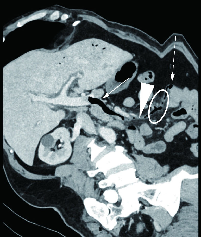
图 1 腹部增强CT示肝门静脉积气及肠系膜上静脉血栓
Figure 1. Abdominal enhanced CT shows hepatic portal vein pneumata and superior mesenteric vein thrombosis
![]()
图 3 CPR重建图像
注:肝门静脉积气(实箭),空肠憩室破口与肠系膜上静脉相通(虚箭)肠系膜上静脉血栓(三角),肠系膜上静脉周围浑浊(环形)。
Figure 3. CPR reconstructed image
![]()
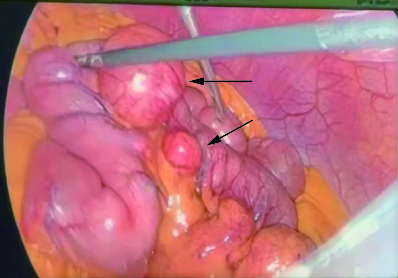
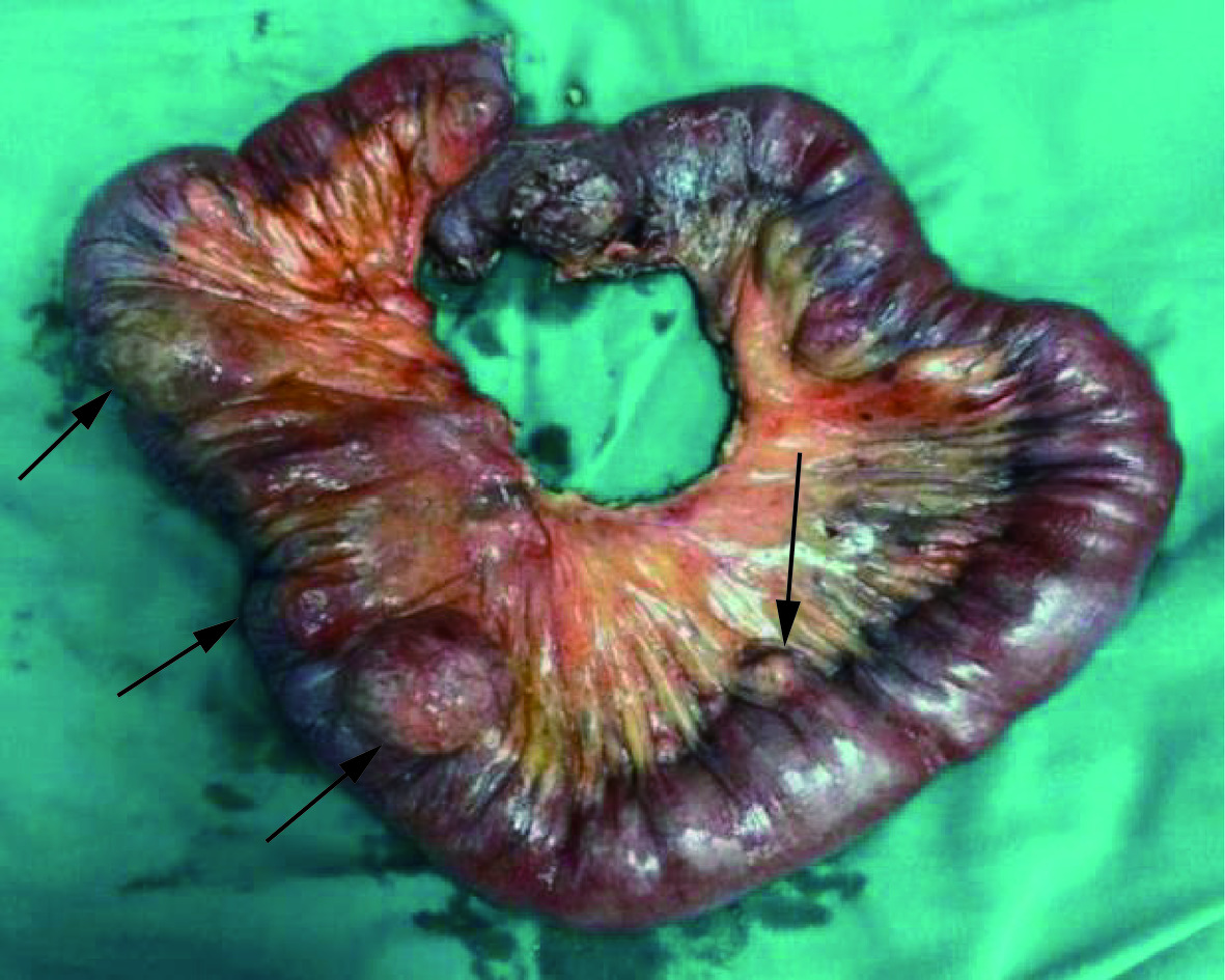
图 4 术中腹腔镜下可见空肠多发憩室(箭)
Figure 4. Multiple jejunal diverticulum is observed under intraoperative laparoscopy (arrow)
-
[1] MAZAHREH T S, ALESHAWI A J, ET AL. Arteriovenous malformations within jejunal diverticulosis: Case report and literature review[J]. BMC Surgery, 2019, 19(1): 70. DOI: 10.1186/s12893-019-0538-0.
[2] HYLAND R, CHALMERS A. CT features of jejunal pathology[J]. Clin Radiol, 2007, 62(12): 1154-1162. DOI: 10.1016/j.crad.2007.05.004.
[3] 黄亮辉, 赵艳平, 刘强. 肝门静脉积气的研究进展[J]. 江西医药, 2019, 54(3): 292-294. DOI: 10.3969/j.issn.1006-2238.2019.3.036. HUANG L H, ZHAO Y P, LIU Q. Research progress on hepatic portal vein gas embolism[J]. Jiangxi Medicine, 2019, 54(3): 292-294. DOI: 10.3969/j.issn.1006-2238.2019.3.036. (in Chinese).
[4] 石爱忠, 王婕妤, 蒋宝国, 等. 肝门静脉积气患者肠坏死与高血压的相关性研究[J]. 临床放射学杂志, 2018, 37(9): 1502-1504. SHI A Z, WANG J Y, JIANG B G, ET AL. Study on the correlation between enteric necrosis and hypertension in patients with hepatic portal vein stasis[J]. Journal of Clinical Radiology, 2018, 37(9): 1502-1504. (in Chinese).
[5] 白杨, 周永坤. 肠系膜上静脉血栓形成致空肠条索状闭锁1例[J]. 疑难病杂志, 2022, 21(06): 644-645. DOI: 10.3969/j.issn.1671-6450.2022.06.019. BAI Y, ZHOU Y K. A case of jejunal atresia due to thrombosis of superior mesenteric vein[J]. Journal of Difficult Diseases, 2022, 21(06): 644-645. DOI: 10.3969/j.issn.1671-6450.2022.06.019. (in Chinese).
[6] 张圣林, 王野, 刘志升. 我国空肠憩室的临床特征[J]. 中国中西医结合外科杂志, 2010, 16(3): 396-397. DOI: 10.3969/j.issn.1007-6948.2010.03.058. ZHANG S L, WANG Y, LIU Z S. Clinical characteristics of jejunal diverticula in China[J]. Chinese Journal of Integrated Chinese and Western Medicine Surgery, 2010, 16(3): 396-397. DOI: 10.3969/j.issn.1007-6948.2010.03.058. (in Chinese).
[7] 张朕, 刘琪, 李通, 等. 小肠巨大憩室1例报道并文献复习[J]. 河南医学研究, 2021, 30(19): 3645-3648. DOI: 10.3969/j.issn.1004-437X.2021.19.066. ZHANG Z, LIU Q, LI T, ET AL. A case report and literature review of giant diverticulum of the small intestine[J]. Henan Medical Research, 2021, 30(19): 3645-3648. DOI: 10.3969/j.issn.1004-437X.2021.19.066. (in Chinese).
[8] LIEBMAN PR, PATTEN MT, MANNY J, ET AL. Hepatic-portal venous gas in adults: Etiology, pathophysiology and clinical signific ance[J]. Annals of Surgery, 1978, 187: 281-287. DOI: 10.1097/00000658-197803000-00012.
[9] KOIZUMI C, MICHIHATA N, ET AL. In-hospital mortality for hepatic portal venous gas: Analysis of 1590 patients using a japanese national inpatient database[J]. World Journal of Surgery, 2018, 42(3): 816-822. DOI: 10.1007/s00268-017-4189-y.
[10] 田慈, 白颐, 马青变, 等. 7例肝门静脉积气的临床特征分析[J]. 北京大学学报(医学版), 2023, 55(4): 743-747. TIAN C, BAI Y, MA Q B, ET AL. Clinical characteristics analysis of 7 cases of hepatic portal gas embolism[J]. Journal of Peking University (Health Sciences), 2023, 55(4): 743-747. (in Chinese).
[11] 刘恩成, 姚远方, 宋磊, 等. 产褥期肠系膜静脉血栓栓塞介入治疗1例[J]. 介入放射学杂志, 2022, 31(1): 32-34. LIU E C, YAO Y F, SONG L, ET AL. A case of mesenteric vein thromboembolism in the puerperium[J]. Journal of Interventional Radiology, 2022, 31(1): 32-3. (in Chinese).
[12] 陈浩, 肖占祥. 急性肠系膜静脉血栓形成的诊疗策略[J]. 中国血管外科杂志(电子版), 2019, 11(1): 7-9. CHEN H, XIAO Z X. Diagnostic and therapeutic strategies for acute mesenteric vein thrombosis[J]. Chinese Journal of Vascular Surgery (Electronic Edition), 2019, 11(1): 7-9. (in Chinese).
[13] 马天翼, 王峰, 金烁, 等. 急性肠系膜缺血早期肠坏死风险因素研究现状[J]. 中国实用外科杂志, 2023, 43(6): 718-720. MA T Y, WANG F, JIN S, et al. Risk factors of early intestinal necrosis in acute mesenteric ischemia [J]. Chinese Journal of Practical Surgery, 2019, 43(6): 718-720. (in Chinese).
[14] EMILE S H, KHAN S M, BARSOUM S H, ET AL. Predictors of bowel necrosis in patients with acute mesenteric ischemia: systematic review and meta-analysis[J]. Updates in Surgery, 2021, 73(1): 47-57. DOI: 10.1007/s13304-020-00857-9.
[15] 赵雪松, 缪飞, 孙菁, 等. CT小肠造影诊断Cronkhite-Canada综合征临床案例分析[J]. CT理论与应用研究, 2023, 32(2): 263-270. DOI: 10.15953/j.ctta.2023.017. ZHAO X S, MIAO F, SUN J, ET AL. The Diagnosis of cronkhite-canada syndrome with CT enterography: A clinical case analysis[J]. CT Theory and Applications, 2023, 32(2): 263-270. DOI: 10.15953/j.ctta.2023.017.
 下载:
下载:


计量
- 文章访问数: 50
- HTML全文浏览量: 14
- PDF下载量: 5


