
Exploring the Influencing Factors of Acute Necrotic Accumulation Outcome
-
摘要:
目的:探究急性坏死性积聚(ANC)转归为包裹性坏死(WON)的独立危险因素及预测效能。方法:回顾性分析53例ANC的CT/MRI特征,根据ANC形成4周后的转归分为WON组与吸收组,采用卡方检验或t检验比较两组在病因、实验室检查方面差异的统计学意义。采用回归法分析影响ANC转归的独立危险因素,并绘制受试者工作特征(ROC)曲线,获得曲线下面积(AUC),评价各危险因素对ANC转归为WON的预测效能。结果:Logistic回归分析显示,坏死体积≥30%与MCTSI>6分的P值均<0.05,OR值分别为9.21、16.04。ROC曲线分析显示,坏死体积≥30%与MCTSI>6分的P值均<0.05,AUC值分别为0.86、0.88。结论:坏死体积≥30%与MCTSI>6分为ANC演变为WON的独立危险因素,且二者预测效能均较显著。
Abstract:ObjectiveTo explore the independent risk factors and predictive efficacy of ANC conversion to encapsulated necrosis (WON). Methods: A Retrospective analysis of CT/MRI features in 53 cases of ANC, divided into the WON and absorption groups, based on the outcome after 4 weeks of ANC formation. The chi square test or t-test were used to compare the statistical significance of differences in etiology and laboratory tests between the two groups. Regression analysis was used to identify independent risk factors affecting the outcome of ANC. Receiver operating characteristic (ROC) curves were used to obtain the area under the curve (AUC) and evaluate the predictive efficacy of each risk factor for the outcome of ANC to WON.
ResultsLogistic regression analysis showed that the P-values for necrotic volume ≥ 30% and MCTSI score > 6 were both < 0.05, with OR values of 9.21 and 16.04, respectively. ROC curve analysis showed that the P-values for necrotic volume ≥ 30% and MCTSI score> 6 were both < 0.05, with AUC values of 0.86 and 0.88, respectively.
ConclusionNecrosis volume ≥ 30% and MCTSI score > 6 points are independent risk factors for the progression of ANC into WON, and the predictive performance of both is significant.
-
Keywords:
- acute necrotic accumulation /
- reversion /
- encapsulated necrosis /
- CT /
- MRI
-
据统计,全球急性胰腺炎(acute pancreatitis,AP)的年发病率为13~45/
100000 [1], 且逐年在上升[2]。目前,AP是患者入院的第二大原因、院内死亡的第五大原因[1]。AP公认的三大病因为胆石症、酗酒及高脂血症[3]。急性坏死性胰腺炎(acute necrotizing pancreatitis,ANP)是指急性胰腺炎伴有胰腺实质和/或胰周组织的坏死。2013年,修订版亚特兰大分类标准(Revised Atlanta classification,RAC)[4]将ANP主要局部并发症分为急性坏死性积聚(acute necrotic collection,ANC)和包裹性坏死( walled-off necrosis,WON)。ANP的预后较差,准确识别ANP并发症是助改善其预后的重要因素之一[5]。ANC不同的转归有不同的预后,WON是ANP患者死亡的危险因素,且WON与ANP不良预后呈正相关[6],因此通过相关因素来早期预测ANC的转归有重要临床意义,对于选择恰当、正确的治疗方法及改善预后亦有重要临床价值。目前,在RAC标准下,对影响ANP局部并发症转归的相关研究鲜有报道。笔者前期研究仅对影响ANC转归的因素做了初步分析[7],并未深入探究ANC转归为WON的独立危险因素及其预测效能,故本文在前期研究结果基础上进一步探究ANC转归为WON的独立危险因素及预测效能。
1. 资料与方法
1.1 研究对象
回顾性分析从2019年1月至2023年2月在北京友谊医院就诊的胰腺炎病人的临床及影像资料。纳入标准:①于本院就诊时为首诊;②CT或MRI显示ANC病变;③每位患者至少进行两次增强CT和/或MRI检查,且检查时间分别为发病2天-4周之间及4周后。排除标准:①患者首诊时合并感染或接受过临床干预;②图像质量差;③有慢性胰腺炎病史或伴有胰腺肿瘤性疾病。符合以上纳排标准者共计53例,其中男性34例,女性19例,年龄范围14~91岁,平均年龄(50.0±16.1)岁。
1.2 影像检查方法
CT检查采用GE Lightspeed 64排CT扫描仪器行腹部平扫及增强检查,扫描范围自膈顶至髂前上棘。扫描参数:管电压120 kV,管电流125~300 mA,螺距0.6~1.25,重建层厚5 mm。增强时,经肘静脉注入对比剂碘海醇(320 mgI/mL)80~100 mL,延迟25 s及70 s采集动脉期及门静脉期图像。
MR检查采用GE Discovery MR750 3.0 T MR仪器行腹部MR平扫及增强检查,扫描范围自膈顶至双肾下极水平。扫描参数:轴面脂肪抑制快速扰相梯度回波,TR 200 ms,TE 2.7 ms,层厚6 mm;轴面单次激发快速自旋回波,TR
1000 ms,TE 80 ms;轴面脂肪抑制快速自旋回波,采用呼吸门控,TR6000 ms,TE 106.5 ms,层厚6 mm;以上扫描层间隔1.5 mm,FOV 380 mm×285 mm。增强扫描采用LAVA序列屏气采集,以0.1 mmol/kg剂量经肘静脉团注对比剂钆喷替酸葡胺(Gd-DTPA),分别延迟25 s、60 s、240 s行轴面脂肪抑制T1 WI动脉期、门静脉期及延迟期扫描。1.3 资料分析
由两位腹部专业的副主任医师及以上的放射科医师双盲法阅片,有不同意见时进行协商。对患者的病因及实验室检查结果进行单独记录。具体记录内容如下:①分析4周后ANC的转归;②记录患者病因,包括胆石症、酗酒、高脂血症;③记录患者发病后三日内实验室检查结果的最高值,包括总淀粉酶、脂肪酶、C反应蛋白(C-reactive protein,CRP)。
1.4 统计学分析
采用SPSS25.0软件对数据进行统计学分析。两组间差异比较时,计量资料采用独立样本t检验(paired samples t-test),计数资料采用卡方检验或Fisher确切概率法。当某个单元格的理论频数大于等于5时,采用皮尔逊卡方检验。当某个单元格的理论频数大于等于1且小于5时,采用连续性校正卡方检验。当某个单元格的理论频数为0时,采用Fisher确切概率法。采用多因素二元logistic逐步回归法来分析ANC转归为WON的独立危险因素,并绘制受试者工作特征(receiver operating characteristic, ROC)曲线,获得曲线下面积(area under the curve,AUC),并以95%置信区间(confidence interval,CI)获得敏感性和特异性,从而来评价各危险因素对ANC转归为WON的预测效能。
2. 结果
2.1 ANC的转归
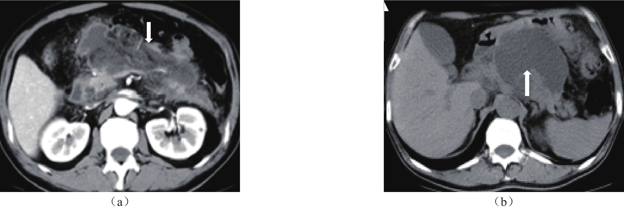
对本组53例ANC发病4周后的变化进行随访,结果显示,53例ANC转归分为两种,即演变为WON与完全吸收。演变为WON者33例(图1、图2),占62.26%,完全吸收者20例,占37.74%。按照ANC转归结局,分为WON组与吸收组进行统计学分析。
![]() 图 1 男,55岁,急性坏死性胰腺炎及4周后ANC转归注:(a)发病后第11天轴面增强CT检查,坏死体积>30%(箭),MCTSI评分8分;(b)发病后3个月第11天轴面CT平扫检查,ANC演变为WON(箭)。Figure 1. Male,55 years old; Acute necrotizing pancreatitis and ANC outcome after 4 weeks
图 1 男,55岁,急性坏死性胰腺炎及4周后ANC转归注:(a)发病后第11天轴面增强CT检查,坏死体积>30%(箭),MCTSI评分8分;(b)发病后3个月第11天轴面CT平扫检查,ANC演变为WON(箭)。Figure 1. Male,55 years old; Acute necrotizing pancreatitis and ANC outcome after 4 weeks![]() 图 2 女,63岁,急性坏死性胰腺炎4周后ANC转归注:发病后第73天,MRI检查轴面增强T1 WI静脉期显示WON的囊壁连续且清晰(箭)。Figure 2. Female,63 years old; ANC outcome after 4 weeks of acute necrotizing pancreatitis
图 2 女,63岁,急性坏死性胰腺炎4周后ANC转归注:发病后第73天,MRI检查轴面增强T1 WI静脉期显示WON的囊壁连续且清晰(箭)。Figure 2. Female,63 years old; ANC outcome after 4 weeks of acute necrotizing pancreatitis2.2 ANC转归的影响因素分析
2.2.1 WON组与吸收组的临床资料比较
笔者前期已对相关影像因素在WON组与吸收组间进行了单因素分析,故本次研究仅分析病因与实验室检查在两组间的差异。统计学分析表明,两组间胆石症、酗酒、高脂血症、总淀粉酶、脂肪酶的差异均无统计学意义(P > 0.05),而CRP值在WON组与吸收组之间的差异具有统计学意义(P < 0.05)(详见表1)。
表 1 WON组与吸收组的影像与临床资料比较Table 1. Comparison of Imaging and Clinical Data between WON Group and Absorption Group影响因素 分组 统计检验 WON组(n=33) 吸收组(n=20) $\chi^2/t $值 P 病因 胆石症 17 12 0.36 0.55 酗酒 4 3 0.09 0.76 高脂血症 11 3 1.31 0.25 实验室检查 总淀粉酶(U/L) 1096.69 1511.36 −0.75 0.46 脂肪酶(U/L) 1094.87 1769.09 −0.10 0.33 CRP(mg/L) 239.06 160.27 2.63 0.01 2.2.2 ANC转归为WON的多因素二元Logistic逐步回归分析
为进一步探究ANC转归为WON的独立危险因素及独立危险因素预测ANC转归为WON的发生概率,以ANC转归结局为因变量,以单因素分析中P<0.05的因素为自变量,进行多因素二元Logistic逐步回归分析。由于坏死是否累及胰腺、坏死累及胰腺部位、坏死体积、MCTSI评分在WON组与吸收组间的差异有统计学意义(笔者前期研究结果),故以上四因素亦考虑为因变量。因变量与自变量赋值情况如下:①ANC转归结局(吸收=0,WON=1);②坏死累及胰腺(否=0,是=1);③坏死累及胰头颈和/或胰体(否=0,是=1);④坏死体积≥30%(否=0,是=1);⑤MCTSI评分>6分(否=0,是=1)。CRP值为连续数值变量,在此不做赋值。结果显示,坏死体积≥30%与MCTSI评分>6分是ANC转归为WON的独立危险因素(P<0.05)(详见表2)。
表 2 ANC转归为WON影响因素的多因素Logistic逐步回归分析结果Table 2. Results of Multi factor Logistic Stepwise Regression Analysis on Factors Influencing the Conversion of ANC to WON因素 β值 SE值 Wald值 OR值 95%CI P值 坏死累及胰腺 19.99 1636.82 0.00 48160.41 0.00~ 0.99 坏死累及胰头颈和/或胰体 −0.76 1.73 0.20 0.47 0.02~13.79 0.66 坏死体积 2.22 1.15 3.76 9.21 0.98~86.88 0.05 MCTSI评分 2.78 1.16 5.75 16.04 1.66~154.92 0.02 CRP(mg/L) 0.00 0.01 0.07 1.00 0.99~1.02 0.79 根据多因素二元Logistic逐步回归结果得到方程Logit(P)=−21.59+2.22(坏死体积是否≥30%)+2.78(MCTSI评分是否>6分),并据此构建预测模型,H-L检验结果
$\chi^2 $ =10.13,P=0.26,表明预测模型与实际情况有非常好的拟合程度。2.2.3 ROC评价预测模型及曲线分析结果
ROC曲线分析结果显示,坏死体积≥30%与MCTSI评分>6分在预测ANC形成4周后演变为WON方面均具有较高的准确性。二者比较,MCTSI评分>6分的曲线下面积更大,为0.88(95%CI:0.75~0.97),较坏死体积≥30%的准确性略高。由于检验变量坏死体积≥30%与MCTSI评分>6分均为二分类变量,故无阈值。当坏死体积为30%时,对应的灵敏度为0.82,特异度为0.90;当MCTSI评分大于6分即MCTSI评分为8分时,对应的灵敏度为0.910,特异度为0.85。详见表3与图3。
表 3 坏死体积≥30%与MCTSI评分>6分的预测效能Table 3. Predictive efficacy of necrotic volume ≥ 30% and MCTSI score > 6 points因素 AUC P值 95%置信区间 敏感性(%) 特异性(%) 折点(%/分) 坏死体积≥30% 0.86 0.00 0.77~0.99 81.82 90 30 MCTSI评分>6分 0.88 0.00 0.75~0.97 90.97 85 8 ![]() 图 3 坏死体积≥30%与MCTSI评分>6分预测ANC演变为WON的ROC曲线Figure 3. ROC curve predicting ANC evolution to WON with necrotic volume ≥ 30% and MCTSI score > 6
图 3 坏死体积≥30%与MCTSI评分>6分预测ANC演变为WON的ROC曲线Figure 3. ROC curve predicting ANC evolution to WON with necrotic volume ≥ 30% and MCTSI score > 63. 讨论
据报道,ANP发生在5%~10%的急性胰腺炎患者[8],其特点是临床病程长,局部并发症发生率高,死亡率高[9]。有文献显示,胰腺坏死的发展和严重程度与AP死亡率的增加直接相关[10]。ANC是指ANP发病4周内的位于胰腺内和/或胰腺外的混有不同数量液体及固体成分的积聚[11]。ANC无包膜或仅有部分包膜[12]。
胰腺坏死的演变是多变的,可随时间推移而消退或进展,并可合并感染[13]。本组研究中共纳入53例ANC患者,其转归为吸收和演变为包裹性坏死,这与相关文献报道的“ANC在形成后的2-4周内,将会逐渐吸收或开始形成包裹[14] ”相一致。本组研究中的53例ANC患者,4周后完全吸收者20例,即37.74%的ANC吸收,62.26%的ANC进展为WON。Manrai等人[15] 研究指出,58.7%的ANC进展为WON;Patra[16] 等人研究结果为41%的ANC会自行吸收,59%进展为WON。本研究结果与以上文献报道结果基本一致。
本研究结果显示ANC坏死体积≥30%与MCTSI>6分为ANC转归为WON的独立危险因素。有文献报道,坏死累及胰腺者更易形成WON,且胰腺坏死面积越大,ANC越易形成WON[17]。胰腺坏死体积越大,累及胰头、颈、体、尾的部位越多,坏死物质及炎性渗出液体越多,越不易吸收。本组研究中,坏死累及胰尾在WON组与吸收组的差异无统计学意义,推测原因可能为胰尾部坏死较胰腺其他部位坏死的炎性渗出及坏死物质更易吸收。坏死体积的预估是有必要的,因为坏死体积的大小对预后有重要意义[18]。Manrai[15]等人研究结果指出,坏死体积>30%是ANC形成WON的重要独立预测因子。与此同时,Sarathi[16]等人研究指出,基线ANC坏死最大直径超过6厘米是ANC形成WON的重要危险因素。由此可见,ANC的坏死体积越大,ANP越严重,ANC转归为WON的可能性越大。
MCTSI与ANP局部并发症的发生发展及死亡发生率有极好的相关性,而且有助于提高诊断AP严重程度的准确率[19],MCTSI评分越高,ANP死亡率越高。研究显示,MCTSI是影响ANC转归的因素之一[20]。ANP越严重,MCTSI评分越高,ANC越易转归为WON,患者预后越差[21]。本研究结果显示,MCTSI>6分为ANC转归为WON的独立危险因素,与文献研究结果一致。
本研究存在一定的局限性。首先,样本量相对较少。其次,影响ANC转归的因素分析可能不够全面,如患者年龄、健康状况等其他因素亦应考虑。
综上所述,坏死体积≥30%、MCTSI>6分为ANC演变为WON的独立危险因素,二者预测ANC演变为WON的准确度均较高。
-
![]()
图 1 男,55岁,急性坏死性胰腺炎及4周后ANC转归
注:(a)发病后第11天轴面增强CT检查,坏死体积>30%(箭),MCTSI评分8分;(b)发病后3个月第11天轴面CT平扫检查,ANC演变为WON(箭)。
Figure 1. Male,55 years old; Acute necrotizing pancreatitis and ANC outcome after 4 weeks
![]()
图 2 女,63岁,急性坏死性胰腺炎4周后ANC转归
注:发病后第73天,MRI检查轴面增强T1 WI静脉期显示WON的囊壁连续且清晰(箭)。
Figure 2. Female,63 years old; ANC outcome after 4 weeks of acute necrotizing pancreatitis
![]()
图 3 坏死体积≥30%与MCTSI评分>6分预测ANC演变为WON的ROC曲线
Figure 3. ROC curve predicting ANC evolution to WON with necrotic volume ≥ 30% and MCTSI score > 6
表 1 WON组与吸收组的影像与临床资料比较
Table 1 Comparison of Imaging and Clinical Data between WON Group and Absorption Group
影响因素 分组 统计检验 WON组(n=33) 吸收组(n=20) $\chi^2/t $值 P 病因 胆石症 17 12 0.36 0.55 酗酒 4 3 0.09 0.76 高脂血症 11 3 1.31 0.25 实验室检查 总淀粉酶(U/L) 1096.69 1511.36 −0.75 0.46 脂肪酶(U/L) 1094.87 1769.09 −0.10 0.33 CRP(mg/L) 239.06 160.27 2.63 0.01  下载: 导出CSV
表 2 ANC转归为WON影响因素的多因素Logistic逐步回归分析结果
Table 2 Results of Multi factor Logistic Stepwise Regression Analysis on Factors Influencing the Conversion of ANC to WON
因素 β值 SE值 Wald值 OR值 95%CI P值 坏死累及胰腺 19.99 1636.82 0.00 48160.41 0.00~ 0.99 坏死累及胰头颈和/或胰体 −0.76 1.73 0.20 0.47 0.02~13.79 0.66 坏死体积 2.22 1.15 3.76 9.21 0.98~86.88 0.05 MCTSI评分 2.78 1.16 5.75 16.04 1.66~154.92 0.02 CRP(mg/L) 0.00 0.01 0.07 1.00 0.99~1.02 0.79
下载: 导出CSV
表 3 坏死体积≥30%与MCTSI评分>6分的预测效能
Table 3 Predictive efficacy of necrotic volume ≥ 30% and MCTSI score > 6 points
因素 AUC P值 95%置信区间 敏感性(%) 特异性(%) 折点(%/分) 坏死体积≥30% 0.86 0.00 0.77~0.99 81.82 90 30 MCTSI评分>6分 0.88 0.00 0.75~0.97 90.97 85 8
下载: 导出CSV
-
[1] LANKISCH P G, APTE M, BANKS P A. Acute pancreatitis[J]. Lancet, 2015, 386(9988): 85-96. DOI: 10.1016/S0140-6736(14)60649-8.
[2] PETROV M S, YADAV D. Global epidemiology and holistic prevention of pancreatitis[J]. Nature Reviews Gastroenterology & Hepatology, 2019, 16(3): 175-184. DOI: 10.1038/s41575-018-0087-5.
[3] HABTEZION A, GUKOVSKAVA A S, PANDOL S J. Acute Pancreatitis: A Multifaceted Set of Organelle and Cellular Interactions[J]. Gastroenterology, 2019, 156(7): 1941-1950. DOI: 10.1053/j.gastro.2018.11.082.
[4] BANKS P A, BOLLEN T L, DERVENIS C, et al. Classification of acute pancreatitis--2012: revision of the Atlanta classification and definitions by international consensus[J]. Gut, 2013, 62(1): 102-111. DOI: 10.1136/gutjnl-2012-302779.
[5] GARDNER T B. Acute Pancreatitis[J]. Ann Intern Med, 2021, 174(2): C17-C32. DOI: 10.7326/AITC202102160.
[6] HUANG J, QU H P, ZHENG Y F, et al. The revised Atlanta criteria 2012 altered the classification, severity assessment and management of acute pancreatitis[J]. Hepatobiliary Pancreat Diseases International, 2016, 15(3): 310-315. DOI: 10.1016/s1499-3872(15)60040-6.
[7] 闫威, 董力宁, 张斌斌, 等. 急性胰腺炎患者坏死性积聚的CT和MRI特征及转归分析[J]. CT理论与应用研究, 2023, 32(01): 113-120. DOI: 10.15953/j.ctta.2022.141. YAN W, DONG L N, ZHANG B B, et al. CT and MRI features and outcome analysis of necrotic accumulation in patients with acute pancreatitis[J]. Research on CT Theory and Application, 2023, 32(01): 113-120. DOI: 10.15953/j.ctta.2022.141.
[8] DIMAIO C J. Management of complications of acute pancreatitis[J]. Current Opinion Gastroenterol, 2018, 34(5): 336-342. DOI: 10.1097/MOG.0000000000000462.
[9] GRASSEDONIO E, TOIA P, LA G L, et al. Role of computed tomography and magnetic resonance imaging in local complications of acute pancreatitis[J]. Gland Surgery, 2019, 8(2): 123-132. DOI: 10.21037/gs.2018.12.07.
[10] TAYDAS O, UNAL E, KARAOSMANOGLU A D, et al. Accuracy of early CT findings for predicting disease course in patients with acute pancreatitis[J]. Japanese journal of radiology, 2018, 36(2): 151-158. DOI: 10.1007/s11604-017-0709-9.
[11] RANA S S, SHARMA R K, GUPTA P, et al. Natural course of asymptomatic walled off pancreatic necrosis[J]. Digestive and Liver Disease, 2019, 51(5): 730-734. DOI: 10.1016/j.dld.2018.10.010.
[12] 闫媛媛, 靳二虎, 张洁, 等. CT和MRI对急性胰腺炎局部并发症的诊断价值研究[J]. CT理论与应用研究, 2018, 27(03): 393-400. DOI: 10.15953/j.1004-4140.2018.27.03.13. YAN Y Y, JIN E H, ZHANG J, et al. Research on the diagnostic value of CT and MRI for local complications of acute pancreatitis[J]. Research on CT Theory and Application, 2018, 27(03): 393-400. DOI: 10.15953/j.1004-4140.2018.27.03.13.
[13] THOENI R F. Imaging of Acute Pancreatitis[J]. Radiologic clinics of North America, 2015, 53(6): 1189-1208. DOI: 10.1016/j.rcl.2015.06.006.
[14] BEZMAREVIC M, VANDIJK S M, VOERMANS R P, ETAL. Management of (Peri)Pancreatic Collections in Acute Pancreatitis[J]. Visceral Medicine, 2019, 35(2): 91-96. DOI: 10.1159/000499631.
[15] MANRAI M, KOCHHAR R, GUPTA V, et al. Outcome of Acute Pancreatic and Peripancreatic Collections Occurring in Patients With Acute Pancreatitis[J]. Annals of Surgery, 2018, 267(2): 357-363. DOI: 10.1097/SLA.0000000000002065.
[16] SARATHI P P, DAS K, BHATTACHARYYA A, et al. Natural resolution or intervention for fluid collections in acute severe pancreatitis[J]. British Journal of Surgery, 2014, 101(13): 1721-1728. DOI: 10.1002/bjs.9666.
[17] 刘建, 李昂, 刘殿刚, 等. CT检查预测急性胰腺炎局部并发症转归的价值[J]. 中华普外科手术学杂志(电子版), 2017, 11(04): 285-288. LIU J, LI A, LIU D G, et al. The value of CT examination in predicting the outcome of local complications in acute pancreatitis[J]. Chinese Journal of General Surgery (Electronic Edition), 2017, 11(04): 285-288.
[18] SUREKA B, BANSAL K, PATIDAR Y, et al. Imaging lexicon for acute pancreatitis: 2012 Atlanta Classification revisited[J]. Gastroenterology report, 2016, 4(1): 16-23. DOI: 10.1093/gastro/gov036.
[19] MEDEROS M A, REBER H A, GIRGIS M D. Acute Pancreatitis: A Review[J]. Journal of the American Medical Association, 2021, 325(4): 382-390. DOI: 10.1001/jama.2020.20317.
[20] ALBERTI P, PANDO E, MATA R, et al. Evaluation of the modified computed tomography severity index (MCTSI) and computed tomography severity index (CTSI) in predicting severity and clinical outcomes in acute pancreatitis[J]. Journal of Digestive Diseases, 2021, 22(1): 41-48. DOI: 10.1111/1751-2980.12961.
[21] YAMAMIYA A, KITAMUTA K, YOSHIDA H, et al. Prediction of the progression of walled-off necrosis in patients with acute pancreatitis on whole pancreatic perfusion CT[J]. Journal of Hepatobiliary Pancreat Sciences, 2020, 27(10): 739-746. DOI: 10.1002/jhbp.803.


计量
- 文章访问数: 22
- HTML全文浏览量: 3
- PDF下载量: 5


