
不同管电压联合器官剂量调制技术对脑灌注图像质量及眼晶体辐射剂量的影响研究
Study on the Impact of Different Tube Voltages Combined with Organ Dose Modulation on Cerebral Perfusion Image Quality and Ocular Lens Radiation Dose
-
摘要:
目的:探讨不同管电压下启用器官剂量调制(ODM)技术在颅脑灌注CT(CTP)中对图像质量及眼晶体辐射剂量的影响。方法:以临床CTP扫描方案为基准,在其他参数相同情况下分别使用5种管电压(70、80、100、120、140 kV)对Catphan模体和一具新鲜离体头颅标本进行3种管电流调制模式的扫描:不使用剂量调制技术(Manual模式)、使用智能管电流调制技术(Smart mA模式)、使用器官剂量调制技术(ODM模式)。在右眼晶体前方相同位置固定长杆电离室,每组参数重复9次扫描,记录平均眼晶体剂量值(Dav)。对Catphan模体中CTP528高对比分辨力模块进行调制传递函数(MTF)的测量。在头颅标本影像中的基底节层面选取尾状核头、豆状核、丘脑作为信号区,选取侧脑室、内囊前肢、内囊后肢作为对应背景区,测算对比度噪声比(CNR)。分割同层面脑组织测量纹理对比度、纹理相关性、差分方差等三项纹理特征参数。对MTF、CNR的比较采用两因素方差分析,对晶体剂量、图像纹理的比较采用多因素非参数方差分析。结果:MTF在不同管电压间差异具有统计学意义,在80 kV以上时其值与管电压呈正相关,在不同管电流调制模式间差异无统计学意义。各信号区与背景区间的CNR在不同管电压间差异均有统计学意义,其中尾状核头-侧脑室的CNR值在120 kV时最大。后两者的CNR值与管电压呈负相关。三者的CNR值在不同管电流调制模式间差异均无统计学意义。各纹理参数与管电流调制模式无关。不同管电压和管电流调制模式下的Dav值差异均存在统计学意义,在管电压120 kV以下时晶体剂量与管电压呈负相关,各管电压下ODM技术均可降低约20%的晶体剂量。结论:使用80至100 kV进行CTP扫描并启用器官剂量调制(ODM)技术可均衡图像的各参数指标,在保证图像质量前提下降低眼晶体辐射剂量,建议临床推广使用。
Abstract:Objective: To investigate the effect of using organ dose modulation (ODM) technology at different tube voltages on image quality and eye lens radiation dose in brain perfusion CT (CTP). Methods: Based on a clinical CTP scanning protocol, five tube voltages (70, 80, 100, 120, and 140 kV) were used to scan a Catphan phantom and a fresh, isolated human head specimen with three tube current modulation modes: without dose modulation technology (Manual mode), with smart tube current modulation technology (Smart mA mode), and with organ dose modulation technology (ODM mode). A long rod ionization chamber was fixed at a consistent position in front of the right eye lens. Each parameter combination was scanned nine times, and the average eye lens dose (Dav) was recorded. The modulation transfer function (MTF) of the CTP528 high-contrast resolution module in the Catphan phantom was measured. In the images of the head specimen, the heads of the caudate nucleus, lenticular nucleus, and thalamus were selected as signal regions at the basal ganglia level, and the lateral ventricle, anterior limb of the internal capsule, and posterior limb of the internal capsule were selected as respective background regions to measure the contrast-to-noise ratio (CNR). The same level of brain tissue was segmented to measure three texture feature parameters: contrast, correlation, and difference variance. Two-factor analysis of variance was used to compare MTF and CNR, and multi-factor non-parametric analysis of variance was used to compare eye lens dose and texture parameters. Results: The differences in MTF across the different tube voltages were statistically significant , with MTF values positively correlated with tube voltage above 80 kV. However, there was no significant difference between different tube-current modulation modes. The CNR of each signal and background region showed significant differences among different tube voltages. The CNR for the caudate nucleus head to lateral ventricle was the largest, at 120 kV. The CNR for the lenticular nucleus/thalamus were negatively correlated with tube voltage. There were no significant differences in CNR among different tube-current modulation modes. No texture parameter was significantly associated with the tube current modulation mode. There were significant differences in Dav values with respect to both different tube voltages and tube current modulation modes. When the tube voltage was below 120 kV, the eye lens dose was negatively correlated with tube voltage, and the ODM technology reduced the eye lens dose by approximately 20% at each tube voltage. Conclusion: Employing 80–100 kV CTP scanning with organ dose modulation technology can balance image parameters and reduce radiation dose to the eye lens while ensuring adequate image quality, as recommended for clinical use.
-
Keywords:
- brain perfusion CT /
- organ dose modulation /
- Lens /
- image quality
-
-
![]()
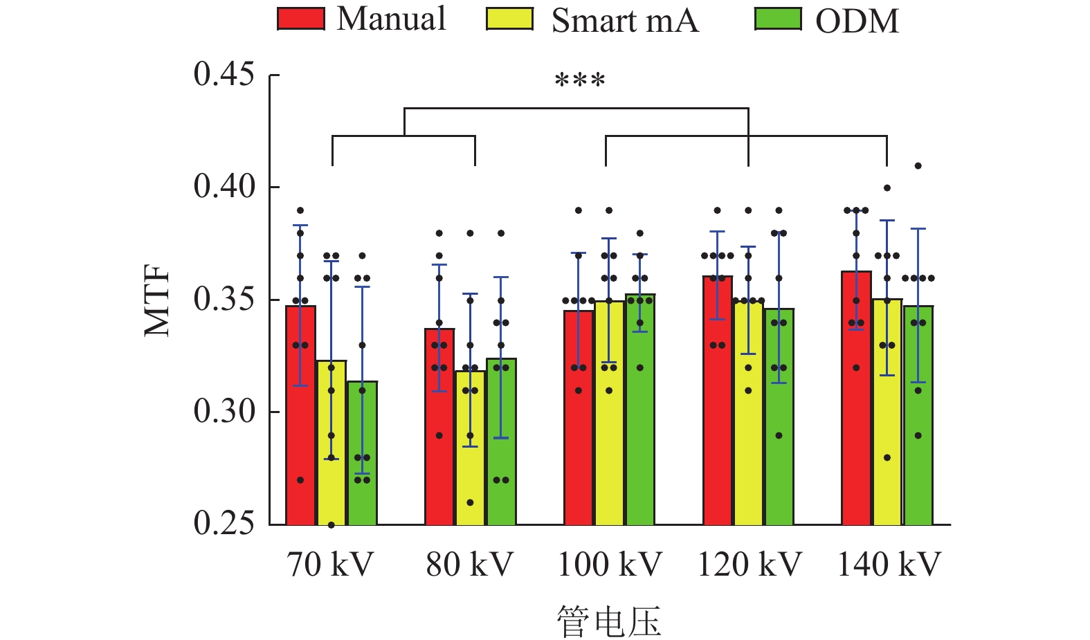
图 1 不同管电压与管电流调制模式下的MTF差异
注: ***代表差异有统计学意义。
Figure 1. MTF differences across different tube voltage and tube current modulation modes
![]()
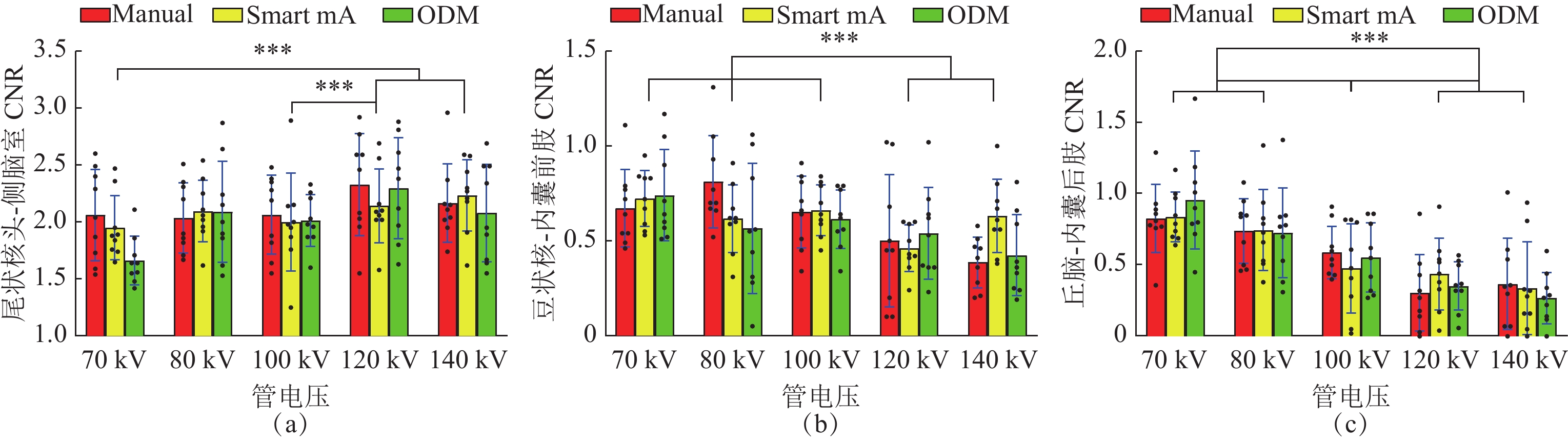
图 2 不同管电压与管电流调制模式下的CNR差异
注: (a)尾状核头-侧脑室 (b)豆状核-内囊前肢 (c)丘脑-内囊后肢 ***代表差异有统计学意义。
Figure 2. CNR differences between different tube voltages and tube current modulation modes
![]()
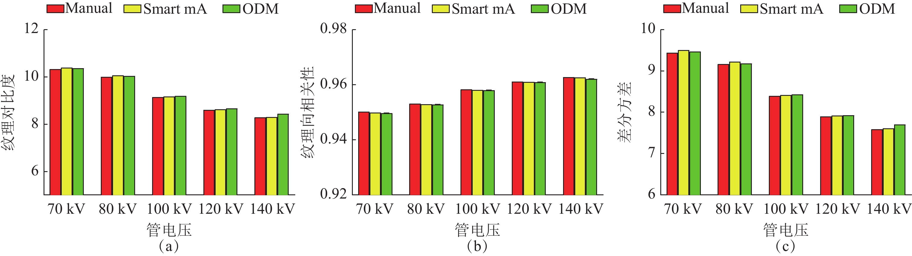
图 3 不同管电压与管电流调制模式下的图像纹理差异
注:a纹理对比度 b纹理相关性 c差分方差
Figure 3. Image texture differences under different modulation modes of tube voltage and tube current
![]()
图 4 不同管电压与管电流调制模式颅脑CTP图像
注:a、b、c 1~5分别为 Manual、 Smart mA、ODM三种管电流调制模式下70~140 kV图像
Figure 4. Brain CTP images at different tube voltages and tube current modulation modes
![]()
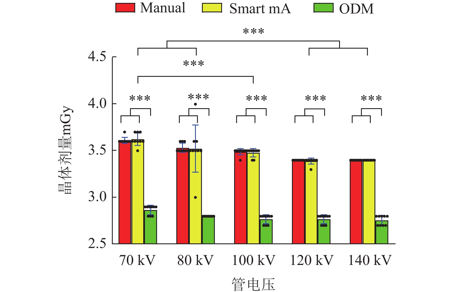
图 5 不同管电压与管电流调制模式下的晶体剂量差异
注: ***代表差异有统计学意义。
Figure 5. Differences in crystal dose across different tube voltage and tube current modulation modes
表 1 不同管电压与管电流调制模式下的MTF值差异性检验
Table 1 MTF value differences across tube voltages and tube current modulation modes
自变量A
(调制模式)自变量B(管电压) F(A) P(A) F(B) P(B) 70 kV 80 kV 100 kV (n=9) 120 kV 140 kV Manual 0.348±0.036 0.338±0.028 0.346±0.026 0.361±0.020 0.363±0.026 Smart mA 0.323±0.044 0.319±0.034 0.350±0.027 0.350±0.024 0.351±0.034 2.59 0.08 4.86 <0.01 ODM 0.314±0.042 0.324±0.036 0.353±0.017 0.347±0.034 0.348±0.034  下载: 导出CSV
表 2 不同管电压与管电流调制模式下的CNR差异性检验
Table 2 CNR differences across different tube voltages and tube current modulation modes
自变量A
(调制模式)自变量B(管电压) F(A) P(A) F(B) P(B) 70 kV 80 kV 100 kV (n=9) 120 kV 140 kV 尾状核头-侧脑室 Manual 2.062±0.401 2.036±0.308 2.063±0.345 2.327±0.445 2.168±0.342 Smart mA 1.950±0.280 2.096±0.272 1.998±0.428 2.143±0.323 2.231±0.312 0.92 0.40 4.02 <0.01 ODM 1.665±0.212 2.092±0.443 2.012±0.229 2.297±0.442 2.080±0.430 豆状核-内囊前肢 Manual 0.671±0.205 0.812±0.244 0.653±0.189 0.501±0.350 0.386±0.135 Smart mA 0.723±0.148 0.617±0.178 0.662±0.136 0.462±0.124 0.632±0.193 0.45 0.64 6.09 <0.01 ODM 0.741±0.243 0.565±0.343 0.613±0.154 0.539±0.241 0.425±0.212 丘脑-内囊后肢 Manual 0.829±0.238 0.741±0.227 0.588±0.184 0.306±0.270 0.368±0.320 Smart mA 0.839±0.177 0.746±0.285 0.477±0.314 0.436±0.254 0.339±0.326 0.02 0.98 21.77 <0.01 ODM 0.957±0.344 0.727±0.315 0.554±0.242 0.353±0.168 0.270±0.179
下载: 导出CSV
表 3 不同管电压与管电流调制模式下的图像纹理差异性检验
Table 3 Inspection of image texture differences under different tube voltage and tube current modulation modes
自变量A
(调制模式)自变量B(管电压) H(A) P(A) H(B) P(B) 70 kV 80 kV 100 kV (n=9) 120 kV 140 kV 纹理对比度 Manual 10.333±0.016 10.011±0.017 9.149±0.015 8.613±0.008 8.289±0.013 Smart mA 10.392±0.011 10.071±0.021 9.179±0.015 8.638±0.010 8.310
(8.310,8.310)3.44 0.18 128.65 <0.01 ODM 10.372±0.014 10.041±0.010 9.206±0.012 8.673±0.011 8.439±0.012 纹理相关性 Manual 0.950±
7.295*10^-50.953±
7.731*10^-50.958±
6.513*10^-50.961
(0.961,0.961)0.963±
5.074*10^-5Smart mA 0.950±
6.736*10^-50.953±
8.652*10^-50.958±
5.539*10^-50.961±
4.051*10^-50.963±
2.216*10^-54.37 0.11 128.65 <0.01 ODM 0.950±
7.065*10^-50.953±
5.342*10^-50.958±
4.265*10^-50.961±
4.479*10^-50.962±
4.871*10^-5差分方差 Manual 9.449±0.016 9.173±0.015 8.400±0.014 7.902±0.006 7.596±0.010 Smart mA 9.506±0.011 9.230±0.020 8.427±0.013 7.923±0.010 7.618
(7.612,7.621)2.94 0.23 128.65 <0.01 ODM 9.474±0.014 9.187±0.008 8.433±0.012 7.934±0.012 7.708±0.013
下载: 导出CSV
表 4 不同管电压与管电流调制模式下的晶体剂量差异性检验
Table 4 Verification of crystal dose differences across different tube voltage and tube current modulation modes
自变量A
(调制模式)自变量B(管电压) H(A) P(A) H(B) P(B) 70 kV 80 kV 100 kV (n=9) 120 kV 140 kV Manual 3.60(3.60,3.60) 3.50(3.50,3.55) 3.45(3.45,3.48) 3.35(3.35,3.38) 3.40(3.35,3.40) Smart mA 3.61±0.06 3.50(3.48,3.55) 3.44±0.05 3.35(3.35,3.40) 3.40(3.35,3.40) 90.46 <0.01 30.74 <0.01 ODM 2.85(2.8,2.85) 2.80(2.78,2.80) 2.75(2.70,2.75) 2.75(2.70,2.75) 2.75(2.70,2.75)
下载: 导出CSV
-
[1] 范丽, 黄劲柏. 大脑中动脉狭窄程度与脑梗死前期CT灌注参数的相关性[J]. CT理论与应用研究, 2020, 29(2): 219-227. DOI: 10.15953/j.1004-4140.2020.29.02.13. FAN L, HUANG J B. Correlation between the Degree of Middle Cerebral Artery Stenosis and CT Perfusion Parameters in the Early Stage of Cerebral Infarction[J]. CT Theory and Applications, 2020, 29(2): 219-227. DOI: 10.15953/j.1004-4140.2020.29.02.13.
[2] 钟立清, 卫军, 常小娜, 等. 128层螺旋CT脑灌注成像对急性脑梗死的诊断价值评价[J]. 现代医用影像学, 2023, 32(2): 256-258. DOI: 10.3969/j.issn.1006-7035.2023.02.014. ZHONG L Q, WEI J, CHANG X N, et al. Evaluation of the diagnostic value of 128-slice spiral ct cerebral perfusion imaging for acute cerebral infarction[J]. Modern Medical Imaging, 2023, 32(2): 256-258. DOI: 10.3969/j.issn.1006-7035.2023.02.014.
[3] ZHANG D, CHRIS H, CAGNON, J, PABLO V, et al. Peak skin and eye lens radiation dose from brain perfusion CT based on Monte Carlo simulation.[J]. American Journal of Roentgenology, 2012, 198(2): 412-7. DOI: 10.2214/AJR.11.7230.
[4] 鲁新亮, 陶建华, 马文涛, 等. 基于不同重建算法的容积再现成像在诊断鼻区线性骨折中的差异研究[J]. CT理论与应用研究, 2024, 33(5): 609-618. DOI: 10.15953/j.ctta.2023.212. LU X L, TAO J H, MA W T, et al. A study on the differences of volume rendering imaging based on different reconstruction algorithms in the diagnosis of linear fractures in the nasal area[J]. Computerized Tomography Theory and Applications, 2024, 33(5): 609-618. DOI: 10.15953/j.cta.2023.212. DOI: 10.15953/j.ctta.2023.212.
[5] 申希平, 祁海萍, 刘小宁, 等. 两因素非参数方差分析在SPSS中的实现[J]. 中国卫生统计, 2013, 30(6): 913-914. SHEN X P, QI H P, LIU X N, et al. Implementation of Two-Factor Nonparametric Analysis of Variance in SPSS[J]. China Health Statistics, 2013, 30(6): 913-914.
[6] 荆梅, 顾欣欣. 颅脑CT灌注成像及磁共振成像在脑梗死患者中的应用[J]. 中国实用神经疾病杂志, 2024, 27(1): 43-47. DOI: 10.12083/SYSJ.230920. JING M, GU X X. Application of brain CT perfusion imaging and magnetic resonance imaging in patients with cerebral infarction [J]. Chinese Journal of practical neurological diseases.
[7] 赵锡鹏, 顾枭成, 陈飞, 等. CT剂量、层厚和射线质对图像高对比度分辨力的影响研究[J]. 中国医学装备, 2023, 20(1): 12-16. DOI: 10.3969/J.ISSN.1672-8270.2023.01.003. ZHAO X P, GU X C, CHEN F, et al. Study on the influence of ct dose, slice thickness, and radiation quality on high-contrast resolution of images[J]. Chinese Medical Equipment, 2023, 20(1): 12-16. DOI: 10.3969/J.ISSN.1672-8270.2023.01.003.
[8] 吕蓉, 陈晨, 胡维娟, 等. CT值与管电流、管电压的关系以及图像噪声与辐射剂量的相关性研究[J]. 实用放射学杂志, 2020, 36(1): 123-127. DOI: 10.3969/j.issn.1002-1671.2020.01.031. LV R, CHEN C, HU W J, et al. Research on the relationship between ct value, tube current, tube voltage, and the correlation between image noise and radiation[J]. Journal of Practical Radiology, 2020, 36(1): 123-127. DOI: 10.3969/j.issn.1002-1671.2020.01.031.
[9] 杨红云, 李海亮, 杨淑慧, 张琳, 闵楠, 朱建国. 五种头颅CT扫描方式受检者眼晶体受照剂量的测量及比较[J]. 中国辐射卫生, 2019, 28(2): 148-151, 154. DOI: 10.13491/j.issn.1004-714x.2019.02.009. YANG H Y, LI H L, YANG S H, et al. Measurement and Comparison of Intraocular Lens Radiation Dose in Subjects Using Five Different Head CT Scanning Methods[J]. China Radiation Health, 2019, 28 (2): 148-151154. DOI: 10.13491/j.issn.1004-714x.2019.02.009.
[10] 孙静坤, 彭刚, 吕发金, 等. 铋屏蔽联合器官管电流调制技术在颅脑CT检查中应用的体模研究[J]. 中华放射医学与防护杂志, 2021, 41(5): 385-389. DOI: 10.3760/cma.j.issn.0254-5098.2021.05.012. SUN J K, PENG G, LV F J, et al. A phantom study on the application of bismuth shielding combined with organ tube current modulation technology in cranial ct examination[J]. Chinese Journal of Radiological Medicine and Protection, 2021, 41(5): 385-389. DOI: 10.3760/cma.j.issn.0254-5098.2021.05.012.
[11] 吴柯薇, 钟朝辉, 王振常, 原媛, 张景东, 崔茹欣. 器官剂量调制技术在头颅CT平扫中的应用[J]. 中国介入影像与治疗学, 2019, 16(8): 491-494. DOI: 10.13929/j.1672-8475.201904002. WU K W, ZHONG C H, WANG Z C, et al. Application of organ dose modulation technology in head ct plain scanning[J]. Chinese Journal of Interventional Imaging and Therapy, 2019, 16(8): 491-494. DOI: 10.13929/j.1672-8475.201904002.
[12] KALRA M K , NAZ N , RIZZO S , et al. Computed tomography radiation dose optimization: scanning protocols and clinical applications of automatic exposure control[J]. Current Problems in Diagnostic Radiology, 2005, 34(5): 171-181. DOI: 10.1067/j.cpradiol.2005.06.002.
[13] 张永县, 牛延涛, 张丽丽, 等. 器官剂量调制技术在胸部CT中降低乳腺辐射剂量的研究[J]. 中华放射学杂志, 2020, 54(6): 587-591. DOI: 10.3760/cma.j.cn112149-20190708-00262. ZHANG Y X, NIU Y T, ZHANG L L, et al. Study on the use of organ dose modulation technology to reduce breast radiation dose in chest CT [j]. Chinese Journal of Radiology, 2020, 54 (6): 587-591. DOI: 10.3760/cma.j.cn112149-20190708-00262.
[14] 张永县, 牛延涛, 张丽丽, 等. 胸部CT探测器宽度、螺距联合器官剂量调制技术对辐射剂量和影像质量影响的模体研究[J]. 中华放射学杂志, 2019, 53(6): 464-469. DOI: 10.3760/cma.j.issn.1005-1201.2019.06.005. ZHANG Y X, NIU Y T, ZHANG L L, et al. Modeling study on the effects of chest ct detector width and pitch combined with organ dose modulation technology on radiation dose and image quality[J]. Chinese Journal of Radiology, 2019, 53(6): 464-469. DOI: 10.3760/cma.j.issn.1005-1201.2019.06.005.
[15] 张永县, 牛延涛, 刘丹丹, 等. 管电压联合器官剂量调制技术对胸部CT辐射剂量和图像质量影响的模体研究[J]. 中华放射医学与防护杂志, 2019, 39(7): 529-533. DOI: 10.3760/cma.j.issn.0254-5098.2019.07.009. ZHANG Y X, NIU Y T, LIU D D, et al. Phantom study on the effect of tube voltage combined with organ dose modulation technology on radiation dose and image quality in chest ct[J]. Chinese Journal Of Radiation Medicine And Protection, 2019, 39(7): 529-533. DOI: 10.3760/cma.j.issn.0254-5098.2019.07.009.

计量
- 文章访问数: 0
- HTML全文浏览量: 0
- PDF下载量: 0


