
Value of Low Tube Voltage Combined with Deep Learning Image Reconstruction Algorithm to Reduce Radiation Dose in Combined Thoracoabdominal Enhanced CT
-
摘要:
目的:探讨在胸腹部联合增强CT扫描中,应用低管电压联合深度学习图像重建算法(DLIR)对降低辐射剂量及图像质量的影响。方法:①模体实验。确定低管电压结合深度学习算法对低对比度分辨力鉴别的可行性。按照不同图像质量参数噪声指数(NI)扫描Catphan 500模体,使用两种扫描条件,优化组扫描参数选择低管电压80 kV结合DLIR进行扫描和图像重建;常规组扫描参数和图像重建算法选择管电压120 kV结合自适应统计迭代重建(ASiR-V),确定优化组条件使用低剂量(NI > 9)时低对比度分辨力相对于常规组使用常规剂量(NI=9)的NI值和有效性。②前瞻性实验。前瞻性收集常规进行胸腹部联合增强CT扫描的患者160例,随机分为低剂量优化组和常规剂量常规组,最终入组149例,低剂量优化组61例,常规剂量常规组88例。根据模体实验的结果确定的低剂量优化组NI优,扫描参数选择优化组条件;常规剂量常规组NI为9,扫描参数和图像重建算法选择常规组条件。记录并计算两组间的辐射剂量并对两组的图像质量进行主、客观评价。结果:低剂量优化组使用NI优=12可以获得常规剂量组NI=9等效的低对比度分辨能力;低剂量优化组的有效剂量(9.56±2.34) mSv低于常规剂量常规组(17.82±5.22) mSv;低剂量优化组的肝脏衰减值、主动脉衰减值显著高于常规剂量常规组,肝脏及主动脉CNR和SNR值显著高于常规剂量常规组,主动脉空间分辨力、肝总动脉空间分辨力、门静脉空间分辨力及小血管/支气管显示情况也均优于常规剂量常规组。 结论:低管电压联合深度学习图像重建算法能够在降低辐射剂量的条件下,仍保证同等甚至更高的胸腹部联合CT 扫描图像质量,为大范围CT扫描辐射剂量的优化提供一个可行方案。
-
关键词:
- 计算机体层摄影 /
- 深度学习图像重建算法 /
- 低管电压 /
- 辐射剂量 /
- 胸腹部联合CT扫描
Abstract:Objective: To investigate the effect of low tube voltage combined with deep learning image reconstruction (DLIR) on radiation dose reduction and maintaining image quality in combined chest and abdominal enhanced CT scans. Methods: (1) Phantom study. To determine the feasibility of combining low tube voltage with deep learning algorithms for low-contrast resolution, Catphan 500 phantoms were scanned under two different conditions. The optimization group used a low tube voltage (80 kV) combined with DLIR for scanning and image reconstruction, while the routine group used a 120 kV tube voltage combined with adaptive statistical iterative reconstruction V (ASiR-V). This study aimed to determine the effectiveness of the optimization group using a low dose (noise index, NI > 9) compared with the routine group using a routine dose (NI=9). (2) Prospective study. A total of 160 patients who underwent routine chest and abdominal enhanced CT scans were prospectively collected and randomly divided into a low-dose optimization group and routine-dose group, with 149 patients ultimately enrolled (61 in the low-dose optimization group and 88 in the routine-dose group). Based on the results of the phantom study, the low-dose optimization group used the optimized condition with NI set to the optimal value, whereas the routine-dose group used the routine condition with NI=9. Radiation doses were recorded and calculated for both groups, and image quality was subjectively and objectively evaluated. Results: The low-dose optimization group using NI=12 achieved an equivalent low-contrast resolution capability to the routine-dose group with NI=9. The effective dose in the low-dose optimization group (9.56±2.34) mSv was significantly lower than that in the routine-dose group (17.82±5.22) mSv. The liver and aorta attenuation values in the low-dose optimization group were significantly higher than those in the routine-dose group, and the CNR and SNR values in the liver and aorta were also significantly higher. The spatial resolution of the aorta, common hepatic artery, and portal vein and the display of small vessels/bronchi were all superior in the low-dose optimization group compared with the routine-dose group. Conclusion: The combination of a low tube voltage and deep learning image reconstruction algorithm can ensure equivalent or even higher image quality while reducing radiation dose, providing a feasible solution for optimizing radiation dose in large-scale CT scans such as the combined thoracoabdominal enhanced CT.
-
-
![]()
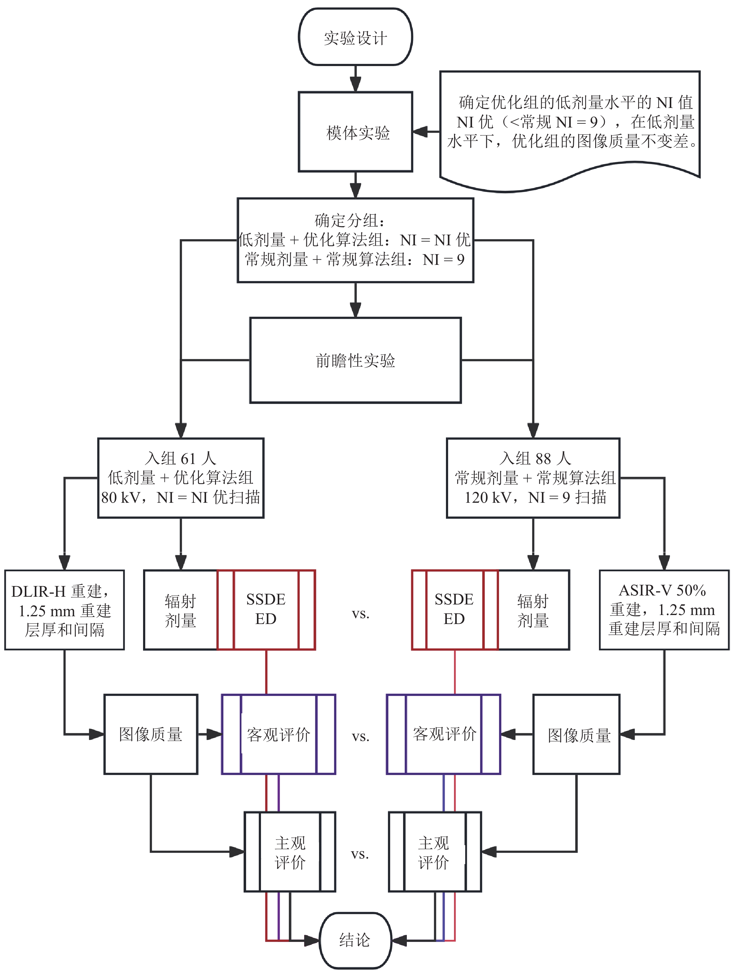
图 1 实验设计流程图,图中给出了病例分组,评价和比较的内容以及结论依据
Figure 1. Flowchart of the experimental design. The chart shows the grouping of cases, content of evaluation and comparison, and the basis for the conclusions
![]()
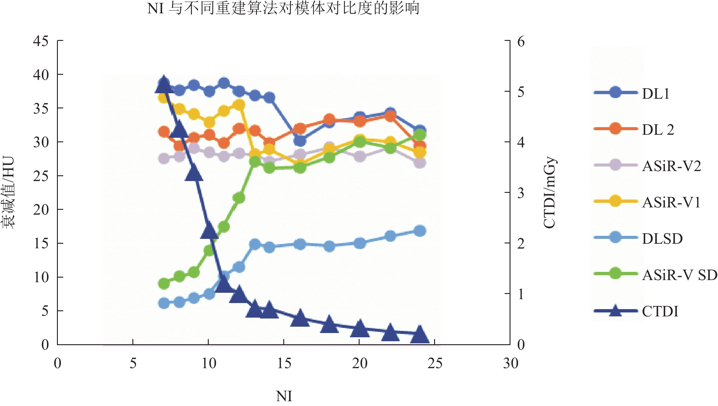
图 2 图中1为CTP515模块中supra-slice 1%密度差的模块,2为CTP515模块中的本底,DL为DLIR重建,ASiR-V为自适应统计迭代重建重建
Figure 2. 1 represents the module with a 1% density difference in the supra-slice of the CTP515 module, and 2 represents the background of the CTP515 module. DL denotes DLIR reconstruction, and ASiR-V denotes Adaptive Statistical Iterative Reconstruction
![]()
图 3 不同体型的胸部以及胸腹的两组分别扫描的对比
Figure 3. Comparison of two groups of separate scans of the chest and chest–abdominal area for different body types
表 1 主观评分标准
Table 1 Subjective scoring criteria
评分 图像噪声 细微结构空间分
辨率(肾上腺)肝总动脉及主动脉
空间分辨力主动脉及肝总
动脉对比度肺裂、小血管/
支气管显示率1分 噪声极明显,图像完全不能诊断 模糊不清 边缘非常模糊 模糊不清 完全不可见 2分 噪声明显,解剖细节重度模糊 隐约可见 低于可接受水平 劣于可接受水平 <25%可见 3分 噪声明显,但可接受 可接受 可接受水平 可接受 25%~75%可见 4分 能观察到噪声 清晰 高于可接受水平 优于可接受水平 >75%可见 5分 几乎无噪声 非常清晰 边缘非常锐利 非常清晰 完全可见  下载: 导出CSV
表 2 两组一般资料与辐射剂量指标比较
Table 2 Comparison of general information and radiation dose indicators between the two groups
项目 组别 统计检验 LD-LV-DLIR RD-HV-ASiRV Z P 例数 61 88 男/女 28/36 47/38 − 0.71 0.48 BMI/(kg/m2) 23.46±2.81 23.95±4.00 −1.84 0.07 年龄/岁 54.77±16.79 52.82±15.12 − 0.71 0.48 CTDIvol/mGy 3.67±1.56 7.39±2.40 3.46 < 0.001 SSDE/mGy 3.72±1.33 7.47±2.02 3.23 < 0.001 校正ED/mSv 9.56±2.34 17.82±5.22 2.79 < 0.001 注:BMI为体重指数;CTDIvol为容积CT剂量指数;SSDE为特定体型的剂量评估;校正ED为校正后的有效剂量。
下载: 导出CSV
表 3 两组间动脉期客观指标比较
Table 3 Comparison of objective indicators in the arterial phase between the two groups
项目 组别 统计检验 LD-LV-DLIR RD-HV-ASiRV Z P 例数 61 88 噪声 8.01±2.57 11.84±2.29 − 7.69 <0.01 肝脏 CNR 25.32(19.72,31.15) 14.37(12.58,16.74) − 8.27 <0.01 SNR 9.63(7.38,11.10) 5.87±1.45 − 7.59 <0.01 主动脉 CNR 65.30±21.33 30.76±7.33 − 9.67 <0.01 SNR 48.79±15.99 21.57±5.55 − 9.85 <0.01 注:CNR为对比噪声比;SNR为信噪比。
下载: 导出CSV
表 4 两组间静脉期客观指标比较
Table 4 Comparison of objective indicators in the venous phase between the two groups
项目 组别 统计检验 LD-LV-DLIR RD-HV-ASiRV Z P 例数 61 88 噪声 7.60(6.40,7.60) 12.51±2.62 − 7.93 <0.01 肝脏 CNR 34.44±10.34 19.45±11.61 − 8.71 <0.01 SNR 19.31±6.09 9.43(8.16,10.54) − 9.00 <0.01 主动脉 CNR 43.58±13.39 21.05±5.89 − 9.14 <0.01 SNR 28.46±9.55 12.41±3.47 − 9.39 <0.01 注:CNR为对比噪声比;SNR为信噪比。
下载: 导出CSV
表 5 两组间腹部动脉期主观指标比较表
Table 5 Comparison of subjective indicators in the abdominal arterial phase between the two groups
项目 组别 统计检验 LD-LV-DLIR RD-HV-ASiRV Z P 例数 61 88 噪声 3(3,3) 2.5(2,3) − 3.71 <0.01 主动脉空间分辨力 5(5,5) 2(2,3.75) − 8.30 <0.01 主动脉对比度 5(5,5) 5(5,5) − 1.90 0.58 肝总动脉空间分辨力 5(5,5) 4(3,4) − 8.08 <0.01 肝总动脉对比度 5(5,5) 5(4,5) − 3.45 0.01
下载: 导出CSV
表 6 两组间腹部静脉期主观指标比较表
Table 6 Comparison of subjective indicators in the abdominal venous phase between the two groups
项目 组别 统计检验 LD-LV-DLIR RD-HV-ASiRV Z P 例数 61 88 噪声 3(3,3) 3(3,4) − 1.99 0.47 细微结构分辨力 3(3,4) 3(3,4) 0.82 0.41 门静脉空间分辨力 4(3,4) 3(3,4) − 3.88 <0.01 门静脉对比度 4(4,5) 4(3,5) − 0.38 0.71
下载: 导出CSV
表 7 两组间胸部主观指标比较表
Table 7 Comparison of subjective indicators in the chest between the two groups
项目 组别 统计检验 LD-LV-DLIR RD-HV-ASiRV Z P 例数 61 88 肺裂 4(3,5) 4(3,5) − 0.25 0.81 小血管/支气管 5(5,5) 4(4,5) − 2.46 0.03
下载: 导出CSV
-
[1] FURTADO C D, AGUIRRE D A, SIRLIN C B, et al. Whole-body CT screening: Spectrum of findings and recommendations in 1192 patients[J]. Radiology, 2005, 237(2): 385-394. DOI: 10.1148/radiol.2372041741.
[2] BRANT-ZAWADZKI M. CT screening: Why I do it[J]. American Journal of Roentgenology, 2002, 179(2): 319-326. DOI: 10.2214/ajr.179.2.1790319.
[3] SCHAUER D A, LINTON O W. National council on radiation protection and measurements report shows substantial medical exposure increase[J]. Radiology, 2009, 253(2): 293-296. DOI: 10.1148/radiol.2532090494.
[4] REHANI M M, HEIL J, BALIYAN V. Multicentric study of patients receiving 50 or 100mSv in a single day through CT imaging-frequency determination and imaging protocols involved[J]. European Radiology, 2021, 31(9): 6612-6620. DOI: 10.1007/s00330-021-07734-y.
[5] JENSEN C T, GUPTA S, SALEH M M, et al. Reduced-dose deep learning reconstruction for abdominal CT of liver metastases[J]. Radiology, 2022, 303(1): 90-98. DOI: 10.1148/radiol.211838.
[6] 刘方韬, 刘隺是, 陈勇, 等. 深度学习重建算法的图像质量体模研究[J]. CT理论与应用研究, 2022, 31(3): 351-356. DOI: 10.15953/j.ctta.2021.061. LIU F T, LIU H S, CHEN Y, et al. Image quality assessment for deep learning image reconstruction algorithm: A phantom study[J]. CT Theory and Applications, 2022, 31(3): 351-356. DOI: 10.15953/j.ctta.2021.061. (in Chinese).
[7] 温德英, 杨杰尹, 汪琴, 等. 深度学习重建算法在上腹部CT成像中的应用[J]. CT理论与应用研究, 2022, 31(3): 329-336. DOI: 10.15953/j.ctta.2021.005. WEN D Y, YANG J Y, WANG Q, et al. Application of deep learning reconstruction algorithm in upper abdomen CT[J]. CT Theory and Applications, 2022, 31(3): 329-336. DOI: 10.15953/j.ctta.2021.005. DOI: 10.15953/j.ctta.2021.005. (in Chinese).
[8] NAM J G, HONG J H, KIM D S, et al. Deep learning reconstruction for contrast-enhanced CT of the upper abdomen: Similar image quality with lower radiation dose in direct comparison with iterative reconstruction[J]. European Radiology, 2021, 31(8): 5533-5543. DOI: 10.1007/s00330-021-07712-4.
[9] LEE S, CHOI Y H, CHO Y J, et al. Noise reduction approach in pediatric abdominal CT combining deep learning and dual-energy technique[J]. European Radiology, 2021, 31(4): 2218-2226. DOI: 10.1007/s00330-020-07349-9.
[10] KAWASHIMA H, ICHIKAWA K, TAKATA T, et al. Performance of clinically available deep learning image reconstruction in computed tomography: A phantom study[J]. Journal of Medical Imaging (Bellingham, Wash), 2020, 7(6): 63503.
[11] LI L, WANG H, SONG J, et al. A feasibility study of realizing low-dose abdominal CT using deep learning image reconstruction algorithm[J]. Journal of X-ray Science and Technology, 2021, 29(2): 361-372.
[12] CAO L, LIU X, LI J, et al. A study of using a deep learning image reconstruction to improve the image quality of extremely low-dose contrast-enhanced abdominal CT for patients with hepatic lesions[J]. British Journal of Radiology, 2021, 94(1118): 20201086. DOI: 10.1259/bjr.20201086.
[13] 唐坤, 曹国全, 李瑞, 等. 低管电压腹部CT扫描对图像质量及辐射剂量影响的体模实验[J]. 中国医学影像技术, 2012, 28(4): 800-804. TANG K, CAO G Q, LI R, et al. Phantom experiment on the influence of low tube voltage abdominal CT scan on image quality and radiation dose[J]. Chinese Journal of Medical Imaging Technology, 2012, 28(4): 800-804. (in Chinese).
[14] AAPM Report No. 204-size-specific dose estimates (SSDE) in pediatric and adult body CT examinations[R]. 2011.
[15] LI B, BEHRMAN R H. Comment on the “report of AAPM TG 204: Size-specific dose estimates (SSDE) in pediatric and adult body CT examinations” [report of AAPM TG 204, 2011][J]. Medical Physics, 2012, 39(7): 4613-4614.
[16] 佘成龙, 苏彤, 沈云, 等. 多模态迭代重建算法对腹部能谱增强CT扫描图像质量及辐射剂量影响[J]. 宁夏医学杂志, 2023, 45(1): 42-44. SHE C L, SU T, SHEN Y, et al. Influence of multi-modal iterative reconstruction algorithm on image quality and radiation dose of abdominal spectral enhanced CT scan[J]. Ningxia Medical Journal, 2023, 45(1): 42-44. (in Chinese).
[17] DELABIE A, BOUZERAR R, PICHOIS R, et al. Diagnostic performance and image quality of deep learning image reconstruction (DLIR) on unenhanced low-dose abdominal CT for urolithiasis[J]. Acta Radiologica (1987), 2022, 63(9): 1283-1292. DOI: 10.1177/02841851211035896.
[18] 王倩, 綦维维, 冯世超, 等. 个体化因素与成人腹部CT客观图像质量的相关性分析[J]. 医学影像学杂志, 2013, 23(3): 437-441. WANG Q, QI W W, FENG S C, et al. Correlation analysis of individual factors and objective image quality of adult abdominal CT[J]. Journal of Medical Imaging, 2013, 23(3): 437-441. (in Chinese).
[19] 綦维维, 安备, 刘卓, 等. 64层螺旋CT的自动管电流调制技术(ATCM)的控制参数和辐射剂量的胸部模体实验研究[J]. 医学影像学杂志, 2014, 24(12): 2169-2174. QI W W, AN B, LIU Z, et al. Experimental study on the control parameters of the automatic tube current modulation technique (ATCM) of 64-slice spiral CT and the radiation dose using a chest phantom[J]. Journal of Medical Imaging, 2014, 24(12): 2169-2174. (in Chinese).
[20] PAPADAKIS A E, DAMILAKIS J. Automatic tube current modulation and tube voltage selection in pediatric computed tomography: A phantom study on radiation dose and image quality[J]. Investigative Radiology, 2019, 54(5): 265-272. DOI: 10.1097/RLI.0000000000000537.
[21] MIYOSHI K, ONODA H, TANABE M, et al. Image quality in dual-source multiphasic dynamic computed tomography of the abdomen: Eevaluating the effects of a low tube voltage (70kVp) in combination with contrast dose reduction[J]. Abdominal Radiology (NY), 2020, 45(11): 3755-3762. DOI: 10.1007/s00261-020-02565-9.
[22] SUN J, LI H, GAO J, et al. Performance evaluation of a deep learning image reconstruction (DLIR) algorithm in “double low” chest CTA in children: A feasibility study[J]. Radiologia Medica, 2021, 126(9): 1181-1188. DOI: 10.1007/s11547-021-01384-2.
[23] 刘建强, 刘晶哲, 綦维维. 100 kV管电压联合全模型迭代重组算法在腹盆联合CT增强扫描成像中的价值[J]. 临床放射学杂志, 2022, 41(8): 1575-1580. LIU J Q, LIU J Z, QI W W. Value of 100 kV tube voltage combined with full model iterative reconstruction algorithm in enhanced CT imaging of the abdomen and pelvis[J]. Journal of Clinical Radiology, 2022, 41(8): 1575-1580. (in Chinese).
[24] 颜利辉, 陈飞, 姚立正, 等. 前置自适应统计迭代重建技术对胸部CT辐射剂量和图像质量的影响: 体模与临床研究[J]. 中国医学影像技术, 2017, 33(3): 468-472. YAN L H, CHEN F, YAO L Z, et al. Influence of pre-adaptive statistical iterative reconstruction technique on radiation dose and image quality of chest CT: Phantom and clinical study[J]. Chinese Journal of Medical Imaging Technology, 2017, 33(3): 468-472. (in Chinese).

计量
- 文章访问数: 126
- HTML全文浏览量: 15
- PDF下载量: 11


