
Value of CCTA Plaque Characterization in Predicting Myocardial Ischemia
-
摘要:
目的:比较缺血组与非缺血组的CCTA斑块特征差异,探求对诊断心肌缺血有价值的斑块特征定性及定量指标,在临床工作中应用,尽早发现可能存在心肌缺血的受检者。方法:连续纳入2022年1月至2024年12月就诊于我院心内科并接受有创冠状动脉造影测量FFR和CCTA扫描的冠心病患者进行回顾性分析。以患者水平分组分析一般资料。以血管水平分组分析CCTA斑块特征信息。斑块分析由两名具有5年以上CCTA诊断经验的放射科医师在未知分组的情况下使用半自动斑块分析软件进行。结果:研究共纳入163例患者,缺血患者组与非缺血患者组的一般资料无统计学显著差异。本研究共纳入253支血管,缺血血管组纳入114支血管,非缺血血管组纳入139支血管。斑块特征定性指标中,缺血血管组的餐巾环征、点状钙化比例高于非缺血血管组,两组的正性重构、低密度斑块指标无统计学显著差异。斑块定量指标中,两组的PL、PB、MLA、MDS、MAS、RI具有统计学显著差异,PV、EI无统计学显著差异。PL、PB、MLA、MDS、MAS、RI诊断斑块所属血管供血区域的心肌缺血的AUC分别为0.672、0.712、0.843、0.830、0.821、0.655,联合检测的AUC为0.844,高于单一指标诊断。结论:CCTA斑块特征分析在预测心肌缺血中具有很大潜力,多种斑块特征定量指标联合诊断对预测心肌缺血具有更高的效能。
Abstract:Objective: To compare the differences in coronary computed tomography angiography (CCTA) plaque characteristics between ischemic and non-ischemic groups and to explore qualitative and quantitative plaque features that are valuable for diagnosing myocardial ischemia. This study aimed to apply these indicators in clinical practice to identify patients with potential myocardial ischemia as early as possible. Methods: A retrospective analysis was conducted on patients with coronary heart disease who underwent invasive coronary angiography for fractional flow reserve (FFR) measurement and CCTA scanning in the cardiology department of our hospital between January 2022 and December 2024. General information was analyzed at the patient level, whereas CCTA plaque characteristics were analyzed at the vessel level. Plaque analysis was performed by two radiologists with more than five years of experience in CCTA diagnosis using semi-automatic plaque analysis software blinded to the patient groups. Results: A total of 163 patients were included in the study, with no statistically significant differences in general information between the ischemic and non-ischemic groups. A total of 253 vessels were included, with 114 vessels in the ischemic and 139 in the nonischemic vessel groups. Among the qualitative plaque characteristics, the napkin-ring sign and punctate calcification were more prevalent in the ischemic than in the nonischemic vessel group. No statistically significant differences were observed in positive remodeling and low-density plaque indicators between the two groups. Among the quantitative plaque characteristics, statistically significant differences in plaque length (PL), plaque burden (PB), minimum lumen area (MLA), minimum diameter stenosis (MDS), maximum area stenosis (MAS), and remodeling index (RI) were observed between the two groups. There were no statistically significant differences in plaque volume (PV) or edge irregularity (EI). The areas under the curve (AUCs) for diagnosing myocardial ischemia in the vascular supply areas of the plaques using PL, PB, MLA, MDS, MAS, and RI were 0.672, 0.712, 0.843, 0.830, 0.821, and 0.655, respectively. The AUC for the combined detection was 0.844, which was higher than that for any single indicator. Conclusion: CCTA plaque characteristic analysis has great potential for predicting myocardial ischemia, and the combined use of multiple quantitative plaque indicators provides higher diagnostic efficacy.
-
Keywords:
- CCTA /
- plaques /
- FFR /
- myocardial ischemia
-
-
![]()
图 2 半自动斑块分析软件操作及结果输出示意图
Figure 2. Schematic of the operation and output of semi-automatic plaque analysis software
![]()
![]()
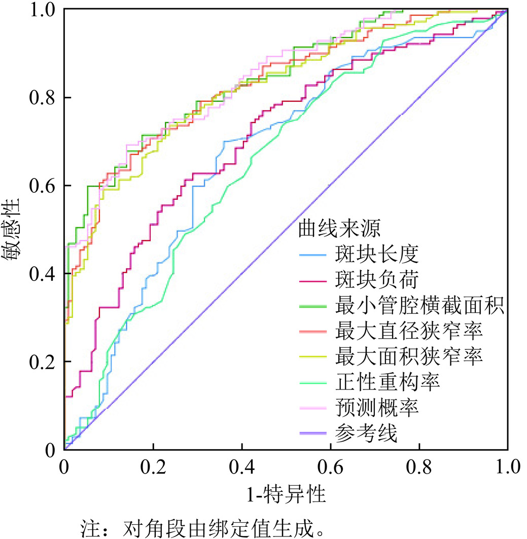
图 4 斑块定量指标诊断心肌缺血的ROC曲线
Figure 4. ROC curve for diagnosing myocardial ischemia using plaque quantification indicators.
表 1 一般资料比较
Table 1 Comparison of clinical data
一般资料 缺血患者组 非缺血患者组 $t/\chi^2 $ P值 n=106 n=57 性别(男/女,n) 77/29 39/18 0.322 0.571 年龄(岁) 63.3±9.5 62.7±10.0 0.354 0.724 BMI(kg/m2) 25.72±3.43 25.51±2.97 2.265 0.681 合并基础疾病[n(%)] 高血压 72(67.92%) 45(78.95%) 2.223 0.136 糖尿病 44(41.51%) 26(45.61%) 0.255 0.614 高血脂 81(76.42%) 42(73.68%) 0.149 0.699 既往史[n(%)] 冠心病家族史 32(30.19%) 12(21.05%) 1.570 0.210 吸烟史 51(48.11%) 31(54.39%) 0.583 0.445 饮酒史 49(46.23%) 25(43.86%) 0.084 0.772  下载: 导出CSV
表 2 斑块特征定性及定量指标比较
Table 2 Comparison of qualitative and quantitative indicators of plaque characteristics
指标 缺血血管组 非缺血血管组 $t/\chi^2 $ P值 n=139 n=114 正性重构[n(%)] 100(71.94%) 72(63.16%) 2.221 0.136 低密度斑块[n(%)] 122(87.77%) 93(81.58%) 1.881 0.170 餐巾环征[n(%)] 25(17.99)% 10(8.77%) 4.460 0.035 点状钙化[n(%)] 57(41.01%) 29(25.44%) 21.592 0.000 PL(mm) 32.09±16.78 22.76±16.46 4.44 0.000 PV(mm3) 622.54±221.78 606.38±219.62 −3.041 0.078 PB(%) 23.00±14.27 12.92±10.96 6.188 0.000 MLA(mm2) 1.14±0.87 2.74±1.66 9.805 0.000 MDS(%) 73.63±17.71 52.07±13.79 10.626 0.000 MAS(%) 84.78±12.66 67.51±14.50 10.102 0.000 RI 1.38±0.28 1.25±0.26 3.525 0.001 EI 0.79±0.27 0.75±0.29 −1.083 0.284
下载: 导出CSV
表 3 斑块特征定性及定量指标对心肌缺血的影响
Table 3 Impact of qualitative and quantitative indicators of plaque characteristics on myocardial ischemia
指标 回归系数 标准误 优势比 95%可信区间 P值 餐巾环征 1.407 0.314 4.086 2.207~7.563 0.000 点状钙化 0.712 0.276 2.037 1.187~3.497 0.010 PL(mm) 0.034 0.008 1.035 1.018~1.051 0.000 PB(%) 0.061 0.011 1.063 1.040~1.087 0.000 MLA(mm2) −1.447 0.204 0.235 0.158~0.351 0.000 MDS(%) 0.093 0.013 1.097 1.070~1.126 0.000 MAS(%) 0.095 0.013 1.100 1.072~1.128 0.000 RI 1.782 0.535 5.939 2.080~16.958 0.001
下载: 导出CSV
表 4 斑块定量指标诊断心肌缺血的效能
Table 4 Efficacy of plaque quantification indicators in diagnosing myocardial ischemia
指标 AUC 标准误 渐进Sig 特异度(%) 灵敏度(%) 95%可信区间 PL(mm) 0.672 0.035 0.000 0.640 0.698 0.604~0.739 PB(%) 0.712 0.032 0.000 0.728 0.612 0.648~0.775 MLA(mm2) 0.843 0.024 0.000 0.863 0.676 0.796~0.889 MDS(%) 0.830 0.025 0.000 0.904 0.619 0.781~0.878 MAS(%) 0.821 0.026 0.000 0.763 0.734 0.771~0.872 RI 0.655 0.035 0.000 0.579 0.662 0.587~0.723 联合预测 0.844 0.023 0.000 0.860 0.691 0.798~0.890
下载: 导出CSV
表 5 斑块定量指标单一和联合预测的性能比较
Table 5 Comparison of predictive performance between single and combined plaque quantification indicators
指标 AUC 面积之间差异 标准误 Z统计 95%CI可信区间 P值 PL(mm) 0.672 0.172 0.034 5.085 0.106~0.239 0.000 PB(%) 0.712 0.132 0.030 4.471 0.074~0.190 0.000 MLA(mm2) 0.843 0.001 0.009 0.120 −0.016~0.019 0.904 MDS(%) 0.830 0.014 0.012 1.229 −0.008~0.037 0.220 MAS(%) 0.821 0.022 0.015 1.503 −0.007~0.052 0.133 RI 0.655 0.188 0.037 5.152 0.117~0.260 0.000 联合预测 0.844
下载: 导出CSV
-
[1] 国家心血管病中心, 中国心血管健康与疾病报告编写组, 胡盛寿. 中国心血管健康与疾病报告2023概要[J]. 中国循环杂志, 2024, 39(07): 625-660. DOI: 10.3969/j.issn.1000-3614.2024.07.001. NATIONAL CENTER FOR CARDIOVASCULAR DISEASES, THE WRITING COMMITTEE OF THE REPORT ON CARDIOVASCULAR HEALTH AND DISEASES IN CHINA, HU S S. Report on cardiovascular health and diseases in China 2023: an updated summary[J]. Chinese Circulation Journal, 2024, 39(07): 625-660. DOI: 10.3969/j.issn.1000-3614.2024.07.001.
[2] 专家组中国冠状动脉血流储备分数测定技术临床路径专家共识. 中国冠状动脉血流储备分数测定技术临床路径专家共识[J]. 中国介入心脏病学杂志, 2019, 27(3): 121-133. DOI: 10.3969/j.issn.1004-8812.2019.03.001. [3] 闫昕, 赵建华. 基于CCTA的冠状动脉周围脂肪组织影像组学研究进展[J]. CT理论与应用研究, 2024, 33(4): 531-538. DOI: 10.15953/j.ctta.2023.179. YAN X, ZHAO JH. Research progress of pericoronary adipose tissue radiomics based on coronary computed tomography angiography[J]. CT Theory and Applications, 2024, 33(4): 531-538. DOI: 10.15953/j.ctta.2023.179.
[4] 胸痛中心专家委员会, 中华医学会心电生理和起搏分会, 中国医师协会心律学专业委员会, 等. 冠状动脉粥样硬化性心脏病猝死防治专家共识(2024)[J]. 中华心血管病杂志(网络版), 2024, 07(1): 1-18. DOI: 10.3760/cma.j.cn116031.2024.1000177. [5] 高扬, 吕滨. 冠状动脉CT血管成像最新临床应用推荐及诊断规范[J]. 中华放射学杂志, 2022, 56(10): 1160-1164. DOI: 10.3760/cma.j.cn112149-20220630-00555. GAO Y, LV B. The latest clinical application recommendation and diagnostic criteria of coronary CT angiography[J]. Chinese Journal of Radiology, 2022, 56(10): 1160-1164. DOI: 10.3760/cma.j.cn112149-20220630-00555.
[6] LEE J M, CHOI G, KOO B, et al. Identification of high-risk plaques destined to cause acute coronary syndrome using coronary computed tomographic angiography and computational fluid dynamics[J]. JACC: Cardiovascular Imaging, 2019, 12(6): 1032-1043. DOI: 10.1016/j.jcmg.2018.01.023.Epub2018Mar14.
[7] 李正腾, 王敏, 潘冬梅, 等. CCTA斑块特征在冠状动脉管腔狭窄程度进展预测及预后的价值研究[J]. CT理论与应用研究, 2025, 34(1): 23-30. DOI: 10.15953/j.ctta.2024.172. LI Z T, WANG M, PAN D M, et al. Predictive Value of CCTA Plaque Characteristics for the Progression and Prognosis of Coronary Artery Stenosis[J]. CT Theory and Applications, 2025, 34(1): 23-30. DOI: 10.15953/j.ctta.2024.172.
[8] 高雪莲, 王瑞, 张宏凯, 等. 冠状动脉CT血管造影影像组学用于冠心病研究进展[J]. 中国医学影像技术, 2024, 40(3): 451-454. DOI: 10.13929/j.issn.1003-3289.2024.03.027. GAO X L, WANG R, ZHANG H K et al. Research progresses of radiomics based on coronary CT angiography in coronary artery disease[J]. Chinese Journal of Medical Imaging Technology, 2024, 40(3): 451-454. DOI: 10.13929/j.issn.1003-3289.2024.03.027.
[9] DESEIVE S, KUPKE M, STRAUB R, et al. Quantified coronary total plaque volume from computed tomography angiography provides superior 10-year risk stratification[J]. European Heart Journal cardiovascular Imaging, 2021, 22(3): 314-321. DOI: 10.1093/ehjci/jeaa228.
[10] JIAN Z, YAO G, GUO H, et al. The impact of baseline calcified plaque volume on coronary rapid plaque progression by serial coronary computed tomography angiography in patients with type 2 diabetes[J]. Annals of Medicine, 2023, 55(1): 2196438. DOI: 10.1080/07853890.2023.2196438.
[11] 盛玉杰, 王询, 王泽静. CT冠状动脉定量在评估冠心病患者心肌缺血诊断中的应用价值[J]. 中国CT和MRI杂志, 2024, 22(5): 103-105. DOI: 10.3969/j.issn.1672-5131.2024.05.033. SHENG Y J, WANG X, WANG Z J. Application value of coronary CT angiography quantification in the diagnosis of myocardial ischemia in patients with coronary heart disease[J]. Chinese Journal of CT and MRI, 2024, 22(5): 103-105. DOI: 10.3969/j.issn.1672-5131.2024.05.033.
[12] 孙俊, 夏花, 江时忠. 冠状动脉CT血管成像定量评估冠心病心肌缺血的效能[J]. 浙江医学, 2022, 44(6): 637-640, 645. DOI: 10.12056/j.issn.1006-2785.2022.44.6.2021-2378. SUN J, XIA H, JIANG S Z. Value of coronary CT angiography in quantitative assessment of myocardial ischemia in patients with coronary heart disease[J]. Zhejiang Medical Journal 2022, 44(6): 637-640, 645. DOI: 10.12056/j.issn.1006-2785.2022.44.6.2021-2378. (in Chinese).
[13] CHUNG C J, JEONG S Y, JEONG J H, et al. Comparison of prophylactic effect of topical alchemilla vulgaris in glycerine versus that of dexamethasone on postoperative sore throat after tracheal intubation using a double-lumen endobronchial tube: a randomized controlled study[J]. Anesthesia and Pain Medicine, 2021, 16(2): 163-170. DOI: 10.17085/apm.20082.
[14] 高艳, 顾慧, 杨世锋, 等. 基于冠状动脉CT血管成像的斑块定量分析及其与心肌缺血损伤的相关性研究[J]. 中华放射学杂志, 2020, 54(2): 7. DOI: 10.3760/cma.j.issn.1005-1201.2020.02.008. GAO Y, GU H, YANG S, et al. Correlation study of coronary plaque quantitative analysis and myocardial ischemic injury based on coronary CT angiography[J]. Chinese Journal Radiology, 2020, 54(2): 7. DOI: 10.3760/cma.j.issn.1005-1201.2020.02.008.
[15] LONG Y, GUO R, JIN K, et al. Analysis of the perivascular fat attenuation index and quantitative plaque parameters in relation to hemodynamically impaired myocardial ischemia[J]. The International Journal of Cardiovascular Imaging, 2024, 40(7): 1455-1463. DOI: 10.1007/s10554-024-03122-x.
[16] BAR S, MAANIITTY T, KIATKITTIKUL P, et al. Incremental prognostic value of artificial intelligence-based automated plaque characterisation on top of ischemia evaluation by CCTA and CCTA/PET[J]. European Heart Journal Cardiovascular Imaging, 2024, 25(Sup1): 1. DOI: 10.1093/ehjci/jeae142.036.
[17] SAITO Y, KOBAYASHI Y, FUJII K, et al. CVIT 2023 clinical expert consensus document on intravascular ultrasound[J]. Cardiovascular Intervention and Therapeutics, 2024, 39(1): 1-14. DOI: 10.1007/s12928-023-00957-4.
[18] LIN A, MANRAL N, MCELHINNEY P, et al. Deep learning-enabled coronary CT angiography for plaque and stenosis quantification and cardiac risk prediction: an international multicentre study[J]. Lancet Digit Health, 2022, 4(4): e256-e265. DOI: 10.1016/S2589-7500(22)00022-X.
[19] KAWASAKI T, KIDOH M, KIDO T et al. Evaluation of significant coronary artery disease based on CT fractional flow reserve and plaque characteristics using random forest analysis in machine learning[J]. Academic Radiology, 2020, 27(12): 1700-1708. DOI: 10.1016/j.acra.2019.12.013.
[20] 朱娜君, 方欣欣, 尹伊君, 等. 心绞痛患者斑块进展危险因素与冠状动脉CT血管成像指标的关系研究[J]. CT理论与应用研究, 2023, 32(2): 217-222. DOI: 10.15953/j.ctta.2022.219. ZHU N J, FANG X X, YIN Y J et al. Risk factors of plaque progression in patients with angina pectoris and their relationships with coronary CT angiography[J]. CT Theory and Applications, 2023, 32(2): 217-222. DOI: 10.15953/j.ctta.2022.219.


计量
- 文章访问数: 35
- HTML全文浏览量: 5
- PDF下载量: 6


