
Study on the Influence of Head and Neck CTA Image Quality under Different Tube Voltages
-
摘要: 目的:研究不同管电压对头颈部CTA图像质量的影响。资料与方法:收集亭林医院2021年9月至2022年1月临床怀疑头颈部血管疾病的患者共67例,其中男37例,女30例,平均年龄(68.94±11.68)岁,根据施加的管电压不同分成3组,将患者分配到A、B、C三组,A组为120 kV 23例、B组为100 kV 24例、C组为80 kV 20例。采用单因素ANOVA检验,比较3组间的主动脉弓、颈内动脉、大脑中动脉M1段以及胸锁乳突肌的CT值、SD值、SNR、CNR,以及图像质量主观评分、TDLP以及ED,采用Kappa检验法,检验两名医师主观评分的一致性。结果:3组间主动脉弓、颈内动脉、大脑中动脉M1段血管CT值及SD值差异有统计学意义;主动脉弓、颈内动脉、大脑中动脉M1段血管CT值及SD值随管电压下降而上升,上升幅度为15%~55%;3组间SNR及CNR差异无统计学意义;3组间的图像质量主观评分差异有统计学意义,B组>A组>C组,以B组评分最高;两名医师主观评分的Kappa=0.80,为高度一致;3组间TDLP以及ED有统计学差异,TDLP以及ED随管电压下降而下降,下降幅度为25% 和39%。结论:根据CT辐射剂量诊断参考水平的要求,在进行头颈部CTA检查时,3种管电压以100 kV为最佳管电压条件。Abstract: Objective: To explore the influence on the image quality of head and neck CTA under different tube voltages. Materials and methods: A total of 67 patients with clinically suspected head and neck vascular diseases in Tinglin hospital from September 2021 to January 2022 were collected as the research objects, including 37 males and 30 females, with an average age of 68.94. According to the applied tube voltage, the patients were randomly assigned into groups A, B and C. There were 23 cases of 120 kV in group A, 24 cases of 100 kV in group B and 20 cases of 80 kV in group C. Single factor ANOVA test was used to compare the CT value, SD value, SNR, CNR of aortic arch, internal carotid artery, M1 segment of middle cerebral artery and sternocleidomastoid muscle among the three groups, and furthermore the subjective score of image quality, TDLP and ED. Kappa test was used to evaluate the consistency of subjective scores of the two physicians. Results: There were statistical differences in CT and SD values of aortic arch, internal carotid artery and M1 segment of middle cerebral artery among the three groups; The CT and SD values of aortic arch, internal carotid artery and M1 segment of middle cerebral artery increased with the decrease of tube voltage, with an increase range of 15%~55%. There was no statistical difference in SNR and CNR among the three groups; There was statistical difference in subjective score of image quality among the three groups; Group B > group A > group C, and group B showed the highest score; the kappa value of the subjective score of the two phsicianss=0. 80, which was highly consistent; There were statistical differences in TDLP and ED among the three groups, TDLP and ED decreased with the decrease of tube voltage, with the decrease ranges of 25% and 39%. Conclusion: According to the requirements of CT radiation dose diagnosis reference level, 100 kV is the best tube voltage condition for head and neck CTA.
-
Keywords:
- head and neck CTA /
- tube voltages /
- image quality
-
-
![]()
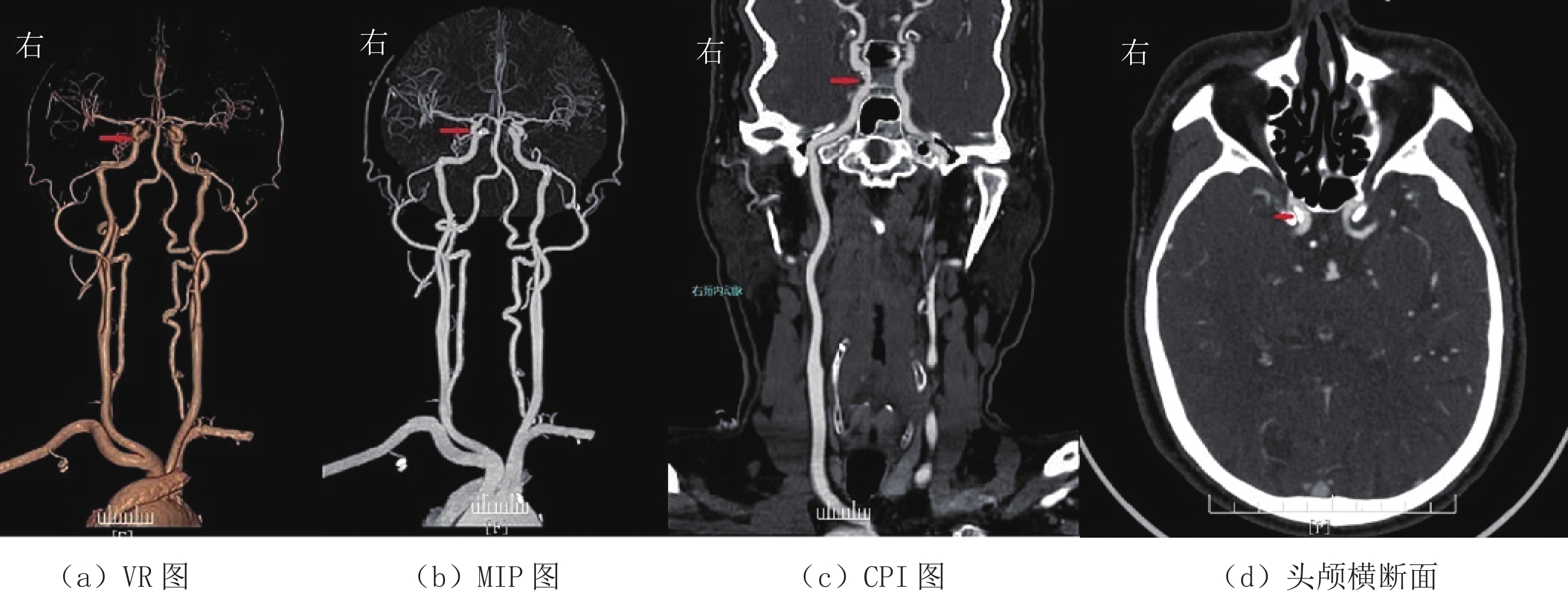
图 1 管电压为120 kV的VR、MIP、CPI及头颅横断面图
(a)~(d)分别为120 kV管电压的VR、MIP、CPI及头颅横断面图,图像质量较好,血管与组织对比较好,解剖结构较清晰,噪声较小,双盲5分值评价法,4分。头颈部CTA诊断:双侧颈内动脉起始部及C6段钙化斑块伴右侧颈内动脉C6段管腔轻度狭窄。
Figure 1. VR、MIP、CPI and head cross-sectional drawing under pipe voltage of 120 kV
![]()
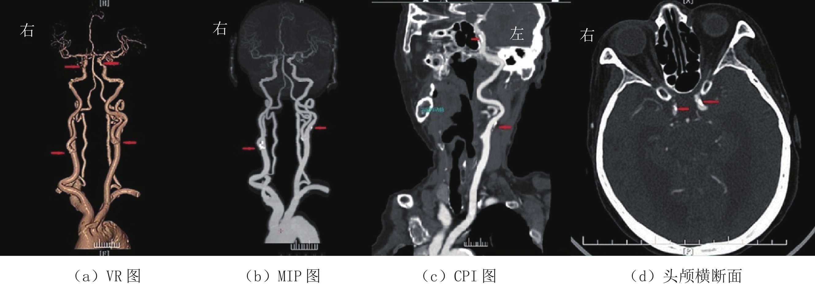
图 2 管电压为100 kV的VR、MIP、CPI及头颅横断面图
(a)~(d)分别为100 kV管电压的VR、MIP、CPI及头颅横断面图,图像质量好,血管与组织对比鲜明,细微解剖结构清晰,噪声小,双盲5分值评价法,5分。头颈部CTA诊断:右侧颈内C6-7段钙化斑块伴管腔轻度狭窄,双侧椎动脉走形迂曲。
Figure 2. VR、MIP、CPI and head cross-sectional drawing under pipe voltage of 100 kV
![]()
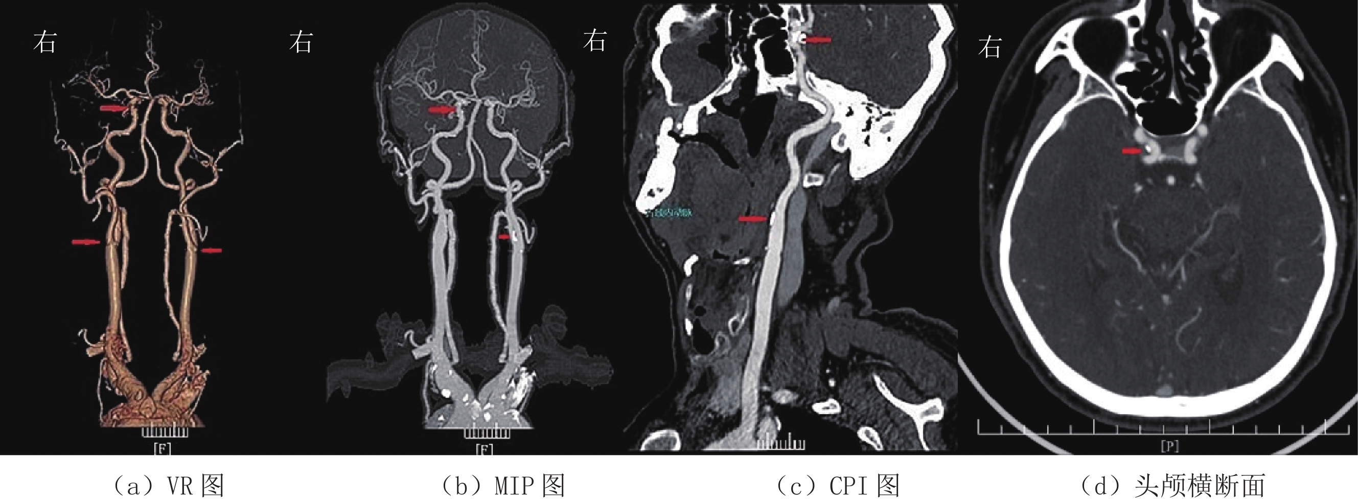
图 3 管电压为80 kV的VR、MIP、CPI及头颅横断面图
(a)~(d)分别为80 kV管电压的VR、MIP、CPI及头颅横断面图,图像质量一般,血管解剖结构显示一般,噪声略大,双盲5分值评价法,3分。头颈部CTA诊断:右侧颈内起始部及C5-6段动脉钙化斑块伴C5-6段管腔轻度狭窄;左侧颈内动脉起始部混合斑块伴局部管腔轻度狭窄;左侧颈内C5-6段钙化斑块伴管腔中度狭窄。
Figure 3. VR、MIP、CPI and head cross-sectional drawing under pipe voltage of 80 kV
表 1 三组病例一般资料、主观评分和辐射剂量之间比较
Table 1 Comparison of general information, subjective score and radiation dose among the three groups
项目 组别 统计检验 A组 B组 C组 F P 男/女 15/8 11/13 11/9 0.88 0.42 年龄/Y 66.83±11.76 69.00±11.59 71.15±10.55 0.78 0.46 身高/mm 165.04±8.67 161.96±6.70 162.65±7.28 1.05 0.36 体重/kg 67.17±12.60 62.04±8.05 62.30±9.16 1.85 0.17 主观评分 4.70±0.47 4.88±0.34 4.40±0.68 4.87 0.01 TDLP/mGy·cm 1169.57±116.07 873.17±56.98 716.70±44.38 180.75 0.00 ED/mGy·cm 9.63±0.96 7.18±0.47 5.90±0.37 180.75 0.00  下载: 导出CSV
表 2 三组病例CT值、SD值、SNR及CNR之间比较
Table 2 Comparison of CT value SD value, SNR and CNR among the three groups
部位 参数 组别 统计检验 A组 B组 C组 F P 升主动脉 血管CT值/HU 296.61±69.92 342.13±76.21 460.50±130.44 17.11 0.00 SD 10.61±3.61 12.92±4.09 16.00±4.15 9.98 0.00 SNR 86.83±38.32 94.69±32.52 97.74±38.14 0.53 0.53 CNR 71.65±34.33 77.03±28.57 84.42±35.46 0.82 0.45 颈动脉分叉 血管CT值/HU 352.13±90.94 429.63±99.42 574.95±136.12 22.77 0.00 SD 5.00±2.83 5.79±6.22 7.40±3.03 4.01 0.02 SNR 103.80±48.11 119.34±45.70 121.33±41.98 1.00 0.38 CNR 88.63±44.04 101.67±41.96 108.00±39.43 1.21 0.31 大脑中动脉
M1段血管CT值/HU 334.21±69.42 385.00±85.53 502.50±110.17 19.94 0.00 SD 5.83±4.72 7.29±6.20 10.80±6.44 4.08 0.02 SNR 97.79±42.20 108.52±47.52 107.70±37.78 0.44 0.64 CNR 82.61±37.93 90.86±43.18 94.37±34.03 0.53 0.59 胸大肌 软组织CT值/HU 52.70±8.43 62.12±6.72 60.55±7.27 10.40 0.00 SD 3.74±0.92 4.08±1.86 5.15±1.93 4.32 0.02
下载: 导出CSV
-
[1] 中华医学会放射学分会. 头颈部CT血管成像扫描方案与注射方案专家共识[J]. 中华放射学杂志, 2019, 52(2): 81-87. Chinese Society of Radiology, Chinese Medical Association. Expert consensus of the head and neck CT angiography scanning and injection protocols[J]. Chinese Journal of Radiology, 2019, 52(2): 81-87. (in Chinese).
[2] YU S, ZHENG J, ZHANG L. Craniocervical computed tomography angiography with adaptive iterative dose reduction 3D algorithm and automatic tube current modulation in patients with different body mass indexes[J]. Medicine, 2018, 97(36): 1−6.
[3] 陈其锋, 张雄彪, 李水连, 等. 多层螺旋CT低剂量对比剂在头颈联合CT血管成像应用的可行性研究[J]. 中国医学装备, 2021,18(5): 61−64. doi: 10.3969/J.ISSN.1672-8270.2021.05.015 CHEN Q F, ZHANG X B, LI S L, et al. The feasibility of the application of low-dose contrast agent of MSCT in the combined CTA on head and neck[J]. China Medical Equipment, 2021, 18(5): 61−64. (in Chinese). doi: 10.3969/J.ISSN.1672-8270.2021.05.015
[4] 黄爱娜, 陆健, 张涛, 等. 256层iCT低剂量扫描联合迭代重建技术在头颈部CTA的应用[J]. 放射学实践, 2018,33(8): 842−846. doi: 10.13609/j.cnki.1000-0313.2018.08.016 HUAN A N, LU J, ZHANG T, et al. Application of 256 iCT low-dosescanning with iterative reconstruction technique in craniocervical arteries[J]. Radiologic Practice, 2018, 33(8): 842−846. (in Chinese). doi: 10.13609/j.cnki.1000-0313.2018.08.016
[5] ELLMANN S, KAMMERER F, ALLMENDINGER T, et al. Dose reduction potential of iterative reconstruction algorithms in neck CTA: A simulation study[J]. Dento Maxillo Facial Radiology, 2016, 45(8): 228−234.
[6] 赵冰辉, 尹伟, 王敏杰. 基于器官的剂量调制技术在头颈部CT联扫中的应用[J]. 中国CT和MRI杂志, 2021,19(11): 38−41. doi: 10.3969/j.issn.1672-5131.2021.11.013 ZHAO B H, YIN W, WANG M J. Application of organ based dose modulation technology in CT head and neck joint scanning[J]. Chinese Journal of CT and MRI, 2021, 19(11): 38−41. (in Chinese). doi: 10.3969/j.issn.1672-5131.2021.11.013
[7] 中华医学会放射学分会质量管理与安全管理学组. CT辐射剂量诊断参考水平专家共识[J]. 中华放射学杂志, 2017,51(11): 817−822. doi: 10.3760/cma.j.issn.1005-1201.2017.11.001 Quality Management and Safety Management Group, Chinese Society of Radiology, Chinese Medical Association. Expert consensus on reference level of CT radiation dose diagnosis[J]. Chinese Journal of Radiology, 2017, 51(11): 817−822. (in Chinese). doi: 10.3760/cma.j.issn.1005-1201.2017.11.001
[8] ROCH P, CÉLIER D, DESSAUD C, et al. Using diagnostic reference levels to evaluate the improvement of patient dose optimisation and the influence of recent technologies in radiography and computed tomography[J]. European Journal of Radiology, 2018, 98(1): 68−74.
[9] 王永胜, 王晨思, 陆浩宇, 等. 个性化造影剂注射方案在提升肺动脉CTA生物应用安全性的价值研究[J]. CT理论与应用研究, 2021,30(6): 777−783. DOI: 10.15953/j.1004-4140.2021.30.06.14. WANG Y S, WANG C S, LU H Y, et al. Study on the value of individualized contrast agent injection scheme in improving the biosafety of pulmonary Artery CTA[J]. CT Theory and Applications, 2021, 30(6): 777−783. DOI: 10.15953/j.1004-4140.2021.30.06.14. (in Chinese).
[10] 陆晓平, 王沄, 徐敏, 等. 100 kVp条件下全模型迭代技术对头颈CTA图像质量的影响[J]. 中国医疗设备, 2021,36(10): 80−95. doi: 10.3969/j.issn.1674-1633.2021.10.019 LU X P, WANG Y, XU M, et al. Evaluation on the effect of FIRST on head and neck CTA image quality at 100 kVp[J]. China Medical Devices, 2021, 36(10): 80−95. (in Chinese). doi: 10.3969/j.issn.1674-1633.2021.10.019
[11] 王贤坤, 邹才盛. 不同管电压对头颈CTA图像质量及辐射剂量的影响分析[J]. 影像研究与医学应用, 2019,3(5): 21−24. doi: 10.3969/j.issn.2096-3807.2019.05.012 WANG X K, ZOU C S. Analysis of image quality and radiation dose of CT angiography under different tube voltages[J]. Journal of Imaging Research and Medical Applications, 2019, 3(5): 21−24. (in Chinese). doi: 10.3969/j.issn.2096-3807.2019.05.012
[12] NI Q Q, CHEN G Z, SCHOEPF U J, et al. Cerebral CTA with low tube voltage and low contrast material volume for detection of intracranial aneurysms[J]. American Journal Neuroradiology, 2016, 37(10): 1774−1780. doi: 10.3174/ajnr.A4803
[13] EBERSBERGER U, TRICARICO F, SCHOEPF U J, et al. CT evaluation of coronary artery stents with iterative image reconstruction: Improvements in image quality and potential for radiation dose reduction[J]. European Radiology, 2013, 23(1): 125−132. doi: 10.1007/s00330-012-2580-5
[14] EISENTOPF J, ACHENBACH S, ULZHEIMER S, et al. Low-dose dual-source CT angiography with iterative reconstruction for coronary artery stent evaluation[J]. JACC: Cardiovascular Imaging, 2013, 6(4): 458−465. doi: 10.1016/j.jcmg.2012.10.023
[15] 刘伟华, 刘登平. 不同管电压下颅脑CT血管造影图像质量及辐射剂量的对照性研究[J]. 实用医学影像杂志, 2018,19(2): 114−117. doi: 10.16106/j.cnki.cn14-1281/r.2018.02.008 LIU W H, LIU D P. Comparative studyon image quality and radiation dose of different tube voltage by cerebral CT angiography[J]. Journal of Pracical Medical Imaging, 2018, 19(2): 114−117. (in Chinese). doi: 10.16106/j.cnki.cn14-1281/r.2018.02.008
[16] 武洪林, 李俊, 李红尧, 等. 双源CT低管电压及低剂量等渗对比剂在头颈部CTA中的初步研究[J]. 医学影像学杂志, 2022,27(7): 1213−1217. WU H L, LI J, LI H Y, et al. Dual source CT angiography with low kVp and low volume of isotonic contrast material for head and neck arteries: A preliminary study[J]. Journal of Medical Imaging, 2022, 27(7): 1213−1217. (in Chinese).
[17] KIDOH M, NAKAURA T, NAKAMURA S, et al. Low-dose abdominal CT: Comparison of low tube voltage with moderate-level iterative reconstruction and standard tube voltage, low tube current with high-level iterative reconstruction[J]. Clinical Radiology, 2013, 68(10): 1008−1015. doi: 10.1016/j.crad.2013.04.008
[18] KAYAN M, DEMIRTAS H, TÜRKER Y, et al. Carotid and cerebral CT angiography using low volume of iodinated contrast material and low tube voltage[J]. Diagnostic and Interventional Imaging, 2016, 97(11): 1173−1179. doi: 10.1016/j.diii.2016.06.005
[19] ERTLWAGNER B B, HOFFMANN R T, BRUNING R, et al. Multi-detector row CT angiography of the brain at various kilovoltage settings[J]. Radiology, 2004, 231(2): 528−535. doi: 10.1148/radiol.2312030543
-
期刊类型引用(11)
1. 王彦淇,张宇,苏有琦,张茜,叶敏. 基于CEEMD的希尔伯特-黄变换算法在森林边界层湍流中的应用. 高原气象. 2025(02): 445-461 .  百度学术
百度学术
2. 宋海龙,叶冬萌,史磊,梁天宇,柴斌,杨松浩,郝治国. 移相控制下的特高压直流断路器失灵保护策略. 高压电器. 2023(05): 19-27 . 百度学术
3. 徐海兵,郭久明. 基于双向GRU模型的网络流量预测的研究. 电子技术应用. 2022(02): 19-22+27 . 百度学术
4. 程江,张旭,涂君,廖春晖. 超声衍射时差法近表面盲区减小算法研究. 传感器与微系统. 2022(06): 48-51+60 . 百度学术
5. 吴传奇,柴晓冬,李立明,郑树彬. 改进希尔伯特-黄变换方法在钢轨波磨检测中的应用. 铁道标准设计. 2021(09): 74-80 . 百度学术
6. 张玲,卞建鹏,郝培旭,李亚敏,孙晓云. 基于CEEMD和ABC-LSTM的电力变压器油中溶解气体预测. 兰州交通大学学报. 2021(05): 58-64+92 . 百度学术
7. 杨本贤,何冰冰,张榆锋,聂建云,姚瑞晗,刘亚杰. 基于EEMD有效成分优选的超声多普勒血流测速法. 云南大学学报(自然科学版). 2021(06): 1107-1116 . 百度学术
8. 李英春,白艺,褚恩亮,朱世刚. 基于CEEMDAN-EDO的行波波头标定算法研究. 中国测试. 2021(12): 98-105 . 百度学术
9. 司友强,呙润华,李梦茹. 基于EMD与EEMD的最优降噪光滑模型在地震波中的应用. CT理论与应用研究. 2020(01): 11-21 . 本站查看
10. 魏香林. 回转齿轮箱故障特征提取的CMF-EEMD方法分析. 机械设计与制造工程. 2020(04): 105-107 . 百度学术
11. 张恒璟,陆帝,文汉江,程鹏飞,崔东东. 一种IGS站高程时间序列分析方法. 遥感信息. 2019(06): 1-5 . 百度学术
其他类型引用(24)
计量
- 文章访问数: 233
- HTML全文浏览量: 51
- PDF下载量: 21
- 被引次数: 35
