
Research Progress of Scattering Artifact Correction in Medical Cone-beam Computed Tomography Imaging Based on Deep Learning
-
摘要: 医用计算机断层扫描成像系统中,X射线与物体相互作用产生的康普顿散射光子严重影响了图像质量,尤其在锥形束计算机断层扫描和多层探测器系统中。目前已有许多散射伪影校正方法,归纳为3类:硬件校正、软件校正、软硬件混合校正方法,但近年随着计算机计算能力的提高以及深度学习在医学图像处理领域的发展,出现了一些新的散射校正方法。本文首先介绍传统校正方法;然后详细介绍基于深度学习方法进行散射伪影校正,并将其分为基于图像域和基于投影域的深度学习方法,以及对不同的深度学习网络在散射伪影校正中的应用进行讨论;最后展望深度学习在多源计算机断层扫描技术中的应用前景。Abstract: In medical computed tomography imaging systems, Compton scattered photons generated by the interaction between X-rays and objects have a serious impact on image quality, especially in cone-beam computed tomography and multi-layer detector systems. Currently, there are many scattering artifact correction methods, which can be classified into three categories: hardware, software, and hybrid software and hardware correction methods. However, with the advances in computing power and development of deep learning in medical image processing, new methods of scattering artifact correction have appeared in recent years. This study first introduces traditional correction methods. Then, a method of scattering artifact correction based on deep learning is described in detail, which is divided into the correction method based on image domain and the correction method based on projection domain. Various deep-learning neural networks for this method are also introduced in detail. Finally, the application prospects of the deep learning method in multi-source computed tomography imaging scattering artifacts were probed .
-
Keywords:
- X-ray optics /
- deep learning /
- Compton scattering /
- cone-beam CT /
- artifact correction
-
-
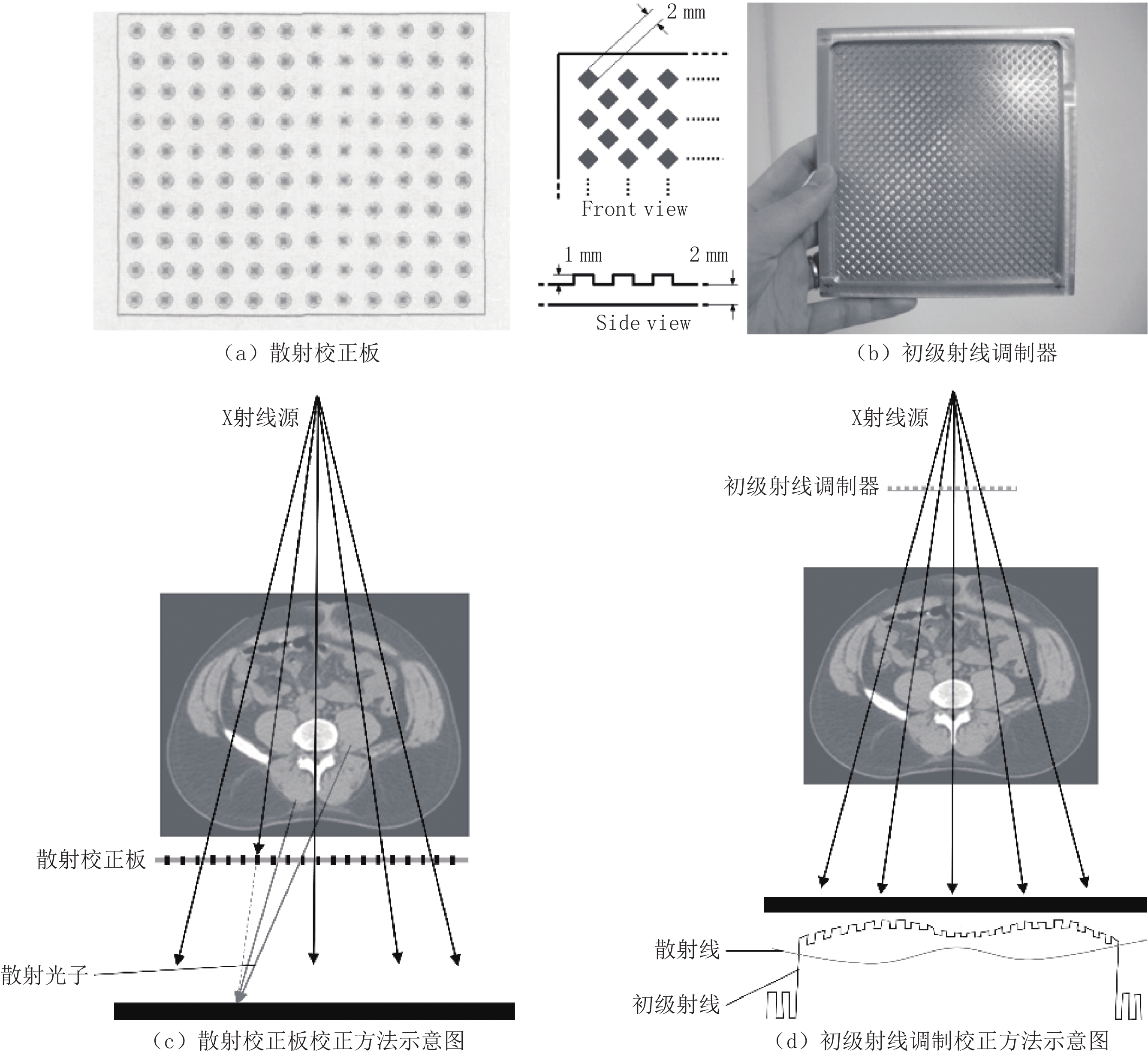
![]()
图 2 软硬件混合校正方法示意图
Figure 2. Schematic diagram of the hybrid correction method of software and hardware
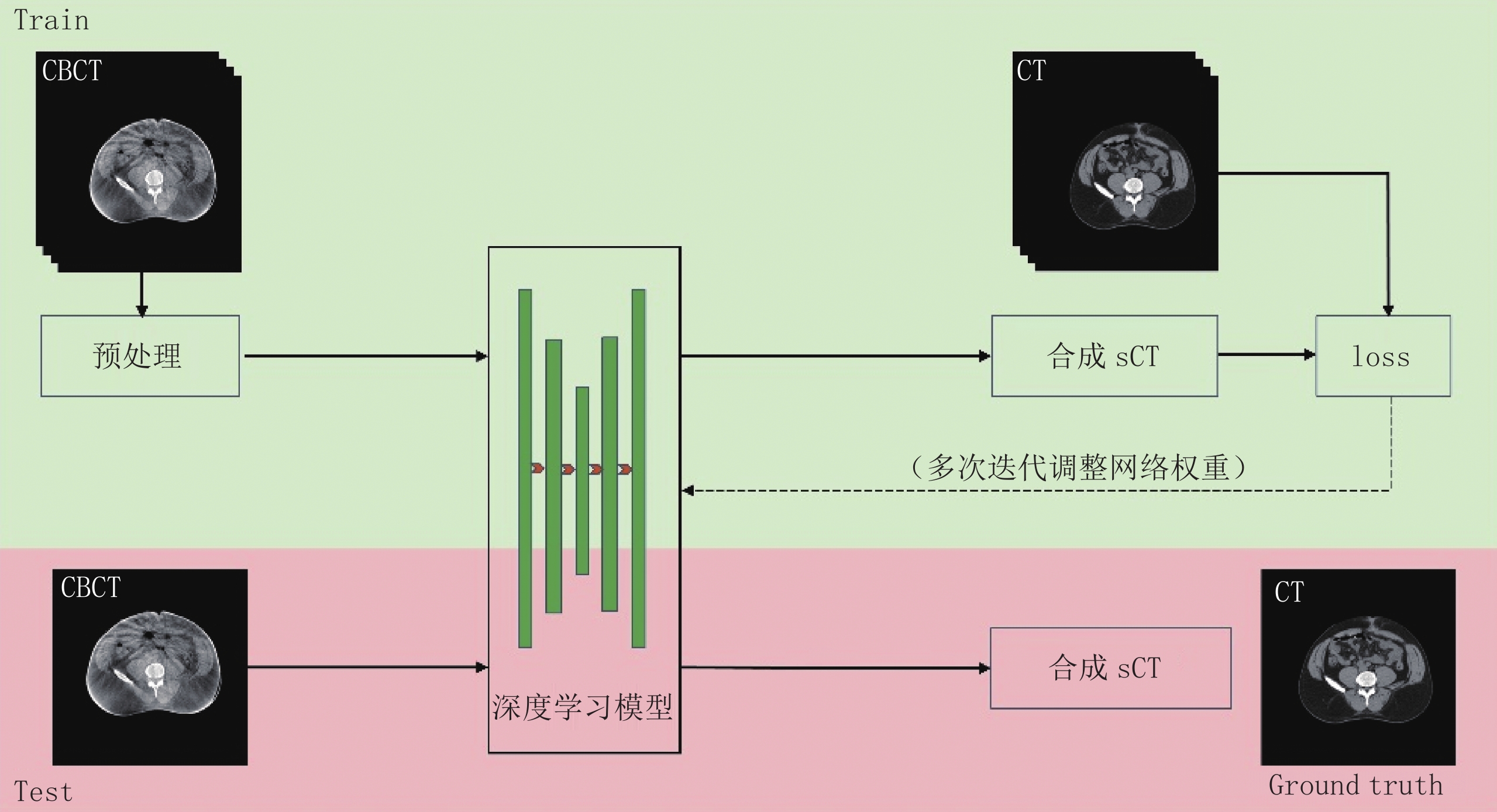
![]()
图 3 深度学习散射伪影校正方法的流程图
Figure 3. Flow diagram of deep learning method for scattering artifact correction
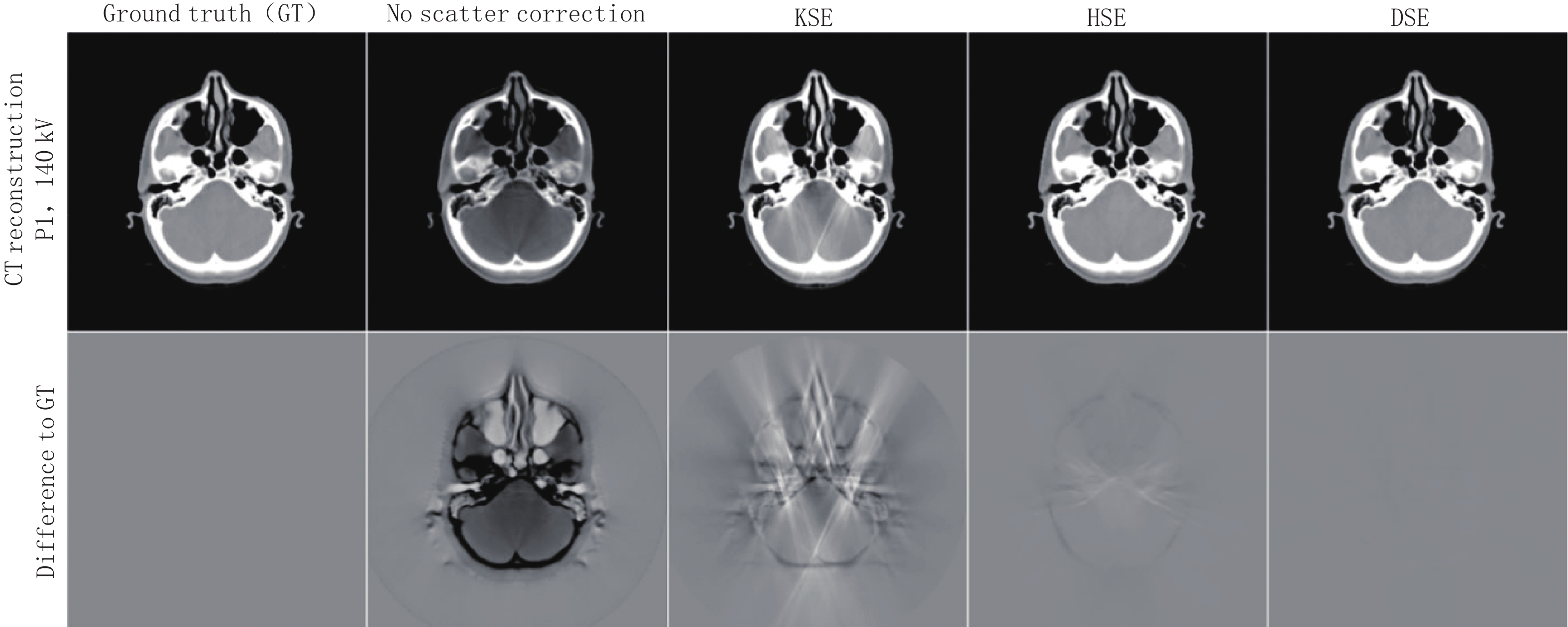
![]()
图 4 卷积核方法、卷积核与MC混合方法和深度学习方法校正散射伪影的对比图
Figure 4. Comparison diagram of kernel-based scatter estimation (KSE), the hybrid scatter estimation (HSE) and deep scatter estimation (DSE) for scattering artifact correction
表 1 基于深度学习神经网络的CBCT散射伪影校正的研究汇总
Table 1 Research summary of CBCT scattering artifact correction based on deep learning neural network
项目 作者 年份 数据模体 数据获取 数据集(训练︰测试/
训练︰验证︰测试)网络 MAE/HU SSIM 其他指标 耗时/s 基
于
投
影
域Hansen等[36] 2018 前列腺癌患者的盆腔 放射治疗PCT和CBCT 15︰7︰8(患者数) U-Net 144.000降至46.000 - ME/HU:138.000降至 -3.000 0.010/投影 Maier等[6] 2018 头部、胸部、盆腔 MC 仿真 22︰6(患者数) U-Net - - ME/HU:278.000降至6.000 0.010/投影 Nomura等[37] 2019 数字几何数字胸部和数字头部模体 Gate的MC仿真 14400︰200︰360 U-Net 头17.900胸29.000 头1.000
胸0.999PSNR/dB:头:37.200
胸:31.7004.800/360投影 Lee等[38] 2019 数字胸部模体 Gate的MC仿真 12960︰360 CNN - MC:0.960
CNN:0.992PSNR/dB:
MC:42.100
CNN:49.7200.017/投影 钟安妮[40] 2020 头颈癌患者的头颈部 gDRR和 gMMC包进行MC仿真 22︰3(患者数)
盆腔模体测试U-Net - 头0.999
盆腔0.999UIQI:头0.994盆腔0.997 头,盆腔:9.050,
9.670/360投影Lalonde等[41] 2020 头颈癌患者的头颈部 MCGPU包仿真患者CBCT投影 29︰9︰10(患者数) U-Net 69.640降至13.410 - ME/HU:-28.610降至 -0.801 0.014/投影 Ma等[22] 2020 头部盆腔 gCTD包进行MC
仿真头部:45︰45
盆腔测试Deep Q-Net - 0.990 PSNR/dB:36.050 dB 1.810/投影 Rusanov等[39] 2021 头胸腹模体和患者头颈部 放疗PCT和CBCT 8004︰1000(头胸腹模体︰患者头颈) U-Net 318.000降至74.000 0.750提升至
0.812CNR:6.690到13.900 20.000/500投影 基
于
图
像
域Kida等[34] 2018 前列腺癌患者的盆腔部 放射治疗PCT和CBCT 14400︰3600 U-Net - 脂肪0.965
肌肉0.969PSNR/dB:
脂肪50.600
肌肉51.30020.000/180切片 Xie等[42] 2018 肺癌患者的胸部 放射治疗PCT和CBCT 15︰5(患者数) Deep CNN - - PSNR/dB:7.889提升至8.823 0.008/切片 Jiang等[43] 2019 患者盆腔 MC-GPU包进行MC仿真 2400︰480︰320 U-NetRNM - 0.950提升至
0.990RMSE/HU:200.000降至20.000 27.000/160切片 Liang等[44] 2019 头颈癌患者头颈部以及一个真实头部模体 放射治疗PCT和CBCT 81(6480)︰9(720)︰20 CycleGAN 69.290 降至29.850 0.730提升至
0.850PSNR/dB:25.280提升至30.650 - 基
于
图
像
域Kurz等[45] 2019 前列腺癌患者的盆
腔部放射治疗PCT和CBCT 25︰8(患者数) CycleGAN 103.000降至87.000 - ME/HU:24.000降至
-6.00010.000/88切片 Harms等[46] 2019 头部盆腔 放射治疗PCT和CBCT 头部︰22︰2
盆腔︰19︰1(患者数)Res-CycleGAN 头部23.800降至13.000
盆腔56.300降至16.100- PSNR/dB:头32.300提升至37.500。盆腔22.200提升至30.700 - Chen等[47] 2020 头颈癌患者头颈部 放射治PCT和CBCT 30(2400)︰7(560)︰7(560) U-Net 44.380降至18.980 0.711提升至0.891 PSNR:27.350提升至33.260 dB - Kida等[48] 2020 前列腺癌患者的盆
腔部放射治PCT和CBCT 16︰4(患者数) CycleGAN - 0.575提升至0.688 CT值:更接近PCT 1.000/32切片 Tien等[49] 2021 乳腺癌患者胸部 放射治PCT和CBCT 12(8706)︰
3(1150)Cycle-Deblur GAN 0.053降至0.024 0.987提升至0.996 PSNR/dB:24.435提升至30.560 0.170/切片 Dong等[50] 2021 前列腺癌患者的盆
腔部放射治疗PCT和CBCT 49︰9(患者数) CycleGAN 49.960降至14.600 0.728提升至0.825 PSNR/dB:26.820提升至32.500 - Qiu等[51] 2021 肺癌患者的胸部 放疗PCT和CBCT 4︰1(20患者) HM-Cycle-GAN 110.000降至66.200 0.850提升至0.910 PSNR/dB:23.000提升至30.300 - Liu等[52] 2021 患者胸部数据 放疗PCT和CBCT 32︰8︰12(患者数) GAN 70.560降至32.700 0.640提升至0.860 PSNR/dB:28.670提升至34.120 <1.000/切片 Park等[53] 2022 患者颌面部骨骼 多层螺旋CT和牙科CBCT 11000,11422(不配对):1100;6831︰1100(配对) GAN - - 骨强度/HU:1162.000提升至1220.000 0.200/切片 Zhang等[54] 2022 头颈癌患者头颈部 放疗PCT和CBCT 90︰30(患者数) Conditional GAN 36.230降至16.750 0.830提升至0.920 PSNR/dB:25.340提升至30.580 0.620/切片 投影
域与
图像
域Iskender
等[55]2021 数字钛棒模体和图像映射的数字胸部模体 MC仿真 27︰3(患者数) DCNN 钛棒35.000降至15.100
胸部35.800降至12.300钛棒0.988提升至0.997。
胸部0.860提升至0.975PSNR/dB:钛棒36.840提升至50.860。胸部26.850提升至37.150 2.000~4.000/重建体积 注:MAPE(mean absolute percentage error)平均绝对百分比误差,ME(mean error)平均误差,MAE(mean absolute error)平均绝对值误差,PSNR(peak signal to noise ratio)峰值
信噪比,SSIM(structured similarity indexing method)结构相似性指数方法,RMSE(root mean square error)均方根误差,UIQI(universal image quality Index)通用图像质量
指数,CNR(contrast-to-noise ratio)对比度噪声比,RNM(residual network modules)残差网络模型。 下载: 导出CSV
-
[1] JOSEPH P M, SPITAL R D. The effects of scatter in X-ray computed tomography[J]. Medical Physics, 1982, 9(4): 464−472. doi: 10.1118/1.595111
[2] 邵义文, 卢文婷, 周凌宏. 锥形束CT系统的散射校正方法分析[J]. 中国医学物理学杂志, 2008,25(3): 634−637. SHAO Y W, LU W T, ZHOU L H. Review of the methods for X-ray scatter correction in cone-beam CT system[J]. Chinese Journal of Medical Physics, 2008, 25(3): 634−637. (in Chinese).
[3] 张峰, 闫镔, 李建新, 等. 工业X-CT散射校正技术综述[J]. CT理论与应用研究, 2009,18(4): 34−43. ZHANG F, YAN B, LI J X, et al. Review of scatter correction on X-ray industrial computed tomography[J]. CT Theory and Applications, 2009, 18(4): 34−43. (in Chinese).
[4] 戎军艳, 刘文磊, 高鹏, 等. 锥束CT散射抑制方法综述[J]. CT理论与应用研究, 2016,25(2): 235−250. DOI: 10.15953/j.1004-4140.2016.25.02.15. RONG J Y, LIU W L, GAO P, et al. The review of scatter suppression methods in cone beam CT[J]. CT Theory and Applications, 2016, 25(2): 235−250. DOI: 10.15953/j.1004-4140.2016.25.02.15. (in Chinese).
[5] MAIER J, SAWALL S, KNAUP M, et al. Deep scatter estimation (DSE): Accurate real-time scatter estimation for X-ray CT using a deep convolutional neural network[J]. Journal of Nondestructive Evaluation, 2018, 37(3): 1−9.
[6] MAIER J, EULIG E, VöTH T, et al. Real-time scatter estimation for medical CT using the deep scatter estimation: Method and robustness analysis with respect to different anatomies, dose levels, tube voltages, and data truncation[J]. Medical Physics, 2019, 46(1): 238−249. doi: 10.1002/mp.13274
[7] HSIEH J. 计算机断层成像技术: 原理、设计、伪像和进展[M]. 张朝宗, 郭志平, 王贤刚, 等, 译. 1版. 北京: 科学出版社, 2006: 22-25. [8] NIU T, ZHU L. Overview of X-ray scatter in cone-beam computed tomography and its correction methods[J]. Current Medical Imaging, 2010, 6(2): 82−89. doi: 10.2174/157340510791268515
[9] ENDO M, TSUNOO T, SATOH K, et al. Performance of cone-beam CT using a flat-panel imager[C]// Medical Imaging 2001: Physics of Medical Imaging. SPIE, 2001: 815-821.
[10] SIEWERDSEN J H, MOSELEY D J, BAKHTIAR B, et al. The influence of antiscatter grids on soft-tissue detectability in cone-beam computed tomography with flat-panel detectors: Antiscatter grids in cone-beam CT[J]. Medical Physics, 2004, 31(12): 3506−3520. doi: 10.1118/1.1819789
[11] KYRIAKOU Y, KALENDER W. Efficiency of antiscatter grids for flat-detector CT[J]. Physics in Medicine & Biology, 2007, 52(20): 6275.
[12] COBOS S F, NIKOLOV H N, POLLMANN S I, et al. Reduction of ring artifacts caused by 2D anti-scatter grids in flat-panel CBCT[C]//Medical Imaging 2020: Physics of Medical Imaging, 2020, International Society for Optics and Photonics: 1131228: 537-543.
[13] 张定华. 锥束CT技术及其应用[M]. 1版. 西安: 西北工业大学出版社, 2010: 142-155. [14] NEITZEL U. Grids or air gaps for scatter reduction in digital radiography: A model calculation[J]. Medical Physics, 1992, 19(2): 475−481. doi: 10.1118/1.596836
[15] RüHRNSCHOPF E P, KLINGENBECK K. A general framework and review of scatter correction methods in X-ray cone-beam computerized tomography. Part 1: Scatter compensation approaches[J]. Medical Physics, 2011, 38(7): 4296−4311. doi: 10.1118/1.3599033
[16] NAIMUDDIN S, HASEGAWA B, MISTRETTA C A. Scatterglare correction using a convolution algorithm with variable weighting[J]. Medical Physics, 1987, 14(3): 330−334. doi: 10.1118/1.596088
[17] 谢世朋, 丁铭晨. 基于自适应点扩散函数的锥束CT散射校正[J]. 中国医学影像技术, 2015,31(11): 1763−1767. doi: 10.13929/j.1003-3289.2015.11.041 XIE S P, DING M C. Scatter correction for cone beam CT using self-adaptive scattered point spread function[J]. Chinese Journal of Medical Imaging Technology, 2015, 31(11): 1763−1767. (in Chinese). doi: 10.13929/j.1003-3289.2015.11.041
[18] BHATIA N, TISSEUR D, LéTANG J. Convolution-based scatter correction using kernels combining measurements and Monte Carlo simulations[J]. Journal of X-ray Science and Technology, 2018, 25(4): 613−628.
[19] ROGERS D. Fifty years of Monte Carlo simulations for medical physics[J]. Physics in Medicine & Biology, 2006, 51(13): R287.
[20] 闫浩, 牟轩沁, 罗涛, 等. 锥束X射线CT投影的蒙特卡罗仿真[J]. 西安交通大学学报, 2008,42(4): 414−417. doi: 10.3321/j.issn:0253-987X.2008.04.007 YAN H, MOU X Q, LUO T, et al. Monte Carlo simulation of cone-beam X-ray computer tomography projection[J]. Journal of Xi’an Jiaotong University, 2008, 42(4): 414−417. (in Chinese). doi: 10.3321/j.issn:0253-987X.2008.04.007
[21] COLIJN A P, BEEKMAN F J. Accelerated simulation of cone beam X-ray scatter projections[J]. IEEE Transactions on Medical Imaging, 2004, 23(5): 584−590.. doi: 10.1109/TMI.2004.825600
[22] MA J, PIAO Z, HUANG S, et al. Monte Carlo simulation fused with target distribution modeling via deep reinforcement learning for automatic high-efficiency photon distribution estimation[J]. Photonics Research, 2021, 9(3): B45−B56. doi: 10.1364/PRJ.413486
[23] XU Y, CHEN Y, TIAN Z, et al. Metropolis Monte Carlo simulation scheme for fast scattered X-ray photon calculation in CT[J]. Optics Express, 2019, 27(2): 1262−1275. doi: 10.1364/OE.27.001262
[24] BAER M, KACHELRIEß M. Hybrid scatter correction for CT imaging[J]. Physics in Medicine & Biology, 2012, 57(21): 6849.
[25] MASLOWSKI A, WANG A, SUN M S, et al. Acuros CTS: A fast, linear Boltzmann transport equation solver for computed tomography scatter. Part I: Core algorithms and validation[J]. Medical Physics, 2018, 45(5): 1899−1913. doi: 10.1002/mp.12850
[26] WANG A, MASLOWSKI A, MESSMER P, et al. Acuros CTS: A fast, linear Boltzmann transport equation solver for computed tomography scatter. Part II: System modeling, scatter correction, and optimization[J]. Medical Physics, 2018, 45(5): 1914−1925. doi: 10.1002/mp.12849
[27] 李双镭, 张丽, 陈志强, 等. 450 keV锥束CT系统的散射校正研究[J]. 核电子学与探测技术, 2006,26(6): 908−908. doi: 10.3969/j.issn.0258-0934.2006.06.059 LI S L, ZHANG L, CHEN Z Q, et al. X-ray scatter correction algorithm for 450 keV cone-beam CT system[J]. Nuclear Electronics & Detection Technology, 2006, 26(6): 908−908. (in Chinese). doi: 10.3969/j.issn.0258-0934.2006.06.059
[28] 唐天旭, 段晓礁, 周志政, 等. 基于散射校正板的锥束微纳CT系统的散射校正[J]. 光学学报, 2019,39(8): 0834001. doi: 10.3788/AOS201939.0834001 TANG T X, DUAN X J, ZHOU Z Z, et al. Scatter correction based on beam stop array for cone-beam micro-computed tomography[J]. Acta Optica Sinica, 2019, 39(8): 0834001. (in Chinese). doi: 10.3788/AOS201939.0834001
[29] GAO H, FAHING R, BENNETT N R, et al. Scatter correction method for X-ray CT using primary modulation: Phantom studies[J]. Medical Physics, 2010, 37(2): 934−946. doi: 10.1118/1.3298014
[30] ZHU L, BENNETT N R, FAHRIG R. Scatter correction method for X-ray CT using primary modulation: Theory and preliminary results[J]. IEEE transactions on medical imaging, 2006, 25(12): 1573−1587.
[31] GAO H, ZHU L, FAHRIG R. Virtual scatter modulation for X-ray CT scatter correction using primary modulator[J]. Journal of X-ray Science and Technology, 2017, 25(6): 1−17.
[32] SCHöRNER K. Development of methods for scatter artifact correction in industrial X-ray cone-beam computed tomography[D]. München: Technische Universität München, 2012.
[33] LITJENS G, KOOI T, BEJNORDI B E, et al. A survey on deep learning in medical image analysis[J]. Medical Image Analysis, 2017, 42: 60−88. doi: 10.1016/j.media.2017.07.005
[34] KIDA S, NAKAMOTO T, NAKANO M, et al. Cone beam computed tomography image quality improvement using a deep convolutional neural network[J]. Cureus, 2018, 10(4): e2548.
[35] XU S, PRINSEN P, WIEGERT J, et al. Deep residual learning in CT physics: Scatter correction for spectral CT[C]//2017 IEEE Nuclear Science Symposium and Medical Imaging Conference (NSS/MIC), October 21-28, 2017, Atlanta, GA, USA. New York: IEEE, 2017: 1-3.
[36] HANSEN D C, LANDRY G, KAMP F, et al. ScatterNet: A convolutional neural network for cone-beam CT intensity correction[J]. Medical Physics, 2018, 45(11): 4916−4926. doi: 10.1002/mp.13175
[37] NOMURA Y, XU Q, SHIRATO H, et al. Projection-domain scatter correction for cone beam computed tomography using a residual convolutional neural network[J]. Medical Physics, 2019, 46(7): 3142−3155. doi: 10.1002/mp.13583
[38] LEE H, LEE J. A deep learning-based scatter correction of simulated X-ray images[J]. Electronics, 2019, 8(9): 944. doi: 10.3390/electronics8090944
[39] RUSANOV B, EBERT M A, MUKWADA G, et al. A convolutional neural network for estimating cone-beam CT intensity deviations from virtual CT projections[J]. Physics in Medicine & Biology, 2021, 66(21): 215007.
[40] 钟安妮. 基于深度卷积神经网络的CT/CBCT图像伪影校正方法研究[D]. 广州: 南方医科大学, 2020: 44-59 ZHONG A N. Artifact correction of CT/CBCT image based on deep convolutional network[D]. Guangzhou: Southern Medical University, 2020: 44-59. (in Chinese).
[41] LALONDE A, WINEY B, VERBURG J, et al. Evaluation of CBCT scatter correction using deep convolutional neural networks for head and neck adaptive proton therapy[J]. Physics in Medicine & Biology, 2020, 65(24): 245022.
[42] XIE S, YANG C, ZHANG Z, et al. Scatter artifacts removal using learning-based method for CBCT in IGRT system[J]. IEEE Access, 2018, 6: 78031−78037. doi: 10.1109/ACCESS.2018.2884704
[43] JIANG Y, YANG C, YANG P, et al. Scatter correction of cone-beam CT using a deep residual convolution neural network (DRCNN)[J]. Physics in Medicine & Biology, 2019, 64(14): 145003.
[44] LIANG X, CHEN L, NGUYEN D, et al. Generating synthesized computed tomography (CT) from cone-beam computed tomography (CBCT) using CycleGAN for adaptive radiation therapy[J]. Physics in Medicine & Biology, 2019, 64(12): 125002.
[45] KURZ C, MASPERO M, SAVENIJE M H, et al. CBCT correction using a cycle-consistent generative adversarial network and unpaired training to enable photon and proton dose calculation[J]. Physics in Medicine & Biology, 2019, 64(22): 225004.
[46] HARMS J, LEI Y, WANG T, et al. Paired cycle-GAN: Based image correction for quantitative cone-beam computed tomography[J]. Medical Physics, 2019, 46(9): 3998−4009. doi: 10.1002/mp.13656
[47] CHEN L, LIANG X, SHEN C, et al. Synthetic CT generation from CBCT images via deep learning[J]. Medical Physics, 2020, 47(3): 1115−1125. doi: 10.1002/mp.13978
[48] KIDA S, KAJI S, NAWA K, et al. Visual enhancement of cone-beam CT by use of cycleGAN[J]. Medical Physics, 2020, 47(3): 998−1010.. doi: 10.1002/mp.13963
[49] TIEN H J, YANG H C, SHUENG P W, et al. Cone-beam CT image quality improvement using cycle-deblur consistent adversarial networks (cycle-deblur GAN) for chest CT imaging in breast cancer patients[J]. Scientific Reports, 2021, 11(1): 1−12. doi: 10.1038/s41598-020-79139-8
[50] DONG G, ZHANG C, LIANG X, et al. A deep unsupervised learning model for artifact correction of pelvis cone-beam CT[J]. Frontiers in Oncology, 2021: 11.
[51] QIU R L, LEI Y, SHELTON J, et al. Deep learning-based thoracic CBCT correction with histogram matching[J]. Biomedical Physics & Engineering Express, 2021, 7(6): 065040.
[52] LIU J, YAN H, CHENG H, et al. CBCT-based synthetic CT generation using generative adversarial networks with disentangled representation[J]. Quantitative Imaging in Medicine and Surgery, 2021, 11(12): 4820. doi: 10.21037/qims-20-1056
[53] PARK H S, JEON K, LEE S H, et al. Unpaired-paired learning for shading correction in cone-beam computed tomography[J]. IEEE Access, 2022, 10: 26140−26148. doi: 10.1109/ACCESS.2022.3155203
[54] ZHANG Y, DING S G, GONG X C, et al. Generating synthesized computed tomography from CBCT using a conditional generative adversarial network for head and neck cancer patients[J]. Technology in Cancer Research & Treatment, 2022, 21: 15330338221085358.
[55] ISKENDER B, BRESLER Y. Scatter correction in X-ray CT by physics-inspired deep learning[J]. arXiv Preprint arXiv: 2103.11509, 2021.
[56] LANDRY G, HANSEN D, KAMP F, et al. Comparing Unet training with three different datasets to correct CBCT images for prostate radiotherapy dose calculations[J]. Physics in Medicine & Biology, 2019, 64(3): 035011.
[57] ROSSI M, CERVERI P. Comparison of supervised and unsupervised approaches for the generation of synthetic CT from cone-beam CT[J]. Diagnostics, 2021, 11(8): 1435. doi: 10.3390/diagnostics11081435
[58] DONG G, ZHANG C, DENG L, et al. A deep unsupervised learning framework for the 4D CBCT artifact correction[J]. Physics in Medicine & Biology, 2022, 67(5): 055012.
[59] ENGEL K J, HERRMANN C, ZEITLER G. X-ray scattering in single- and dual-source CT[J]. Medical Physics, 2008, 35(1): 318−332.
[60] GONG H, YAN H, JIA X, et al. X-ray scatter correction for multi-source interior computed tomography[J]. Medical Physics, 2017, 44(1): 71−83. doi: 10.1002/mp.12022
[61] GONG H, LI B, JIA X, et al. Physics model-based scatter correction in multi-source interior computed tomography[J]. IEEE Transactions on Medical Imaging, 2017, 37(2): 349−360.
[62] PIVOT O, FOURNIER C, TABARY J, et al. Scatter correction for spectral CT using a primary modulator mask[J]. IEEE Transactions on Medical Imaging, 2020, 39(6): 2267−2276. doi: 10.1109/TMI.2020.2970296
[63] ERATH J, VöTH T, MAIER J, et al. Deep learning-based forward and cross-scatter correction in dual-source CT[J]. Medical Physics, 2021, 48(9): 4824−4842. doi: 10.1002/mp.15093
-
期刊类型引用(2)
1. 张科,张春晓. 基于深度残差网络的儿科肺炎辅助诊断算法. 中国医疗设备. 2022(09): 42-46+56 .  百度学术
百度学术
2. 周丽媛,赵启军,高定国. 基于注意力引导深度纹理特征学习的复杂背景藏药材切片图像识别. 世界科学技术-中医药现代化. 2022(12): 4825-4832 . 百度学术
其他类型引用(5)
计量
- 文章访问数: 1160
- HTML全文浏览量: 324
- PDF下载量: 254
- 被引次数: 7


