
Application of Energy Spectrum Purification Combined with Iterative Reconstruction Algorithm in Low-dose CT Examination of Patients with Secondary Pulmonary Tuberculosis
-
摘要:
目的:探讨基于能谱纯化结合迭代重建算法在继发性肺结核患者低剂量CT检查中的应用。方法:选取2019年4月至6月武汉市肺科医院确诊的70例继发性肺结核患者作为研究对象。初检采用常规剂量组:管电压110 kV,参考管电流52 mAs,滤波反投影(FBP)重建图像;复检采用低剂量组(能谱纯化):管电压Sn 110 kV,参考管电流73 mAs,正弦图迭代重建(SAFIRE)。比较两组图像主观评分、升主动脉CT值、图像噪声、SNR及辐射剂量差异。结果:两组图像主观评分相比无差异,满足临床诊断要求。两组升主动脉CT值无差异;低剂量组图像SNR低于常规剂量组。低剂量组ED(0.36±0.15)mSv与常规剂量组(2.35±0.73)mSv相比降低约84.7%。结论:能谱纯化结合迭代重建算法在继发性肺结核患者检查中能够显著降低患者所受辐射剂量,同时图像质量也能满足临床诊断要求。
Abstract:Objective: To explore the application of energy spectrum purification combined with iterative reconstruction algorithm in low-dose CT examination of patients with secondary pulmonary tuberculosis. Methods: 70 patients with secondary pulmonary tuberculosis diagnosed in Wuhan Pulmonary Hospital from April to June 2019 were selected as the research subjects. We performed initial detection on group of conventional dose: tube voltage of 110 kV, reference tube current 52 mAs, FBP image reconstruction; then we performed reinspection on the low-dose group (spectral filtration): tube voltage Sn of 110 kV, reference tube current of 73 mAs, SAFIRE level 3 image reconstruction. Subjective image score, CT value of ascending aorta, image noise, SNR and radiation dose were compared between the two groups. Results: There was no statistically significant difference in subjective scores between the two groups, which met the requirements of clinical diagnosis. there was no significant difference in CT value between the two groups; image SNR of the low-dose group was lower than that of the conventional dose group; the ED (0.36±0.15) mSv in the low dose group was 84.7% lower than that in the conventional dose group (2.35±0.73) mSv. Conclusion: Energy spectrum purification combined with iterative reconstruction algorithm can significantly reduce the radiation dose applied in patients with secondary pulmonary tuberculosis while the image quality can meet the requirements of clinical diagnosis.
-
临床上,结核病为一种结核分枝杆菌造成的慢性传染性疾病,会对多种脏器造成侵袭,其中肺结核的感染发生率最高,临床最为常见[1]。多年来,临床上普遍采用X线胸片检查来诊断和治疗评估肺结核,但是在诊断肺结核活动性方面有较大地局限性[2]。随着医疗影像技术不断发展,CT技术由此诞生,它具有密度分辨力高,无层面以外的结构干扰,能早期发现肺内微小病变的特点。
目前,CT正在逐步取代X线片,成为临床诊断和判断肺结核的首选检查[3]。不过CT检查带来的辐射剂量可能会引发一定的致癌风险[4],更重要的是肺结核患者在诊断及治疗过程中经常需要多次复查CT,这就可能使致癌风险大大提高。如何在不影响图像诊断的前提下降低CT辐射剂量成为各大医院临床工作者研究的重点。文献报道证实采用迭代重建技术在低剂量CT扫描时可显著降低图像噪声,提高图像质量[5-6]。以往大多数医院研究低剂量CT扫描采用降低管电压或管电流技术的方法,而对于采用能谱纯化技术的方法来研究低剂量CT扫描却鲜有报道。
本研究旨在探讨能谱纯化结合迭代重建算法在继发性肺结核患者检查中能够在降低辐射剂量的同时满足图像诊断要求的方法,以期给临床及放射医师提供参考。
1. 对象与方法
1.1 一般资料
回顾性收集2019年4月至6月武汉市肺科医院确诊的70例继发性肺结核患者的临床影像资料,其中男43例,女27例,年龄20~75岁,平均年龄为(44.63±11.29)岁。
排除标准:①体质量指数(body massindex,BMI)≥24 kg/m2者;②孕妇及哺乳期者;③无法配合屏气者。本研究通过武汉市肺科医院医学伦理委员会审核批准,所有受检者均签署知情同意书。
1.2 方法
采用西门子公司SOMATONM go.NOW 16排螺旋CT扫描仪。嘱患者平躺于CT扫描床,双手举过头顶,头先进,吸气后屏住自肺尖至肺底扫描。常规剂量组管电压110 kV,参考管电流52 mAs,采用滤波反投影(filtered back projection,FBP)重建图像;低剂量组(能谱纯化技术):管电压Sn 110 kV,参考管电流73 mAs,采用正弦图迭代重建(sinogram affirmed iterative reconstruction,SAFIRE)3级重建图像。其余扫描参数均采用CARE Dose 4D技术,准直器宽度16×0.7 mm,螺距1.5,转速0.8 s/r,矩阵512×512,层厚层距10 mm,重建层厚2 mm,增量1 mm,肺窗卷积核60。
1.3 图像质量评价及辐射剂量
主观评分:由两名具有10年以上资质的放射主治医师采用双盲法对两组胸部图像质量进行评分,如发现评分结果不一致,经高级职称专家评定,给出最终评分。采用评分标准[7]:图像质量优秀,可用于诊断为5分;图像质量良好,可用于诊断为4分;图像质量有瑕疵,不影响诊断为3分;图像质量欠佳,影响诊断为2分;图像质量差,不能诊断为1分;最终评分≥3分的图像满足临床诊断要求。
客观评价:选择肺动脉分叉层面,在升主动脉上选取面积约80 mm2感兴趣区(region of interest,ROI)测量CT值和标准差(standard deviation,SD),ROI尽量避开血管壁,连续测量3次,取其平均值;背景噪声(background noise,BN)用升主动脉噪声水平SD的平均值来表示;信噪比(signal to noise rations,SNR)=SI升主动脉/BN。
辐射剂量:记录每位患者检查完毕后CT机显示的辐射剂量参数,包括容积CT剂量指数(CT dose index volume,CTDIvol)和剂量长度乘积(dose length product,DLP)。根据有效剂量(effective dose,ED)=剂量长度乘积(DLP)×k,k为胸部剂量因子,取值0.014 mSv/mGy·cm[8]。
1.4 统计学分析
采用SPSS 17.0统计学软件进行数据分析,计量资料以均数±标准差表。采用配对样本t检验对两组升主动脉CT值、图像噪声、SNR及辐射剂量进行比较;采用Mann-Whitney U检验对两组图像主观评分进行比较,以P<0.05为差异有统计学意义。两名放射医师对两组图像主观评分一致性采用Kappa检验来评价(Kappa≥0.75表示一致性很好)。
2. 结果
2.1 两组图像主观评分比较
两名放射医师对两组图像主观评价一致性很好。两组图像评分结果≥3分,评分为5分居多,满足临床诊断要求;低剂量组图像主观评分与常规剂量组相比差异无统计学意义(表1、图1和图2)。
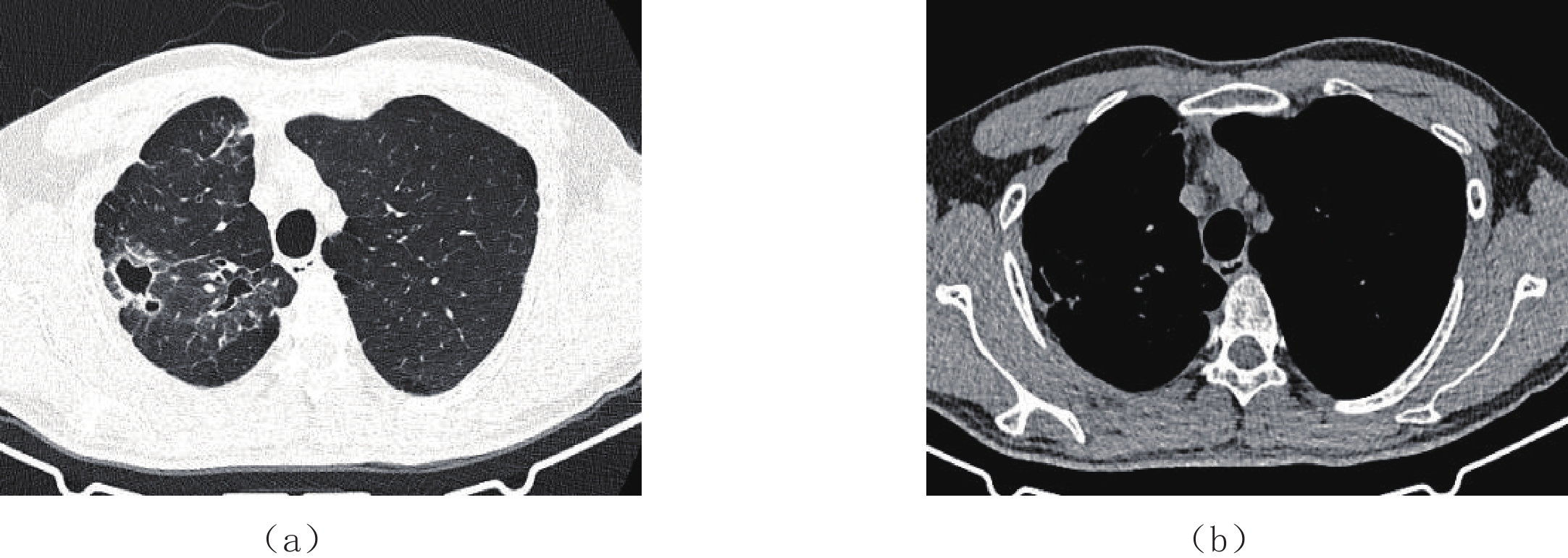
表 1 两组图像主观评分比较Table 1. Comparison of subjective image score between the two groups组别 5分 4分 3分 2分 1分 常规剂量组(n=70) 48 20 2 0 0 低剂量组(n=70) 46 21 3 0 0 ![]() 图 1 图像质量主观评价常规剂量组,患者男,36岁,继发性肺结核。管电压110 kV,参考管电流52 mAs,FBP重建,图像主观评分5分(图像质量优秀,可用于诊断)。Figure 1. Subjective evaluation of image quality
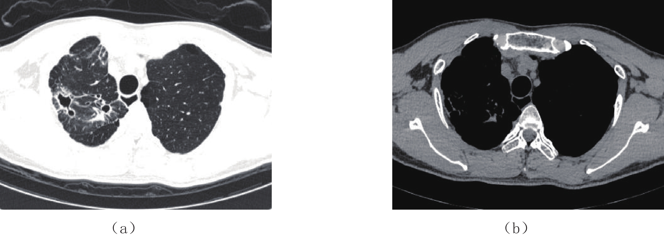
图 1 图像质量主观评价常规剂量组,患者男,36岁,继发性肺结核。管电压110 kV,参考管电流52 mAs,FBP重建,图像主观评分5分(图像质量优秀,可用于诊断)。Figure 1. Subjective evaluation of image quality![]() 图 2 图像质量主观评价低剂量组,患者男,36岁,继发性肺结核。管电压Sn 110 kV,参考管电流73 mAs,SAFIRE 3级重建,图像主观评分4分(图像质量良好,可用于诊断)。Figure 2. Subjective evaluation of image quality
图 2 图像质量主观评价低剂量组,患者男,36岁,继发性肺结核。管电压Sn 110 kV,参考管电流73 mAs,SAFIRE 3级重建,图像主观评分4分(图像质量良好,可用于诊断)。Figure 2. Subjective evaluation of image quality2.2 两组图像客观评价比较
两组升主动脉CT值差异无统计学意义;低剂量组图像噪声高于常规剂量组,差异有统计学意义;低剂量组SNR低于与常规剂量组,差异有统计学意义(表2)。
表 2 两组图像客观评价比较Table 2. Comparison of objective image evaluation between the two groups组别 升主动脉 CT 值/HU 噪声 SNR 常规剂量组(n=70) 47.56±2.27 19.31±2.73 2.50±0.38 低剂量组(n=70) 48.16±2.51 27.81±2.12 1.74±0.15 t -1.206 -20.445 14.404 P 0.234 <0.001 <0.001 2.3 两组辐射剂量比较
低剂量组辐射剂量(CTDIvol、DLP、ED)均明显低于常规剂量组,差异有统计学意义,其中低剂量组ED较常规剂量组降低约84.7%(表3)。
表 3 两组辐射剂量比较Table 3. Comparison of radiation dose between the two groups组别 CTDIvol/mGy DLP/(mGy·cm) ED/mSv 常规剂量组(n=70) 4.66±1.35 167.79±51.95 2.35±0.73 低剂量组(n=70) 0.72±0.17 25.85±10.58 0.36±0.15 t 19.809 18.554 18.555 P <0.001 <0.001 <0.001 3. 讨论
近些年肺结核的发病率逐渐提升,考虑与环境质量逐渐下降、吸烟率逐渐增高有关,临床对于肺结核的及时、准确诊断对于临床确定科学治疗方案具有重要指导意义[9]。临床上诊断肺结核多采用影像学检查,以X线胸片居多,这是因X线胸片检查经济实惠、普及性高,但X线胸片密度分辨率低,肺部细微病变无法显示,且影像重叠多;而胸部CT具有良好的密度分辨率,能发现肺部细微病变,不会出现影像重叠,因此胸部CT检查成为临床筛查和诊断肺结核的主要影像学手段。
由于肺结核属于慢性呼吸系统疾病,患者治疗时间长,同一名患者经常需要多次复查CT,而CT检查过程中会产生电离辐射,对受检者带来辐射损害,特别对于那些特殊人群如未婚青年、育龄妇女应尽量避免受到过多的剂量照射,因此控制CT辐射剂量至关重要。以往大部分研究学者采用降低管电压或管电流的方法来达到降低辐射剂量的目的[10-11],而本研究低剂量组采用西门子公司独创研制的能谱纯化技术,该技术原理是用特殊锡滤过板放置于X线球管前方,将X线的辐射能量频谱图中低能量部分的截止能量提高,从而使X线能谱变窄,能量增加,使之更接近CT成像理论要求的光子能量的均一性,有效提高X线的利用率,从而减少患者所受辐射剂量[12-13]。
赵朝祥等[14]研究表明采用管电压Sn 100 kV进行肺部结节扫描较管电压100 kV相比,辐射剂量降低约87.1%;Haubenreisser等[15]研究显示在成人胸部CT扫描中采用Sn 100 kV所产生的辐射剂量与100 kV相比降低约90%。本研究结果显示低剂量组有效剂量(0.36±0.15) mSv显著低于常规剂量组(2.35±0.73) mSv,降低约84.7%,与以上研究结果相近。
低剂量CT扫描虽显著降低患者所受辐射剂量,但同时也增加图像噪声,图像分辨率下降,导致图像质量无法达到临床诊断要求。传统的FBP是目前临床应用较为广泛的一种重建算法,它对重建数据要求较高,通过对每组投影值进行校准、滤波、加权及反投影[16],但是当辐射剂量降低时,人体成像所需的X线光子数目会减少,图像噪声就会相应地增加。因此低剂量扫描采用FBP重建算法来保证图像质量的方法是不可行的。
近几年,随着计算机硬件和软件不断提升以及迭代算法不断优化,迭代重建算法所具有降低噪声和保证图像质量的特点而逐渐被临床及放射专家认可。有关研究表明在儿内科患儿胸部CT检查中应用ASIR技术可在确保图像质量的同时降低辐射剂量[17];256层iCT IMR重建技术在肺动脉CTA检查中图像质量能够满足临床诊断需求,同时有效地降低患者辐射剂量[18]。
本研究采用正弦图确定迭代重建算法(sinogram affirmed iterative reconstruction,SAFIRE),它属于近年来出现的一种新型的基于原始数据的重建算法,该算法可以将原始数据中的噪声投射到图像中,利用反复插入方法去除噪声,通过多次迭代来去除伪影、减少噪声,提高图像分辨率[16,19];同时结合人体肺组织的特性,属于含气器官,本身与周围软组织具有天然的对比度,而且X线衰减与人体组织密度成正比,相较而言肺组织比其它组织对X线衰减的影响明显减弱,从而为低剂量扫描奠定了基础。本研究结果显示低剂量组图像SNR虽低于常规剂量组,但低剂量组图像主观评分与常规剂量组相比差异无统计学意义,依然满足临床诊断要求。
本研究的局限性:①纳入的研究对象较少,未对BMI≥24 kg/m2的肥胖患者进行研究;②未对SAFIRE迭代重建等级进行研究。
总之,能谱纯化结合迭代重建算法所获图像质量满足临床诊断要求,可用于临床筛查和诊断肺结核,同时显著降低患者CT检查时所受辐射剂量,该技术值得临床及放射医师借鉴。
-
![]()
图 1 图像质量主观评价
常规剂量组,患者男,36岁,继发性肺结核。管电压110 kV,参考管电流52 mAs,FBP重建,图像主观评分5分(图像质量优秀,可用于诊断)。
Figure 1. Subjective evaluation of image quality
![]()
图 2 图像质量主观评价
低剂量组,患者男,36岁,继发性肺结核。管电压Sn 110 kV,参考管电流73 mAs,SAFIRE 3级重建,图像主观评分4分(图像质量良好,可用于诊断)。
Figure 2. Subjective evaluation of image quality
表 1 两组图像主观评分比较
Table 1 Comparison of subjective image score between the two groups
组别 5分 4分 3分 2分 1分 常规剂量组(n=70) 48 20 2 0 0 低剂量组(n=70) 46 21 3 0 0  下载: 导出CSV
表 2 两组图像客观评价比较
Table 2 Comparison of objective image evaluation between the two groups
组别 升主动脉 CT 值/HU 噪声 SNR 常规剂量组(n=70) 47.56±2.27 19.31±2.73 2.50±0.38 低剂量组(n=70) 48.16±2.51 27.81±2.12 1.74±0.15 t -1.206 -20.445 14.404 P 0.234 <0.001 <0.001
下载: 导出CSV
2 两组图像客观评价比较
组别 升主动脉 CT 值/HU 噪声 SNR 常规剂量组(n=70) 47.56±2.27 19.31±2.73 2.50±0.38 低剂量组(n=70) 48.16±2.51 27.81±2.12 1.74±0.15 t -1.206 -20.445 14.404 P 0.234 <0.001 <0.001
下载: 导出CSV
3 两组辐射剂量比较
组别 CTDIvol/mGy DLP/(mGy·cm) ED/mSv 常规剂量组(n=70) 4.66±1.35 167.79±51.95 2.35±0.73 低剂量组(n=70) 0.72±0.17 25.85±10.58 0.36±0.15 t 19.809 18.554 18.555 P <0.001 <0.001 <0.001
下载: 导出CSV
表 3 两组辐射剂量比较
Table 3 Comparison of radiation dose between the two groups
组别 CTDIvol/mGy DLP/(mGy·cm) ED/mSv 常规剂量组(n=70) 4.66±1.35 167.79±51.95 2.35±0.73 低剂量组(n=70) 0.72±0.17 25.85±10.58 0.36±0.15 t 19.809 18.554 18.555 P <0.001 <0.001 <0.001
下载: 导出CSV
-
[1] 李易霞, 张国庆, 张冠男, 等. CT检查在活动性肺结核治疗中的应用价值[J]. 中国医药导报, 2012,9(10): 113−115. doi: 10.3969/j.issn.1673-7210.2012.10.055 LI Y X, ZHANG G Q, ZHANG G N, et al. The value of CT examination in the treatment of active pulmonary tuberculosis[J]. China Medical Journal, 2012, 9(10): 113−115. (in Chinese). doi: 10.3969/j.issn.1673-7210.2012.10.055
[2] 候准, 杨梓, 尤小芳, 等. Karl迭代技术降低64排CT肺结核患者胸部扫描辐射剂量的临床研究[J]. 临床放射学杂志, 2020,39(4): 778−781. HOU Z, YANG Z, YOU X F, et al. Clinical study of Karl iterative technique for reducing radiation dose of 64-slice CT chest scan in patients with pulmonary tuberculosis[J]. Journal of Clinical Radiology, 2020, 39(4): 778−781. (in Chinese).
[3] 吕平欣, 李多, 骆宝建. CT评价肺结核病灶活动性的作用[J]. 中华放射学杂志, 2013,47(1): 87−89. doi: 10.3760/cma.j.issn.1005-1201.2013.01.022 LV P X, LI D, LUO B J, et al. CT evaluation of the role of pulmonary tuberculosis focal activity[J]. Chinese Journal of Radiology, 2013, 47(1): 87−89. (in Chinese). doi: 10.3760/cma.j.issn.1005-1201.2013.01.022
[4] Brenner D J, Hall E J. Computed tomography-an increasing source of radiation exposure[J]. The New England Journal of Medicine, 2007, 357: 2277−2284. doi: 10.1056/NEJMra072149
[5] 何欣, 张景东, 杨正汉, 等. 前置自适应统计迭代重建技术对超低剂量CT图像质量影响的模型研究[J]. 实用放射学杂志, 2019,35(1): 110−113. doi: 10.3969/j.issn.1002-1671.2019.01.028 HE X, ZHANG J D, YANG Z H, et al. A model study on the influence of pre-adaptive statistical iterative reconstruction technique on image quality of ultra low dose CT[J]. Journal of Practical Radiology, 2019, 35(1): 110−113. (in Chinese). doi: 10.3969/j.issn.1002-1671.2019.01.028
[6] 杜恒鑫, 候阳, 王一, 等. 全模型迭代重建技术对低剂量CT冠状动脉支架显示的初步研究[J]. 中国临床医学影像杂志, 2019,30(1): 57−60. DU H X, HOU Y, WANG Y, et al. A preliminary study on low-dose CT coronary stent display using full model iterative reconstruction technique[J]. Chinese Journal of Clinical Medical Imaging, 2019, 30(1): 57−60. (in Chinese).
[7] 侯钦国, 施楠楠, 宋凤祥, 等. 胸部低剂量CT辅助诊断新型冠状病毒肺炎[J]. 中国医学影像技术, 2020,36(3): 411−414. HOU Q G, SHI N N, SONG F X, et al. Low-dose chest CT assisted in the diagnosis of COVID-19[J]. Chinese Medical Imaging Technology, 2020, 36(3): 411−414. (in Chinese).
[8] LELL M, HINKMANN F, ANDERS K, et al. High-pitch electrocardiogram-triggered computed tomography of the chest: Initial results[J]. Investigative Radioliology, 2009, 44(11): 728−733. doi: 10.1097/RLI.0b013e3181b9df7e
[9] 袁吉欣, 张喜平, 梁矿立, 等. 低剂量螺旋CT活动性肺结核扫描与常规剂量CT的对照分析[J]. 现代医用影像学, 2013,22(2): 138−140. YUAN J X, ZHANG X P, LIANG K L, et al. Comparison of low-dose spiral CT scan with conventional CT for active pulmonary tuberculosis[J]. Modern Medical Imaging, 2013, 22(2): 138−140. (in Chinese).
[10] 彭德华, 杨勤顺, 周鹏, 等. 16层CT低管电压联合迭代重建技术在胸部检查中的应用[J]. 中国医学装备, 2020,17(2): 13−16. doi: 10.3969/J.ISSN.1672-8270.2020.02.004 PENG D H, YANG Q S, ZHOU P, et al. The application of low tube voltage combined with iterative reconstruction of 16-slice CT in chest examination[J]. Chinese medical equipment, 2020, 17(2): 13−16. (in Chinese). doi: 10.3969/J.ISSN.1672-8270.2020.02.004
[11] 宋婷妮, 李万江, 李雳, 等. ASIR-V结合低管电流技术在头颅能谱CT血管成像扫描中的临床应用[J]. 实用放射学杂志, 2020,36(7): 1141−1145. doi: 10.3969/j.issn.1002-1671.2020.07.032 SONG T N, LI W J, LI L, et al. Clinical application of ASIR-V combined with low tube current technique in cranial energy spectrum CT angiography[J]. Journal of Applied Radiology, 2020, 36(7): 1141−1145. (in Chinese). doi: 10.3969/j.issn.1002-1671.2020.07.032
[12] 王绍芳, 彭成东, 胡军武, 等. CT低剂量扫描技术在新型冠状病毒肺炎检查中的应用[J]. 中国医疗设备, 2020,35(6): 22−25. doi: 10.3969/j.issn.1674-1633.2020.06.007 WANG S F, PENG C D, HU J W, et al. Application of CT low-dose scanning technique in the detection of COVID-19[J]. Chinese Medical Equipment, 2020, 35(6): 22−25. (in Chinese). doi: 10.3969/j.issn.1674-1633.2020.06.007
[13] 刘炜, 刘婧娟, 薛华丹, 等. 第3代双源CT能谱纯化技术在儿童胸部CT检查中的可行性[J]. 中国医学科学院学报, 2017,39(1): 21−27. LIU W, LIU J J, XUE H D, et al. Feasibility of the third generation dual-source CT spectral filtration technique in children chest CT examination[J]. Journal of Chinese Academy of Medical Sciences, 2017, 39(1): 21−27. (in Chinese).
[14] 赵朝祥, 余鑫, 陆泓宇, 等. 第3代双源CT能谱纯化大螺距扫描技术在肺部结节筛查中的运用价值[J]. 贵州医药, 2019,43(9): 1462−1464. doi: 10.3969/j.issn.1000-744X.2019.09.049 ZHAO C X, YU X, LU H Y, et al. The application value of the third generation dual-source CT spectral filtration and large pitch scanning technology in pulmonary nodules screening[J]. Guizhou Medicine, 2019, 43(9): 1462−1464. (in Chinese). doi: 10.3969/j.issn.1000-744X.2019.09.049
[15] HAUBENREISSER H, MEYER M, SUDARSKI S, et al. Unenhanced third-generation dual-source chest CT using a tin filter for spectral shaping at 100 kVp[J]. European Journal of Radiology, 2015, 84(8): 1608−1613. doi: 10.1016/j.ejrad.2015.04.018
[16] 马桂娜, 江奇琦, 韩萍, 等. SAFIRE重建联合CARE kV技术在主动脉CT低剂量成像中的临床应用[J]. 临床放射学杂志, 2018,37(10): 1755−1759. MA G N, JIANG Q Q, HAN P, et al. Clinical application of SAFIRE reconstruction combined CARE kV technology in low-dose aortic CT imaging[J]. Journal of Clinical Radiology, 2018, 37(10): 1755−1759. (in Chinese).
[17] 胡光玉, 童和平, 叶永灿, 等. 自适应统计迭代重建技术在儿内科患儿胸部CT检查中的应用[J]. 中国CT和MRI杂志, 2020,18(11): 59−62. doi: 10.3969/j.issn.1672-5131.2020.11.019 HU G Y, TONG H P, YE Y C, et al. The application of adaptive statistical iterative reconstruction technique in pediatric chest CT examination[J]. Chinese Journal of CT and MRI, 2020, 18(11): 59−62. (in Chinese). doi: 10.3969/j.issn.1672-5131.2020.11.019
[18] 张征宇, 王希明, 胡春洪, 等. 低辐射剂量联合IMR重建在肺动脉CTA中的应用研究[J]. 临床放射学杂志, 2017,36(10): 1504−1507. ZHANG Z Y, WANG X M, HU C H, et al. Low radiation dose combined with IMR reconstruction in pulmonary artery CTA[J]. Journal of Clinical Radiology, 2017, 36(10): 1504−1507. (in Chinese).
[19] 杨斌, 赵卫, 雷静, 等. 智能最佳kV技术联合SAFIRE迭代重建在体检人群胸部CT检查中的图像质量评估[J]. 实用放射学杂志, 2014,30(1): 138−142. doi: 10.3969/j.issn.1002-1671.2014.01.032 YANG B, ZHAO W, LEI J, et al. Intelligent optimal kV technology combined with SAFIRE iterative reconstruction for image quality assessment in chest CT examination of physical examination population[J]. Journal of Practical Radiology, 2014, 30(1): 138−142. (in Chinese). doi: 10.3969/j.issn.1002-1671.2014.01.032
计量
- 文章访问数: 332
- HTML全文浏览量: 110
- PDF下载量: 23


