
Diagnostic Value of Enhanced CT Combined with Serum Hmam Level in Detection of Axillary Lymph Node Metastasis of Breast Cancer
-
摘要:
目的:探讨增强CT联合血清人乳腺珠蛋白(hMAM)水平检测对乳腺癌腋窝淋巴结转移的诊断价值。方法:选取2017年6月至2020年6月之间我院就诊并行手术治疗的乳腺癌患者100例,所有患者术前均行增强CT,并检测血清hMAM水平,以术后病理结果为标准,比较增强CT、血清hMAM水平单独检查及联合检查对腋窝淋巴结转移的检出率,分析其与病理结果的一致性,进一步分析增强CT联合血清hMAM水平对乳腺癌腋窝淋巴结转移的诊断价值。结果:血清hMAM和病理结果共同诊断为腋窝淋巴结转移阳性的患者51例(51%),血清hMAM和病理结果共同诊断为腋窝淋巴结转移阴性的患者12例(12%),一致率81.5%,Kappa检验一致性良好;增强CT和病理结果共同诊断为腋窝淋巴结转移阳性的患者55例(55%),增强CT和病理结果共同诊断为腋窝淋巴结转移阴性的患者13例(13%),一致率82.3%,Kappa检验一致性良好;增强CT联合血清hMAM检查和病理结果共同诊断为腋窝淋巴结转移阳性的患者61例(61%),增强CT联合血清hMAM检查和病理结果共同诊断为腋窝淋巴结转移阴性的患者16例(16%),一致率90.4%,Kappa检验一致性良好;增强CT和血清hMAM对淋巴结转移检出的灵敏度分别78.0% 和76.5%,特异度分别69.5% 和70.3%,差异不具有显著性,增强CT联合血清hMAM对淋巴结转移检出的灵敏度、特异度明显高于两者单独检查,差异具有统计学意义。结论:增强CT联合血清hMAM水平对乳腺癌腋窝淋巴结转移具有较高的诊断价值,和病理结果比较一致性高,乳腺癌术前综合检查、全面分析,有利于疾病的诊断,对于手术方式的选择提供依据。
Abstract:Objective: To investigate the diagnostic value of enhanced CT combined with serum human mammaryglobin (hMAM) level in detection of axillary lymph node metastasis of breast cancer. Methods: 100 patients with breast cancer treated in our hospital from June 2017 to June 2020 were selected. All patients underwent enhanced CT before operation, and the serum hMAM level was detected. We further analyzed the diagnostic value of enhanced CT combined with serum hMAM level in axillary lymph node metastasis of breast cancer by comparing the consistency between the results of the detection rate of axillary lymph node metastasis obtained respectively by enhanced CT, serum hMAM level, combined examination and the postoperative pathological results which was taken as the standard. Results: 51 patients (51%) were diagnosed as positive axillary lymph node metastasis by serum hMAM and pathological results while 12 patients (12%) with negative axillary lymph node metastasis were diagnosed by both serum hMAM and pathological results, with the consistency rate of 81.5%. Kappa test showed great consistency, 55 cases (55%) were diagnosed as positive axillary lymph node metastasis by enhanced CT and pathological results while 13 cases (13%) with negative axillary lymph node metastasis were diagnosed by enhanced CT and pathology, with the consistency rate of 82.3%. Kappa test showed great consistency, There were 61 cases (61%) with positive axillary lymph node metastasis diagnosed by enhanced CT combined with serum hMAM examination and pathological results while 16 cases (16%) with negative axillary lymph node metastasis diagnosed by enhanced CT combined with serum hMAM examination and pathological results, with the consistency rate of 90.4%. Kappa test showed great consistency, The sensitivity of enhanced CT and serum hMAM were respectively 78.0% and 76.5% while the specificity were respectively 69.5% and 70.3%, which showed no significant difference. The sensitivity and specificity of enhanced CT combined with serum hMAM in detecting lymph node metastasis were significantly higher than those of the two alone. Conclusion: The method of enhanced CT combined with serum hMAM level hold high diagnostic value for axillary lymph node metastasis of breast cancer, and show high consistency with the pathological results. Preoperative comprehensive examination and comprehensive analysis of breast cancer are conducive to the diagnosis of the disease and can provide basis for the selection of surgical methods.
-
Keywords:
- enhanced CT /
- serum hMAM /
- breast cancer /
- axillary lymph node metastasis
-
-
![]()
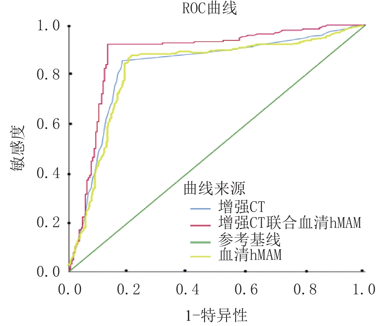
图 2 增强CT和血清hMAM对淋巴结转移灵敏度及特异度ROC曲线图
Figure 2. ROC curve of sensitivity and speci-ficity of enhanced CT and serum hMAM for lymph node metastasis
表 1 血清hMAM和病理结果比较
Table 1 Comparison between serum hMAM and pathological results
病理 血清 hMAM 一致率/% K P 阳性/例 阴性/例 腋窝淋巴结阳性 51 27 81.5 0.753 0.000 腋窝淋巴结阴性 10 12  下载: 导出CSV
表 2 血清hMAM和不同大小的阳性腋窝淋巴结比较
Table 2 Comparison between serum hMAM and positive axillary lymph nodes of different sizes
腋窝淋巴结大小/cm hMAM/(ng/mL) F P <1 8.9±4.12 1.590 0.043 1~2 9.3±4.23 >2 9.8±4.18
下载: 导出CSV
2 血清hMAM和不同大小的阳性腋窝淋巴结比较
腋窝淋巴结大小/cm hMAM/(ng/mL) F P <1 8.9±4.12 1.590 0.043 1~2 9.3±4.23 >2 9.8±4.18
下载: 导出CSV
表 3 增强CT结果和病理结果比较
Table 3 Comparison between enhanced CT results and pathological results
病理 增强CT 一致率/% K P 阳性/例 阴性/例 腋窝淋巴结阳性 55 23 82.3 0.755 0.000 腋窝淋巴结阴性 9 13
下载: 导出CSV
表 4 增强CT联合血清hMAM结果和病理结果比较
Table 4 Comparison between enhanced CT combined with serum hMAM results and pathological results
病理 增强 CT 联合血清 hMAM 一致率/% K P 阳性/例 阴性/例 腋窝淋巴结阳性 61 17 90.4 0.757 0.000 腋窝淋巴结阴性 6 16
下载: 导出CSV
4 增强CT联合血清hMAM结果和病理结果比较
病理 增强 CT 联合血清 hMAM 一致率/% K P 阳性/例 阴性/例 腋窝淋巴结阳性 61 17 90.4 0.757 0.000 腋窝淋巴结阴性 6 16
下载: 导出CSV
5 增强CT和血清hMAM对淋巴结转移灵敏度及特异度对比(
$\bar x \pm s $ )组别 增强 CT 血清 hMAM 增强 CT 联合血清 hMAM t P 灵敏度/% 78.0 76.5 88.7 6.32 0.061 特异度/% 69.5 70.3 83.5
下载: 导出CSV
表 5 增强CT和血清hMAM对淋巴结转移灵敏度及特异度对比(
$\bar x \pm s $ )Table 5 Comparison of sensitivity and specificity between enhanced CT and serum hMAM for lymph node metastasis (
$\bar x \pm s $ )组别 增强 CT 血清 hMAM 增强 CT 联合血清 hMAM t P 灵敏度/% 78.0 76.5 88.7 6.32 0.061 特异度/% 69.5 70.3 83.5
下载: 导出CSV
-
[1] 田英, 杨碎胜, 张志艳. 新辅助化疗对乳腺癌患者血清hMAM水平影响的观察[J]. 中华肿瘤防治杂志, 2013,12: 939−941, 948. TIAN Y, YANG S S, ZHANG Z Y. The effect of neoadjuvant chemotherapy on serum hMAM levels in breast cancer patients[J]. Chinese Journal of Cancer Prevention and Treatment, 2013, 12: 939−941, 948. (in Chinese).
[2] 崔俊涛. CT与动态增强磁共振成像在诊断乳腺癌患者腋窝淋巴结有无转移中的对比研究[J]. 现代诊断与治疗, 2017,28(23): 4406−4407. doi: 10.3969/j.issn.1001-8174.2017.23.056 [3] 沈春华, 朱昆喜. 钼靶(MG)、计算机断层扫描(CT)与动态增强磁共振成像(DCE-MRI)对乳腺癌患者腋窝淋巴结有无转移的诊断价值[J]. 影像研究与医学应用, 2018,20: 55−56. doi: 10.3969/j.issn.2096-3807.2018.14.029 [4] ZHAO S, YANG H, ZHANG M, et al. Circulating tumor cells (CTCs) detected by triple-marker EpCAM, CK19, and hMAM RT-PCR and their relation to clinical outcome in metastatic breast cancer patients[J]. Cell Biochem Biophys. 2013, 65(2): 263-273. DOI: 10.1007/s12013-012-9426-2.
[5] 刘奕仕, 黄冬玲, 邱其良. 彩色多普勒超声和CT增强扫描对乳腺癌腋窝淋巴结转移的早期诊断价值比较[J]. 中国医药科学, 2020,10(9): 169−171. doi: 10.3969/j.issn.2095-0616.2020.09.050 LIU Y S, HUANG D L, QIU Q L. Comparison of color doppler ultrasound and CT enhanced scan in the early diagnosis of axillary lymph node metastasis of breast cancer[J]. Chinese Medical Science, 2020, 10(9): 169−171. (in Chinese). doi: 10.3969/j.issn.2095-0616.2020.09.050
[6] 高洋, 张文海, 李建一, 等. 增强CT对乳腺癌腋窝淋巴结术前评估的价值[J]. 中华内分泌外科杂志, 2013,7(5): 364−367. doi: 10.3760/cma.j.issn.1674-6090.2013.05.004 GAO Y, ZHANG W H, LI J Y, et al. Preoperative assessment value of enhanced CT for axillary lymph node in breast cancer[J]. Chinese Journal of Endocrine Surgery, 2013, 7(5): 364−367. (in Chinese). doi: 10.3760/cma.j.issn.1674-6090.2013.05.004
[7] FABISIEWICZ A, KULIK J, KOBER P, et al. Detection of circulating breast cancer cells in peripheral blood by a two-marker reverse transcriptase-polymerase chain reaction assay[J]. Acta Biochimica Polonica, 2004, 51(3): 747−755. doi: 10.18388/abp.2004_3559
[8] CANESSA P A, MANTA C, FERRO P, et al. Clinical relevance of human mammaglobin mRNA in pleural effusion from patients undergoing thoracoscopy: A pilot study[J]. The International Journal of Biological Markers, 2012, 27(2): e99−e104. DOI: 10.5301/JBM.2012.9305.
[9] 徐海声, 张娟. 血清hMAM对乳腺癌患者新辅助化疗疗效的评估价值[J]. 实用癌症杂志, 2014,(1): 10−12. doi: 10.3969/j.issn.1001-5930.2014.01.004 XU H S, ZHANG J. Evaluation value of serum hMAM on the efficacy of neoadjuvant chemotherapy in breast cancer[J]. Practical Journal of Cancer, 2014, (1): 10−12. (in Chinese). doi: 10.3969/j.issn.1001-5930.2014.01.004
[10] 张伟晶. CT与动态增强磁共振成像在诊断乳腺癌患者腋窝淋巴结有无转移中的对比研究[J]. 现代医用影像学, 2017,6: 1738−1740. [11] LV X, FENG X, HAO X, et al. Anti-hMAM monoclonal antibodies evaluated in breast and non-breast tissues for differential diagnosis implication[J]. Tumori Journal, 2016, (3): 264−269. DOI: 10.5301/tj.5000496.
[12] NGUYEN H M, DAO M Q. Detection of human mammaglobin mRNA in breast cancer cells among Vietnamese women[J]. Breast Cancer (Dove Med Press), 2019, 11: 143−150. DOI: 10.2147/BCTT.S193777.
[13] 姚岚, 张殿龙, 孟佳佳, 等. 超声、增强CT与增强MRI在乳腺癌腋窝淋巴结转移中的诊断价值比较[J]. 中国现代医生, 2019,57(7): 94−98, 169. YAO L, ZHANG D L, MENG J J, et al. Comparison of diagnostic value of ultrasound, enhanced CT and enhanced MRI in axillary lymph node metastasis of breast cancer[J]. Chinese Modern Doctor, 2019, 57(7): 94−98, 169. (in Chinese).
[14] SHEN C, HU L, XIA L, et al. The detection of circulating tumor cells of breast cancer patients by using multimarker (Survivin, hTERT and hMAM) quantitative real-time PCR[J]. Clinical Biochemistry, 2009, 42(3): 194−200. DOI: 10.1016/j.clinbiochem.2008.10.016.
-
期刊类型引用(8)
1. 李海芬,邓亚云,李洪来,戴林,汪凤勃. 超声测定PI、RI、Vmax结合血清sE-cadherin、CYFRA21-1对乳腺癌良恶性及淋巴结转移的诊断价值. 生物医学工程与临床. 2024(01): 56-62 .  百度学术
百度学术
2. 黄小妹. 乳腺癌及腋窝淋巴结转移诊断中增强CT扫描的作用与影像学特征分析. 影像研究与医学应用. 2024(08): 171-173 . 百度学术
3. 熊文明,梁云,潘丽. MSCT增强扫描对非特殊型乳腺癌腋窝淋巴结转移的诊断价值. 贵州医药. 2024(08): 1289-1291 . 百度学术
4. 张江华,王海峰,张克俭,贾国洪. 血清hMAM、HER2、UBE2C联合检测对乳腺癌腋窝淋巴结转移的诊断价值. 中国现代普通外科进展. 2024(11): 856-860 . 百度学术
5. 杨灵敏. 分析超声诊断乳腺癌腋窝淋巴结转移的影像学表现及其临床效果. 中国医药指南. 2023(09): 106-108 . 百度学术
6. 修超,倪东贺. 多模态影像组学辅助诊断早期乳腺癌的效能评价. 北华大学学报(自然科学版). 2023(02): 227-230 . 百度学术
7. 李文周,曹永峰,张玉. 增强CT联合超声弹性成像鉴别乳腺小结节(直径≤2cm)性质的价值探究. 中国CT和MRI杂志. 2023(07): 95-96 . 百度学术
8. 陆兴练. 乳腺癌诊断技术的研究进展. 大医生. 2022(18): 123-126 . 百度学术
其他类型引用(0)


计量
- 文章访问数: 292
- HTML全文浏览量: 163
- PDF下载量: 15
- 被引次数: 8


