
The Study of Application of Different Pitch Combined with ASIR in Low-dose Chest CT Screening on COVID-19
-
摘要: 目的:探讨2019冠状病毒病(COVID-19)患者使用大螺距联合自适应统计迭代重建技术行低剂量扫描的可行性,比较辐射剂量及图像质量的差异。方法:收集我院确诊为COVID-19的病例共91例作为低剂量组,同时选取同期于我院行新冠肺炎筛查的91例未确诊为COVID-19的患者作为常规剂量组。低剂量组使用大螺距扫描联合自适应统计迭代重建算法(ASIR);常规剂量组使用常规螺距扫描,使用滤波反投影法(FBP)进行重建。记录两组辐射剂量并对两组病例的图像质量进行主观及客观评分,比较各组图像质量与辐射剂量的差别。结果:低剂量及常规剂量两种方法的辐射剂量有统计学差异低剂量组辐射剂量明显低于常规剂量组(ED降低了52%);低剂量组与常规剂量组的图像质量评分无统计学差异;低剂量组与常规剂量组的客观评价无统计学差异。结论:应用大螺距联合ASIR重建技术对COVID-19患者行低剂量扫描可以明显降低辐射剂量而图像质量无明显下降。
-
关键词:
- 自适应统计迭代重建技术 /
- 新型冠状病毒肺炎 /
- 图像质量 /
- 辐射剂量
Abstract: Objective: To explore the feasibility of low-dose scanning for COVID-19 patients by using the method of high-pitch combined with ASIR, and compare the diffrence of radiation dose and image quality. Methods: 91 patients diagnosed with COVID-19 in our hospital were collected as the low-dose group while 91 cases of undiagnosed COVID-19 patients who were screened for COVID-19 in our hospital during the same period were selected as the conventional dose group. High-pitch combined with ASIR was performed on the low-dose group while conventional dose group was scanned by common pitch and reconstructed by FBP. In order to compare the differnce of the image quality and radiation dose between the two groups, we recorded the radiation dose and performed subjective and objective evaluation of image quality of the two groups.Results: There was statistical differences in radiation dose between the two groups, and the radiation dose of the low-dose group was significantly lower than that of the conventional dose group (the ED was reduced by about 52%). There was no statistical difference in the image quality scores between the two groups; There was no statistical difference in the objective evaluation of images between the two groups . Conclusion: Low dose scanning with high-pitch combined with ASIR for COVID-19 patients can evidently reduce the radiation dose without significant decrease in image quality.-
Keywords:
- ASIR /
- COVID-19 /
- image quality /
- radiation dose
-
-
![]()
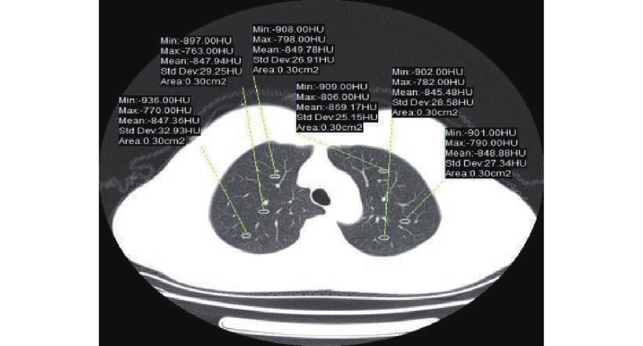
图 4 肺窗上测量肺实质CT值及SD值的方法
Figure 4. The method of measuring CT and SD values of lung parenchyma on lung window
![]()
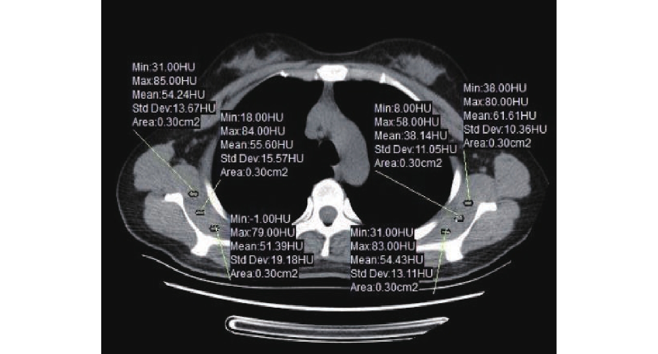
图 5 纵膈窗上测量胸壁肌肉CT值的方法
Figure 5. The method of measuring CT values of chest wall muscle on mediastinum window
表 1 图像质量主观评分标准
Table 1 The criterion of subjective scoring for image quality
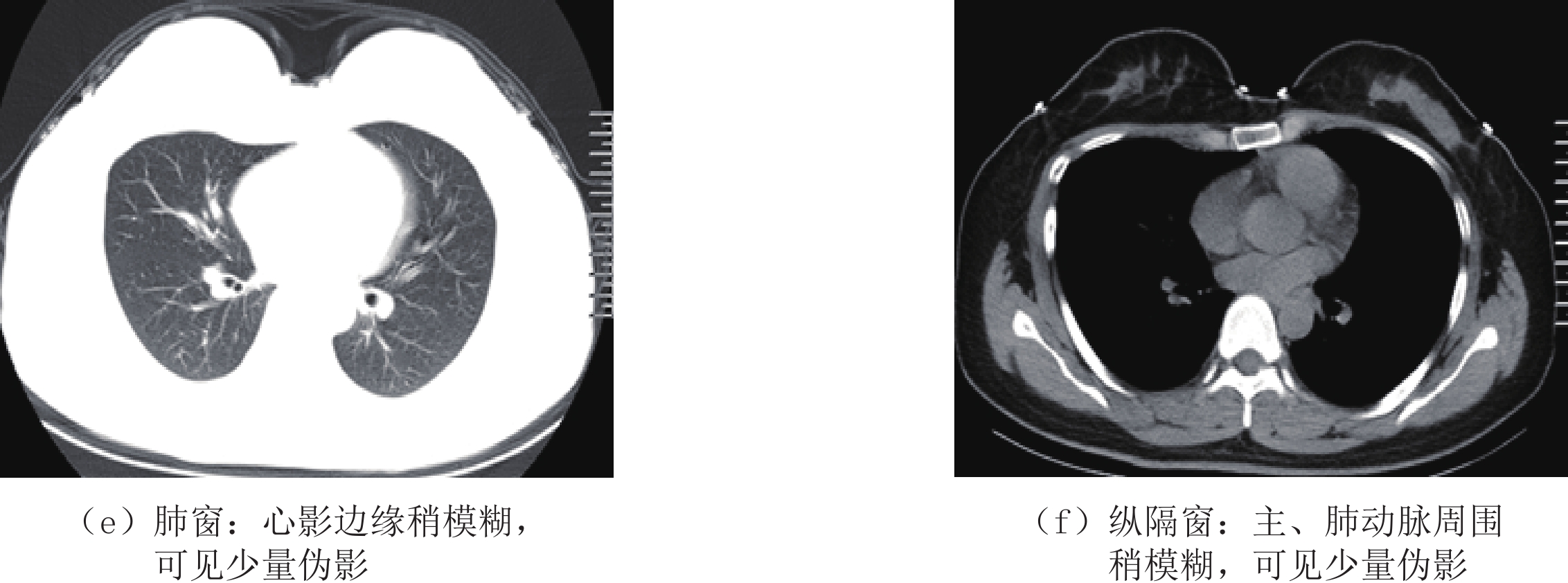
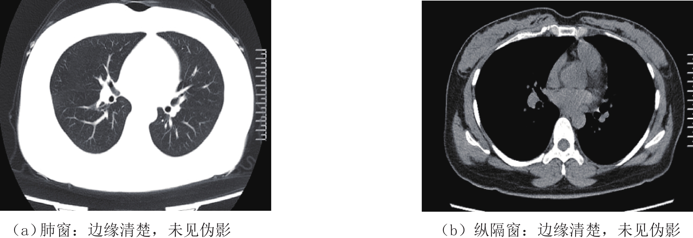
等级 图像质量 0分 未能显示正常结构 1分 大量伪影,正常结构中断 2分 边缘模糊,可见中等量伪影 3分 边缘稍模糊,可见少量伪影 4分 边缘稍模糊,未见伪影 5分 边缘清楚,未见伪影  下载: 导出CSV
表 2 两组CTDIvol、DLP、ED比较
Table 2 The Comparison of CTDIvol, DLP and ED values between two groups
分组及检验 CTDIvol/mGy DLP/mGy·cm ED/mSv 低剂量组 3.90±0.92 129.78±26.21 1.82±0.37 常规剂量组 7.82±2.37 271.73±82.85 3.80±1.16 Z -10.529 -10.897 -10.883 P <0.001 <0.001 <0.001
下载: 导出CSV
表 3 两组主观评价评分频数
Table 3 The frequency of subjective evaluation of the two groups
分组 主观评分 平均值 统计检验 5分 4分 3分 2分 1分 Z P 低剂量组 16(17.6%) 71(78.0%) 4(4.4%) 0 0 4.13±0.45 -0.963 0.336 常规剂量组 21(23.1%) 67(73.6%) 3(3.3%) 0 0 4.20±0.48
下载: 导出CSV
表 4 两组间客观评价结果
Table 4 The objective evaluation between the two groups
分组及检验 CNR SNR 低剂量组 28.83±2.95 30.62±3.10 常规剂量组 28.98±2.48 30.69±2.46 Z -0.851 -0.673 P 0.395 0.501
下载: 导出CSV
-
[1] World Health Organization Press Conference. The World Health Organization (WHO) has officially named the disease caused by the novel coronavirus as COVID-19[EB/OL].(2020-02-11)[2021-02-01]. https://www.who.int/emergencies/diseases/novel-coronavirus-2019.
[2] HOEHL S, RABENAU H, BERGER A, et al. Evidence of SARS-CoV-2 infection in returning travelers from Wuhan, China[J]. The New England Journal of Medicine, 2020, 382(13): 1278−1280. doi: 10.1056/NEJMc2001899
[3] 国家卫生健康委. 新型冠状病毒感染的肺炎诊疗方案(试行第六版)[EB/OL]. (2020-02-19)[2021-02-01]. http://www.nhc.gov.cn/yzygj/s7653p/202002/8334a8326dd94d329df351d7da8aefc2/files/b218cfeb1bc54639af227f922bf6b817.pdf. [4] WAN Y, SHANG J, GRAHAM R, et al. Receptor recognition by the novel coronavirus from Wuhan: An analysis based on Decade-Long structural studies of SARS coronavirus[J]. Journal of Virology, 2020, 94(7): e00127−20.
[5] LI W, MOORE M J, VASILIEVA N, et al. Angiotensin-converting enzyme 2 is a functional receptor for the SARS coronavirus[J]. Nature, 2003, 426(6965): 450−454. doi: 10.1038/nature02145
[6] HUANG C, WANG Y, LI X, et al. Clinical features of patients infected with 2019 novel coronavirus in Wuhan, China[J]. Lancet, 2020, 395(10223): 497−506. doi: 10.1016/S0140-6736(20)30183-5
[7] ZHU N, ZHANG D, WANG W, et al. A Novel Coronavirus from patients with pneumonia in China, 2019[J]. The New England Journal of Medicine, 2020, 382(8): 727−733. doi: 10.1056/NEJMoa2001017
[8] WANG D, HU B, HU C, et al. Clinical characteristics of 138 hospitalized patients with 2019 novel coronavirus-infected pneumonia in Wuhan, China[J]. Journal of The American Medical Association, 2020, 323(11): 1061−1069. doi: 10.1001/jama.2020.1585
[9] SONG F, SHI N, SHAN F, et al. Emerging 2019 novel coronavirus (2019-nCoV) pneumonia[J]. Radiology, 2020, 297(3): E346. doi: 10.1148/radiol.2020209021
[10] PAN F, YE T, SUN P, et al. Time course of lung changes at chest CT during recovery from coronavirus disease 2019 (COVID-19)[J]. Radiology, 2020, 295(3): 715−721. doi: 10.1148/radiol.2020200370
[11] 宋娟, 王成伟, 李勇. 双低扫描技术联合自适应统计迭代重建技术在能谱CT冠状动脉成像中的应用研究[J]. 实用放射学杂志, 2015,(3): 467−472. DOI: 10.3969/j.issn.1002-1671.2015.03.031. SONG J, WANG C W, LI Y. Applied research on double low scanning technique in gem spectrum CT coronary artery angio-graphy[J]. Journal of Practical Radiology, 2015, (3): 467−472. DOI: 10.3969/j.issn.1002-1671.2015.03.031. (in Chinese).
[12] PRAKASH P, KALRA M K, DIGUMARTHY S R, et al. Radiation dose reduction with chest computed tomography using adaptive statistical iterative reconstruction technique: Initial experience[J]. Journal of Computer Assisted Tomography, 2010, 34(1): 40−45. doi: 10.1097/RCT.0b013e3181b26c67
[13] 张丽, 于红, 刘士远, 等. 迭代重建技术对低剂量肺部平扫CT图像质量的影响[J]. 中华放射学杂志, 2013,47(4): 316−320. DOI: 10.3760/cma.j.issn.1005-1201.2013.04.006. ZHANG L, YU H, LIU S Y, et al. Radiation dose reduction with chest CT using iterative reconstruction technique[J]. Chinese Journal of Radiology, 2013, 47(4): 316−320. DOI: 10.3760/cma.j.issn.1005-1201.2013.04.006. (in Chinese).
[14] HU X H, DING X F, WU R Z, et al. Radiation dose of non-enhanced chest CT can be reduced 40% by using iterative reconstruction in image space[J]. Clinical Radiology, 2011, 66(11): 1023−1029. doi: 10.1016/j.crad.2011.04.008
[15] SINGH S, KALRA M K, GILMAN M D, et al. Adaptive statistical iterative reconstruction technique for radiation dose reduction in chest CT: A pilot study[J]. Radiology, 2011, 259(2): 565−573. doi: 10.1148/radiol.11101450
[16] 张皓, 刘明, 华海琴, 等. 肺CT低剂量成像及体重指数相关性研究[J]. 医学影像学杂志, 2017,27(9): 1681−1685. ZHANG H, LIU M, H UA H Q, et al. Corrletion study on lung low-dose CT imaging and body mass index[J]. Journal of Medical Imaging, 2017, 27(9): 1681−1685. (in Chinese).
[17] 汤芳, 王晓芹, 栾进, 等. 新型冠状病毒肺炎的流行病学研究进展[J]. 武警医学, 2020,31(3): 272−276. doi: 10.3969/j.issn.1004-3594.2020.03.025 [18] AI T, YANG Z, HOU H, et al. Correlation of chest CT and RT-PCR testing for coronavirus disease 2019 (COVID-19) in China: A report of 1014 cases[J]. Radiology, 2020, 296(2): E32−E40. doi: 10.1148/radiol.2020200642
[19] FANG Y, ZHANG H, XIE J, et al. Sensitivity of chest CT for COVID-19: Comparison to RT-PCR[J]. Radiology, 2020, 296(2): E115−E117. doi: 10.1148/radiol.2020200432
[20] 国家卫生健康委. 新型冠状病毒肺炎诊疗方案(试行第五版 修正版)[EB/OL]. (2020-02-08)[2021-02-01]. http://www.gov.cn/zhengce/zhengceku/2020-02/09/5476407/files/765d1e65b7d1443081053c29ad37fb07.pdf. [21] 马琼, 石秀东, 陆阳, 等. 新型冠状病毒肺炎临床及影像学研究进展[J]. 中国临床医学, 2020,27(1): 23−26. DOI: 10.12025/j.issn.1008-6358.2020.20200340. MA Q, SHI X D, LU Y, et al. Research progress on clinical and imaging study of novel coronavirus pneumonia[J]. Chinese Journal of Clinical Medicine, 2020, 27(1): 23−26. DOI: 10.12025/j.issn.1008-6358.2020.20200340. (in Chinese).
[22] 汪锴, 康嗣如, 田荣华, 等. 新型冠状病毒肺炎胸部CT影像学特征分析[J]. 中国临床医学, 2020,27(1): 27−31. DOI: 10.12025/j.issn.1008-6358.2020.20200169. WANG K, KANG S R, TIAN R H, et al. CT characteristic appearances of patients with novel coronavirus pneumonia[J]. Chinese Journal of Clinical Medicine, 2020, 27(1): 27−31. DOI: 10.12025/j.issn.1008-6358.2020.20200169. (in Chinese).
[23] 闵锐. 医源性电离辐射损伤及其生物医学防护[J]. 辐射防护通讯, 2013,(3): 8−15. DOI: 10.3969/j.issn.1004-6356.2013.03.002. MIN R. Iatrogenic ionizing radiation damage and its medical and biological protection[J]. Radiation Protection Bulletin, 2013, (3): 8−15. DOI: 10.3969/j.issn.1004-6356.2013.03.002. (in Chinese).
[24] 王伯胤. CT影像质量评价和质量控制[J]. 中华放射学杂志, 2001,35(8): 634−635. doi: 10.3760/j.issn:1005-1201.2001.08.020 [25] NAIDICH D P, MARSHALL C H, GRIBBIN C, et al. Low-dose CT of the lungs: Preliminary observations[J]. Radiology, 1990, 175(3): 729−731. doi: 10.1148/radiology.175.3.2343122
[26] 张巍, 郭玉林. 低剂量螺旋CT扫描技术的临床应用[J]. 医学影像学杂志, 2006,16(8): 861−864. DOI: 10.3969/j.issn.1006-9011.2006.08.027. ZHANG W, GUO Y L. The clinical application of the low-dose CT examination[J]. Journal of Medical Imaging, 2006, 16(8): 861−864. DOI: 10.3969/j.issn.1006-9011.2006.08.027. (in Chinese).
[27] 陈伟, 李文政, 唐友林. 螺旋CT图像噪声的评价[J]. 放射学实践, 2003,18(7): 535−536. DOI: 10.3969/j.issn.1000-0313.2003.07.029. CHEN W, LI W Z, TANG Y L. Assessment of image noise in spiral CT[J]. Radiologic Practice, 2003, 18(7): 535−536. DOI: 10.3969/j.issn.1000-0313.2003.07.029. (in Chinese).
[28] 袁颖, 钟朝辉, 吴天棋, 等. 自动管电流调节技术下螺距对胸部CT图像质量及辐射剂量影响的体模研究[J]. 中国医疗设备, 2019,34(1): 74−77. DOI: 10.3969/j.issn.1674-1633.2019.01.020. YUAN Y, ZHONG Z H, WU T Q, et al. Study on the effect of pitch on the quality of chest CT image and radiation dose under automatic tube current regulation[J]. China Medical Devices, 2019, 34(1): 74−77. DOI: 10.3969/j.issn.1674-1633.2019.01.020. (in Chinese).
[29] 江时淦, 洪春凤, 王豪, 等. 眼眶部低剂量螺旋CT扫描参数的优化[J]. 放射学实践, 2013,28(2): 210−213. DOI: 10.3969/j.issn.1000-0313.2013.02.026. JIANG S G, HONG C F, WANG H, et al. The research of the relativity between the scan parameters optimization and radiation dose at orbital helical CT[J]. Radiologic Practice, 2013, 28(2): 210−213. DOI: 10.3969/j.issn.1000-0313.2013.02.026. (in Chinese).
[30] FLICEK K T, HARA A K, SILVA A C, et al. Reducing the radiation dose for CT colonography using adaptive statistical iterative reconstruction: A pilot study[J]. American Journal of Roentgenology, 2010, 195(1): 126−131. doi: 10.2214/AJR.09.3855
[31] 束宏敏, 李小虎, 宋建, 等. 自适应统计迭代重建技术对泌尿系结石低剂量CT图像质量的影响[J]. 中国医学影像学杂志, 2016,24(2): 148−152. DOI: 10.3969/j.issn.1005-5185.2016.02.017. SHU H M, LI X H, SONG J, et al. Effect of adaptive statistical iterative reconstruction on image quality of urinary calculi with low-dose CT[J]. Chinese Journal of Medical Imaging, 2016, 24(2): 148−152. DOI: 10.3969/j.issn.1005-5185.2016.02.017. (in Chinese).
[32] 浦仁旺, 刘义军, 刘静红, 等. 低管电压结合ASIR重建对腹部CT图像质量的影响: 体模研究[J]. 实用放射学杂志, 2015,31(2): 296−299. DOI: 10.3969/j.issn.1002-1671.2015.02.032. PU R W, LIU Y J, LIU J H, et al. Impact of low tube voltage with ASIR on abdominal CT image quality: A phantom study[J]. Journal of Practical Radiology, 2015, 31(2): 296−299. DOI: 10.3969/j.issn.1002-1671.2015.02.032. (in Chinese).
[33] 唐慧, 贺太平, 燕洋洋, 等. ASIR算法联合自动管电流技术在胸部低剂量CT扫描中的应用[J]. 实用放射学杂志, 2018,34(1): 109−113. DOI: 10.3969/j.issn.1002-1671.2018.01.030. TANG H, HE T P, YAN Y Y, et al. The application of ASIR combined with automatic tube current modulation in low-dose chest CT screening[J]. Journal of Practical Radiology, 2018, 34(1): 109−113. DOI: 10.3969/j.issn.1002-1671.2018.01.030. (in Chinese).
[34] 艾娜娜, 宋振, 翟艳慧, 等. 大螺距联合自适应迭代重建技术对胸部能谱成像影响的体模研究[J]. 放射学实践, 2020,35(5): 619−623. DOI: 10.13609/j.cnki.1000-0313.2020.05.010. AI N N, SONG Z, ZHAI Y H, et al. The effect of high-pitch combined adaptive iterative reconstruction on chest energy spectrum imaging: A phantom study[J]. Radiologic Practice, 2020, 35(5): 619−623. DOI: 10.13609/j.cnki.1000-0313.2020.05.010. (in Chinese).
-
期刊类型引用(2)
1. 袁婷婷,万胜洪. MRI磁敏感加权成像技术在颅内海绵状血管瘤中的诊断效能及影像学征象分析. 医学信息. 2024(10): 105-108 .  百度学术
百度学术
2. 李丽,阮玖根,高阳,肖琼. 磁敏感加权成像在颅内海绵状血管瘤诊断中的应用价值分析. 现代诊断与治疗. 2022(17): 2535-2537 . 百度学术
其他类型引用(0)

计量
- 文章访问数: 1255
- HTML全文浏览量: 231
- PDF下载量: 28
- 被引次数: 2


