
CT Findings and Differential Diagnosis of Primary Testicular Tumors
-
摘要: 目的:分析睾丸原发肿瘤的CT影像特征。方法:回顾性分析46例经病理证实的睾丸肿瘤CT影像资料,结合其他资料对病灶做综合评估。结果:所有肿瘤均为单侧发病。精原细胞瘤24例,其中16例可见不均匀轻度分隔样强化;内胚窦瘤10例,8例可见多发迂曲、增粗供血血管;畸胎瘤4例,可见斑点状、不规则钙化,2例可见脂肪;混合性生殖细胞肿瘤2例,增强扫描明显不均匀强化,可见迂曲、增粗血管影,未见钙化及脂肪;平滑肌瘤2例,边界清晰,密度均匀,增强扫描轻度均匀强化;弥漫大B细胞淋巴瘤3例,2例边界清晰,密度均匀,增强扫描中度强化,1例局部边界不清,增强不均匀强化;恶性间质细胞瘤1例,可见粗大钙化,实性成分中度强化,伴腹膜后淋巴结及双肺转移。结论:对于不同类型睾丸肿瘤CT有较好的鉴别能力,能为临床治疗方案提供较为准确的依据。Abstract: Objective: To analyze the imaging features of primary testicular tumor. Methods: The CT imaging data of 46 cases of testicular tumor confirmed by pathology were retrospectively reviewed. Combined with the other data, the location, attenuation, size, shape, edge, and enhancement patterns of lesions were comprehensively analyzed. Result: All 46 cases of testicular tumor were unilateral. There were 24 cases of seminoma, among whom 16 cases showed uneven mild septal enhancement; 10 cases of endodermal sinus tumor, and 8 cases showed multiple tortuous and thickened blood vessels; 4 cases of teratoma, spot or irregular calcification; 2 cases of mixed germinoma, obvious heterogenous enhancement with no calcification or fat; 2 cases of leiomyoma with clear border, homogeneous density and nhomogeneous enhancement in enhanced scan; 3 cases of diffuse large B cell lymphoma, 2 cases showed clear boundary, uniform density and homogeneous enhancement, 1 case of patchy low density area, with unclear boundary, persistent enhancement, the low density area showed no enhancement; 1 case of malignant mesenchymal cell tumor with thick calcification and moderate enhancement of solid components, accompanied by retroperitoneal lymph node and lung metastasis. Conclusion: CT can distinguish the imaging features of different types of testicular tumors, and have high value in the diagnosis and differential diagnosis of testicular tumors. Thus CT can provide a more accurate basis for clinical treatment.
-
Keywords:
- tomography /
- testicular /
- tumor
-
睾丸肿瘤发病率低,约占男性全身肿瘤的1%~2%[1]。睾丸由不同组织构成,其肿瘤来源分类较多,各类型的肿瘤在诊断、治疗及预后均有所差异。因此,借助影像学检查提高术前准确率,对于临床治疗方案及患者预后意义重大。
超声、CT和MRI是诊断睾丸肿瘤的常用影像学方法。超声简便、快捷,常作为睾丸病变的首选检查方法;MRI软组织分辨率高,且无辐射,能更好地显示肿瘤内部结构特点;但由于预约及检查时间较长,故临床在超声诊断提示为睾丸肿瘤性病变时,多采用CT作为进一步鉴别诊断的方法。因此掌握睾丸肿瘤的CT表现对鉴别诊断具有重要意义。
1. 资料与方法
收集本院及高要区中医院2012年6月至2021年6月经病理证实的睾丸肿瘤46例,年龄范围1~66岁,平均36.7岁。临床症状无特异性表现,首诊以无痛性肿块为主,少部分下腹部隐痛就诊;病程数天至半年不等;所有患者均无隐睾病史。
采用东芝公司Aquilion 64排螺旋CT,所有病例均行平扫和增强扫描,范围包完盆腔及会阴。电压140 kV,电流250~300 mA,层厚5 mm。使用非离子型对比剂(碘帕醇,上海博莱科有限公司,规格:100 mL),注射速率2.5 mL/s。应用高压注射器静脉注射对比剂后25 s和60 s行增强扫描。对本组病例的CT图像进行综合评判,包括发病年龄、肿瘤大小、形态、境界、密度、强化方式、淋巴结转移及血行转移,结合大体病理标本对照分析。
2. 结果
2.1 病理结果
46例睾丸肿瘤中,生殖细胞瘤肿瘤40例,占87%,其中精原细胞瘤24例(图1(a)~图2(c)),占52%,非精原细胞瘤16例,包括内胚窦瘤10例(图2(a)~(c)),畸胎瘤4例(图3(a)和图2(b)),混合性生殖细胞瘤2例(其中1例主要成分为胚胎性癌(图4(a)~图4(c)),少许成分为绒毛膜上皮癌及精原细胞瘤,1例为精原细胞瘤及胚胎性癌)。其他肿瘤类型包括平滑肌瘤2例,弥漫大B细胞淋巴瘤3例,恶性间质细胞瘤1例(图5(a)~图5(c))。
![]() 图 4 男,48岁,左侧睾丸混合性生殖细胞肿瘤Figure 4. Male, 48 years old, Mixed germ cell tumor of left testis
图 4 男,48岁,左侧睾丸混合性生殖细胞肿瘤Figure 4. Male, 48 years old, Mixed germ cell tumor of left testis![]() 图 5 男,40岁,右侧睾丸恶性间质细胞瘤Figure 5. Male, 40 years old, Malignant Leydig cell tumor of right testis
图 5 男,40岁,右侧睾丸恶性间质细胞瘤Figure 5. Male, 40 years old, Malignant Leydig cell tumor of right testis2.2 CT表现
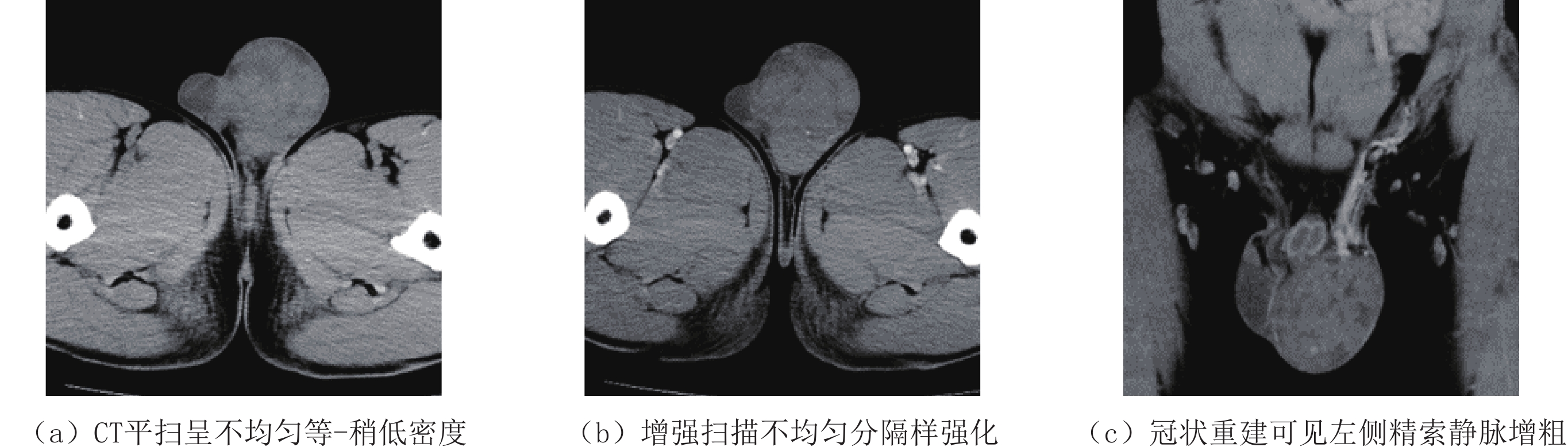
精原细胞瘤24例。发病年龄22~51岁,平均42.5岁;直径5~14 cm;右侧16例,左侧8例。CT表现为阴囊内类圆形、边界清楚的软组织肿块,平扫呈等低密度,CT值约20~35 HU,增强扫描呈轻度强化,CT值增加10~25 HU。16例出现分隔样强化,病灶内可见不同程度的更低密度未强化坏死区。12例患者出现腹膜后或盆腔多发淋巴结转移,但均未见血性转移。
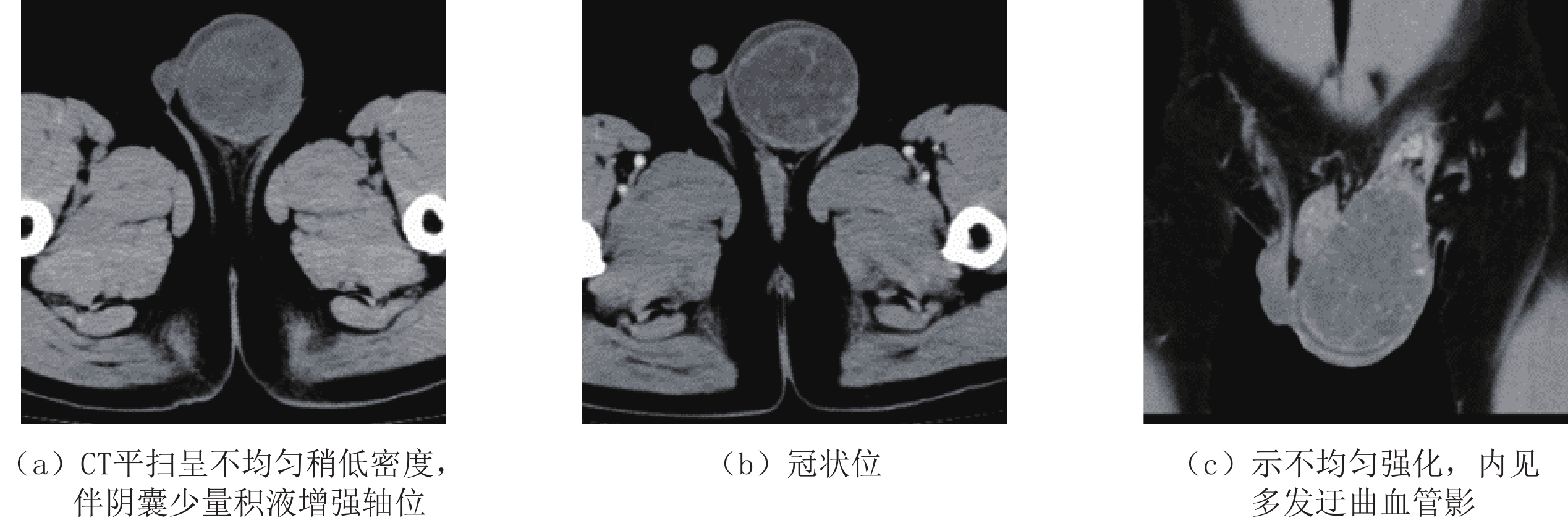
内胚窦瘤10例,左右侧各5例。发病年龄6个月至5岁,平均年龄16个月;直径3~6 cm。CT平扫密度不均匀,可见低密度液化坏死区;4例合并出血,增强扫描呈絮状或斑片状不均匀明显强化;CT值增加约45~60 HU,瘤内均可见多发迂曲、增粗血管影。
4例畸胎瘤,均为良性畸胎瘤。右侧3例,左侧1例;年龄1~20岁;肿瘤直径3~7 cm。CT平扫表现为边界清楚的等低混杂密度肿块,CT值平均约17~28 HU,4例可见斑点状、片状钙化,2例可见脂肪密度影;增强扫描病灶呈不均匀轻度强化,CT值增加15~20 HU。
2例混合性生殖细胞瘤。发病年龄分别为32岁和48岁;均位于左侧;直径分别为10 cm和13 cm。CT平扫可见出血、坏死,边界不清,增强扫描明显不均匀强化,瘤内可见迂曲血管影。2例均出现腹膜后淋巴结转移,未见血行转移。
平滑肌瘤2例。年龄分别为62岁和64岁;直径均为4 cm。CT平扫边界清楚、密度均匀,增强轻度均匀强化。
淋巴瘤3例。年龄60~72岁;直径5~7 cm;左侧2例,右侧1例。2例边界清楚,密度均匀,增强均匀强化,CT值25~40 HU;1例右侧壁边界局部不清,增强病灶大部分呈轻至中度不均匀强化,局部边界不清区域显著强化。
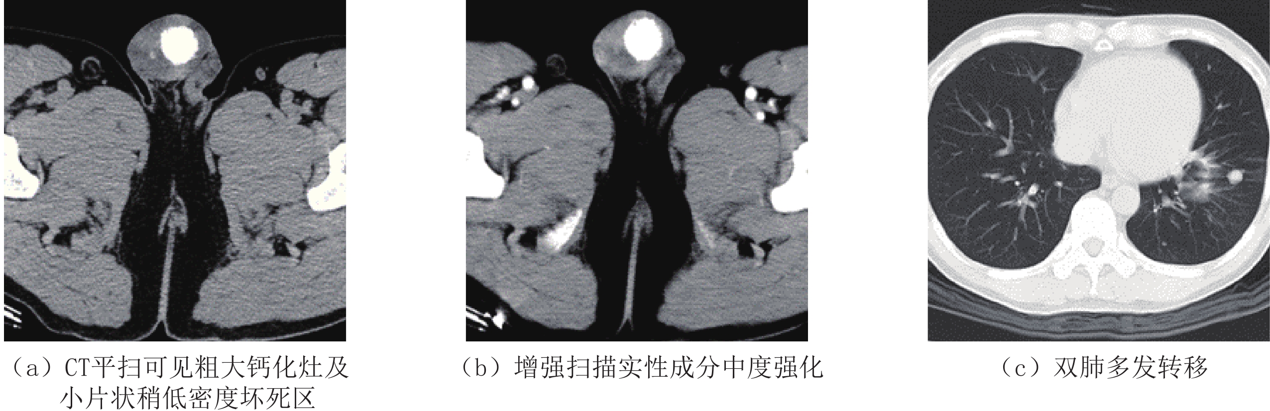
恶性间质细胞瘤1例。年龄40岁,直径4.5 cm。CT平扫右侧睾丸类圆形肿块,边界不清,密度不均,可见片状粗大钙化,实性成分平扫CT值22 HU,增强扫描CT值57 HU;可见多发淋巴结转移及双肺转移。
2.3 睾丸肿瘤性病变超声与CT对比分析
将经病理证实的46例睾丸肿瘤CT诊断结果与超声诊断结果相比较,结果显示:CT平扫加增强扫描方案对睾丸各类型肿瘤的诊断准确率较超声诊断具有较大的优势,尤其是在混合型生殖细胞瘤及淋巴瘤的诊断准确率上,CT诊断效能较超声的诊断效能具有明显的优势。导致这一结果的可能原因,一方面可能是CT平扫加增强扫描方案能够提供更丰富的影像学信息,并且具有一定的影像学特征;另一方面由于CT诊断报告通常由两位不同级别的医师书写,而超声诊断报告多数是由一位医师独立完成,因此前者的工作模式能够有效提高睾丸肿瘤的诊断符合率。各类型睾丸肿瘤两者诊断符合率具体数据见表1。
表 1 睾丸肿瘤超声与CT诊断符合率(%)Table 1. Coincidence rate of ultrasonography and CT in diagnosis of testicular tumors检查方法 精原细胞瘤
(n=24)内胚窦瘤
(n=10)畸胎瘤
(n=4)混合型生殖细胞瘤(n=2) 平滑肌瘤
(n=2)淋巴瘤
(n=3)恶性间质细胞瘤
(n=1)CT符合率 92 80 100 100 0 66.7 0 超声符合率 75 60 80 0 0 33.3 0 3. 讨论
3.1 临床概述
睾丸肿瘤较为少见,其中生殖细胞类肿瘤占90%~95%,而非生殖细胞类肿瘤仅占5%~10%[2]。本组46例睾丸肿瘤,其中生殖细胞类肿瘤占89%(41/46),与文献基本相符。根据细胞的分化程度,生殖细胞类肿瘤可分为精原细胞瘤和非精原细胞瘤两大类。非精原细胞瘤包括内胚窦瘤、畸胎瘤,胚胎性癌等,其他少见睾丸肿瘤主要为淋巴瘤、白血病浸润等。本组病例基本涵盖以上病理类型。
睾丸肿瘤的发病主要有几个特点:①青春期前、后肿瘤的发生率、组织学类型差别较大;②睾丸肿瘤发生在青春期后约占 60%~70%;③生殖细胞瘤约占儿童睾丸肿瘤的90%,而成人最多见睾丸肿瘤则为精原细胞瘤,其发病高峰约为30~45岁,而60岁以上的老年男性则应重点考虑淋巴瘤[3]。
睾丸肿瘤的临床表现常无特异性,多以无痛性包块就诊。肿瘤较大时压迫精索静脉,可引起局部胀痛。在实验室检查方面,3个肿瘤标志物,即甲胎蛋白(AFP)、绒毛膜促性腺激素(HCG)和胎盘碱性磷酸酶(PLAP)对部分肿瘤的诊断和鉴别具有十分重要的意义。当AFP>1000 ng/mL时,对内胚窦瘤有诊断意义。
本组10例内胚窦瘤,其中8例出现AFP明显升高,术前均准确诊断。HCG则对含有合体滋养层细胞一类肿瘤具有诊断意义,例如绒毛膜癌、胚胎癌及含有以上成分的混合性生殖细胞肿瘤,HCG均可升高。本组2例AFP及HCG同时升高,术前均准确诊断为混合型生殖细胞瘤。当HCG>1000 mlU/mL,绒癌或含有绒癌成分的混合性生殖细胞肿瘤可能性大;当HCG>2000 mlU/mL,对绒癌有诊断意义。80%非精原细胞瘤性生殖细胞瘤患者将有以上两种肿瘤标志物中的一种或两种升高,而精原细胞瘤仅5% HCG阳性。PLAP在胎盘合体滋养层细胞可正常表达,故对生殖细胞瘤的诊断有一定帮助,但无特异性,精原细胞瘤中的50%患者PALP可升高。
概而言之,常见睾丸肿瘤中,内胚窦瘤的AFP阳性且显著升高;绒癌的HCG明显升高;胚胎癌的HCG和AFP都阳性,但都不是很高,本组1例胚胎性癌生化指标未见异常;精原细胞瘤的HCG和AFP都不升高,但PLAP可升高。
3.2 CT表现及鉴别诊断
3.2.1 精原细胞瘤
睾丸生殖细胞类肿瘤中最常见,占所有生殖细胞源性肿瘤的40%~50%[4]。本组40例生殖细胞肿瘤中精原细胞瘤24例,占60%。精原细胞瘤CT平扫表现为等或稍低密度,可见包膜,部分病例包膜不完整,病理证实包膜为睾丸的白膜,当肿瘤在睾丸内生长时并无包膜,当肿瘤增大体积超过睾丸时,肿瘤与睾丸共用同一被膜,故影像上肿瘤多表现为边界清楚,有包膜的软组织肿块。由于病灶体积较小及CT分辨率限制,本组24例其中仅14例可见包膜。
增强扫描肿瘤呈轻度强化,CT值增加一般不超过25 HU,可能与青春期后形成的血睾屏障有关,它阻隔了血液循环内的细胞毒性物质渗透到曲细精管内的保护层,保持了内环境的相对稳定,对比剂不能通过未被破坏的血睾屏障,故正常睾丸组织基本不强化[5]。因此通过肿瘤的强化程度可能提示血睾屏障的破坏程度,间接反映肿瘤的恶性程度,这种推测还有待扩大样本量的进一步研究。
本组24例增强扫描均为轻-中度强化(图1(b)),与既往报道基本一致。增强后肿瘤实体部分见分隔样强化,部分学者认为此征象是精原细胞瘤的特征性表现,病理基础主要是肿瘤内含有纤维血管分隔[5-6]。本组仅16例出现上述征象,可能与CT软组织分辨率相对较低有关。肿瘤中心多见小灶状囊变、坏死,另有文献报道肿瘤内可见不规则钙化[7],本组24例均未见钙化。淋巴结主要是该肿瘤的转移通路,以腹膜后及盆腔转移多见,本组中有6例患者出现腹膜后或盆腔多发淋巴结转移,但均未见远处转移。
3.2.2 内胚窦瘤
内胚窦瘤又称卵黄囊瘤,成人主要发生在性腺器官,儿童则好发于性腺外组织,睾丸相对少见,却是小儿睾丸肿瘤中最常见的病理类型。在低龄男童中90% 的睾丸肿瘤是生殖细胞源性,其中2/3为内胚窦瘤[8]。Kaplan报告269例小儿睾丸肿瘤中,内胚窦瘤占67%,常在2岁以内发病,平均年龄17个月[9]。本组10例其中8例发病年龄小于2岁,平均年龄16个月,与文献报道相符。
血清AFP显著升高是其重要生物学特性,但应注意,尚有10% 的患儿AFP并不升高,本组8例血清AFP明显增高。CT表现睾丸肿块密度不均匀,常见低密度液化坏死,部分肿瘤合并出血,增强扫描呈絮状或斑片状不均匀强化,早期以周边强化明显(图2(b)),延迟扫描持续强化。其病理学特点主要包括以下几种形式:
①内胚窦样结构:细胞围绕血管呈放射状排列,外周则呈环状排列;②腺泡状结构:细胞排列成腺泡状,间质内见丰富肿瘤小血管;③疏松网状结构:肿瘤细胞排列成疏松的网状或筛状,形成大小不等的网眼或囊性裂隙[10]。
以上肿瘤细胞结构特点以及间质富含血管的组织成分决定了内胚窦瘤的强化特点,再结合患者发病年龄及AFP结果,可明显提高术前诊断准确率。
3.2.3 畸胎瘤
新版WHO分类中纯型畸胎瘤根据患者年龄划分为青春期前型和青春期后型,前者临床表现良性,为避免产生歧义,青春期后型畸胎瘤宜注明“恶性”,青春期前型则标注为“良性”[11]。
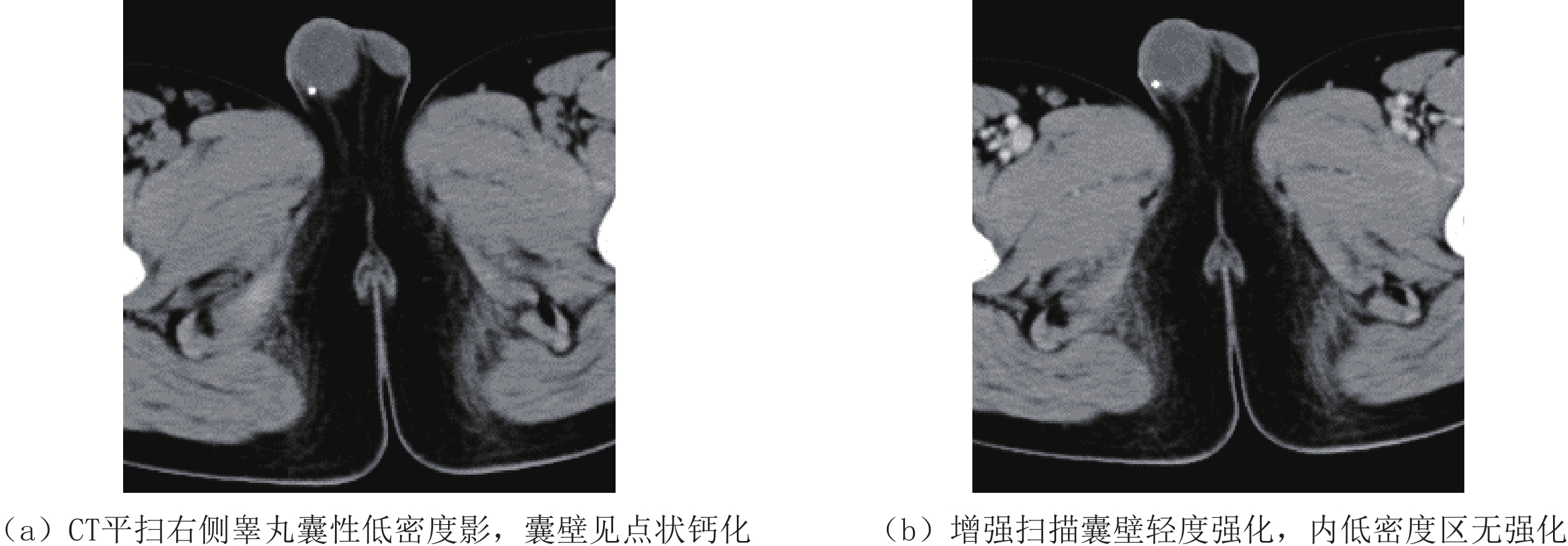
典型畸胎瘤由于存在3个胚层成分,影像上与其他睾丸肿瘤鉴别不难,瘤内脂肪和(或)钙化是较为特征性的鉴别点。若畸胎瘤内仅伴有钙化而无脂肪成分时,应注意与精原细胞瘤伴钙化相鉴别。畸胎瘤内钙化一般较多且大,而精原细胞瘤钙化小而少。恶性畸胎瘤发病年龄通常偏大,故青春期前CT上见到含有钙化和(或)脂肪成分的肿瘤应该首先诊断为良性畸胎瘤(图3(a))。良性畸胎瘤有时仅表现为单囊或多囊性病变,不伴钙化和脂肪,此时密度均匀,囊壁厚薄均匀,增强扫描仅囊壁呈轻-中度强化(图3(b))。典型的畸胎瘤内能见到牙齿及不规则的骨骼影,但本组4例均没有见到牙齿或骨骼,均为不规则钙化灶。
3.2.4 混合性生殖细胞肿瘤
凡是含2种或2种以上类型生殖细胞瘤者都统称为混合性生殖细胞肿瘤[12],以畸胎瘤和胚胎性癌组合最为常见,约占25%,40% 的病例可见灶状卵黄囊瘤[13]。最常见的肿瘤成分是胚胎性癌。年龄对于混合性生殖细胞肿瘤的诊断有一定的帮助,多见于20~35岁男性[14]。胚胎性癌及卵黄囊的恶性程度较高,生长速度较快,故肿块体积较大,肿瘤易发生缺血坏死。
文献报道睾丸混合性生殖细胞瘤患者血AFP和HCG升高的分别占60% 和55%,局灶性出血和坏死常见[15]。CT表现出血、大片坏死的混杂密度,有时可见脂肪,且AFP及HCG升高时应考虑到混合性生殖细胞瘤的诊断。
3.2.5 平滑肌瘤
睾丸平滑肌瘤发生率极低,1972年由Alber等[16]命名并报道以来,多以个案报道形式发表,截至目前,尚未见大样本影像学资料报道。发病人群以中老年为主,以左侧多见。虽然在新版WHO分类中[1],睾丸平滑肌瘤归为其他非特异性间质肿瘤,但其组织学起源仍存在争议。①睾丸的白膜组织;②曲细精管细胞;③多能干细胞。睾丸平滑肌瘤多位于睾丸上下两极,与白膜关系密切,故笔者认为其来源于睾丸白膜的可能性大,但其确切来源尚有待免疫组织化学和分子生物学的技术进一步发展。
目前,睾丸平滑肌瘤的影像表现多以超声个案报道,均质的低回声或包含钙化而表现为不均匀回声[17]。本组2例平滑肌瘤年龄超过60岁且密度均匀,增强轻度均匀强化,与淋巴瘤鉴别困难。
3.2.6 淋巴瘤
睾丸淋巴瘤主要为非霍奇金淋巴瘤,尽管发病率较低,却是60岁以上老年人最常见的睾丸恶性肿瘤,在小于30岁的年轻人中罕见[18]。最常见的病理类型是弥漫大B细胞淋巴瘤,占所有睾丸淋巴瘤的50%~80%,少见类型有Burkitt淋巴瘤、滤泡性淋巴瘤及NK/T淋巴瘤等。CT表现为一侧或双侧睾丸类圆形等密度肿块,境界清楚,密度均匀,这种影像特点是由于淋巴细胞弥漫浸润于睾丸实质所致,增强后多呈均匀性持续强化。如果肿块较大,中央可发生坏死。有文献报道[19]8例睾丸原发性淋巴瘤CT表现为增强扫描均匀性强化,强化CT值高于25 HU,低于45 HU,并可见肿块内部小血管穿行,认为小血管穿行是睾丸淋巴瘤的特征之一。
本组3例影像表现与文献报道基本相符,其中1例病灶右侧壁边界不清,增强大部分病灶呈轻至中度不均匀强化,局部边界不清区域显著强化,病理证实为肿瘤侵犯白膜,结合血清标志物,可与睾丸平滑肌瘤、纤维瘤等良性非生殖细胞肿瘤相鉴别。
3.2.7 恶性间质细胞瘤
本例误诊为精原细胞瘤,回顾性分析不符合精原细胞瘤的依据主要有以下几点。
(1)精原细胞瘤钙化少见,本组24例精原细胞瘤均未见钙化。睾丸粗大钙化通常被认为是睾丸良性病变的特征,但最近有研究认为睾丸粗大钙化与恶性肿瘤相关[20]。最早曾有作者使用“烧毁性肿瘤(burned-out tumor)”描述睾丸粗大钙化与睾丸恶性肿瘤之间的关系,是指睾丸恶性肿瘤的原发灶在未经治疗情况下出现自发性消退,可能与机体的免疫机制有关,而粗大钙化则是原发部位肿瘤出现自发性消退后的残留表现。
(2)由于血睾屏障存在,精原细胞瘤增强扫描多呈轻度强化,CT值增加一般不超过25 HU,而本例增强中度强化,CT值增加约35 HU。
(3)精原细胞瘤可出现淋巴结转移,但远处转移少见,本例出现双肺多发转移。
3.3 睾丸肿瘤性病变超声与CT对比分析
超声是睾丸病变的首选检查方法,文献报道[14]超声检查对睾丸肿瘤诊断率为85%,特异度为44.4%。本组睾丸常见肿瘤的CT诊断符合率高于超声。结合临床表现及相关实验室检查,CT检查可明显提高睾丸肿瘤的定性准确率,对睾丸肿瘤的鉴别具有重要价值。
4. 结论
综上所述,睾丸肿瘤的影像学表现具有不同特点,CT能很好地显示出睾丸肿瘤的影像特征,在肿瘤的诊断中有较高的实用价值,结合临床资料及肿瘤标志物有助于鉴别,此外还可以显示淋巴结及远处转移情况,为肿瘤的分期和临床制定治疗方案提供参考。
-
![]()
图 4 男,48岁,左侧睾丸混合性生殖细胞肿瘤
Figure 4. Male, 48 years old, Mixed germ cell tumor of left testis
![]()
图 5 男,40岁,右侧睾丸恶性间质细胞瘤
Figure 5. Male, 40 years old, Malignant Leydig cell tumor of right testis
表 1 睾丸肿瘤超声与CT诊断符合率(%)
Table 1 Coincidence rate of ultrasonography and CT in diagnosis of testicular tumors
检查方法 精原细胞瘤
(n=24)内胚窦瘤
(n=10)畸胎瘤
(n=4)混合型生殖细胞瘤(n=2) 平滑肌瘤
(n=2)淋巴瘤
(n=3)恶性间质细胞瘤
(n=1)CT符合率 92 80 100 100 0 66.7 0 超声符合率 75 60 80 0 0 33.3 0  下载: 导出CSV
-
[1] MOCH H, HUMPHREY P A, ULBRIGHT T M, et al. WHO classification of tumors of the urinary system and male genital organ[M]. Lyon: IARC Press, 2016.
[2] ULBRIGHT T M, YOUNG R H. Tumors of the testis and adjacent structures (AFP atlas of tumor pathology series 4)[M]. Sliver Spring (Maryland): American Registry of Pathology, 2013.
[3] HUYGHE E, MATSUDA T, THONNEAU P. Increasing incidence of testicular cancer worldwide: A review[J]. The Journal of Urology, 2003, 170(1): 5−11. doi: 10.1097/01.ju.0000053866.68623.da
[4] 曹登峰. 睾丸生殖细胞肿瘤的病理形态和免疫组织化学标志物[J]. 中华病理学杂志, 2014,43(11): 776−781. doi: 10.3760/cma.j.issn.0529-5807.2014.11.016 CAO D F. Pathomorphology and immunohistochemical markers of testicular germ cell tumors[J]. Chinese Journal of Pathology, 2014, 43(11): 776−781. (in Chinese). doi: 10.3760/cma.j.issn.0529-5807.2014.11.016
[5] UENO T, TANAKA Y O, NAGATA M, et al. Spectrum of germ cell tumors: From head to toe[J]. Radiographics, 2004, 24(2): 387−404. doi: 10.1148/rg.242035082
[6] 王建明, 赵双全, 宋世军, 等. 睾丸良恶性肿块的CT、MRI表现及鉴别诊断[J]. 中国中西医结合影像学杂志, 2019,17(2): 143−146. doi: 10.3969/j.issn.1672-0512.2019.02.009 WANG J M, ZHAO S Q, SONG S J, et al. CT and MRI features and differential diagnosis of benign and malignant testicular mass lesions[J]. Chinese Imaging Journal of Integrated Traditional and Western Medicine, 2019, 17(2): 143−146. (in Chinese). doi: 10.3969/j.issn.1672-0512.2019.02.009
[7] 贾承晔, 张晓琴, 杨署, 等. 睾丸实性肿块的CT和MRI表现特征[J]. 中国医学影像技术, 2017,33(6): 929−932. JIA C H, ZHANG X Q, YANG S, et al. CT and MRI features of testicular solid lesions[J]. Chinese Journal of Medical Imaging Technology, 2017, 33(6): 929−932. (in Chinese).
[8] 段大兵. 小儿睾丸原发性卵黄囊瘤的CT及MRI表现[J]. 国际医药卫生导报, 2019,(5): 737−739. DUAN D B. CT and MRI findings of primary yolk SAC tumor of testis in children[J]. International Medicine and Health Guidance News, 2019, (5): 737−739. (in Chinese).
[9] KAPLAN G W, CROMIE W C, KELALIS P P, et al. Prepubertal yolk sac testicular tumors: Report of the testicular tumor registry[J]. Journal of Urology, 1988, 140(3): 1109−1112.
[10] 杨小英, 王娅宁, 雷延成. 小儿睾丸内胚窦瘤CT诊断[J]. 医学影像学杂志, 2013,23(7): 1098−1100. doi: 10.3969/j.issn.1006-9011.2013.07.036 YANG X Y, WANG Y N, LEI Y C. CT diagnosis of endodermal sinus tumor of testis in children[J]. Journal of Medical Imaging, 2013, 23(7): 1098−1100. (in Chinese). doi: 10.3969/j.issn.1006-9011.2013.07.036
[11] 吕炳建, 程亮. 睾丸生殖细胞肿瘤分类和诊断进展[J]. 中华病理学杂志, 2017,46(6): 435−438. doi: 10.3760/cma.j.issn.0529-5807.2017.06.021 LV B J, CHENG L. Progress in classification and diagnosis of testicular germ cell tumors[J]. Chinese Journal of Pathology, 2017, 46(6): 435−438. (in Chinese). doi: 10.3760/cma.j.issn.0529-5807.2017.06.021
[12] BAHRAMI A, RO J Y, AYALA A G. An overview of testicular germ cell tumors[J]. Archives of Pathology & Laboratory Medicine, 2007, 131(8): 1267−1280.
[13] ZHANG C F, LIU C, SHI Q L, et al. Clinicopathological analysis of testicular mixed germ cell tumor[J]. National Journal of Andrology, 2011, 23(4): 336−341.
[14] WILDEMBERG L, NETO L V, TABOADA G F, et al. Sellar and suprasellar mixed germ cell tumor mimicking a pituitary adenoma[J]. Pituitary, 2011, 14(4): 345−350. doi: 10.1007/s11102-008-0161-z
[15] 陶立新, 龙德云, 张福刚. 睾丸混合性生殖细胞瘤4例MRI表现[J]. 武警医学, 2014,25(8): 827−829. doi: 10.3969/j.issn.1004-3594.2014.08.022 TAO L X, LONG D Y, ZHANG F G. MRI findings of mixed germinoma of testis: A report of 4 cases[J]. Medical Journal of the Chinese People's Armed Police Force, 2014, 25(8): 827−829. (in Chinese). doi: 10.3969/j.issn.1004-3594.2014.08.022
[16] 李立民, 孙二琳, 顾兴洲, 等. 睾丸平滑肌瘤2例报告并文献复习[J]. 临床泌尿外科杂志, 2015,30(6): 556−558. LI L M, SUN E L, GU X Z, et al. Leiomyoma of testis: A report of 2 cases and review of literature[J]. Journal of Clinical Urology, 2015, 30(6): 556−558. (in Chinese).
[17] 刘学琳, 张建蕾. 睾丸平滑肌瘤超声表现1例[J]. 中国医学影像技术, 2020,36(1): 147. LIU X L, ZHANG J L. Ultrasonic manifestations of testicular leiomyoma: Case report[J]. Chinese Journal of Medical Imaging Technology, 2020, 36(1): 147. (in Chinese).
[18] 胡兴荣, 朱鑫, 贵丹, 等. 超声、CT与MRI对原发性睾丸淋巴瘤诊断价值的临床研究[J]. 影像科学与光化学, 2020,38(6): 994−999. doi: 10.7517/issn.1674-0475.200304 HU X R, ZHU X, GUI D, et al. The clinical study of diagnostic value in primary testicular lymphoma by methods of ultrasound, CT and MRI[J]. Imaging Science and Photochemistry, 2020, 38(6): 994−999. (in Chinese). doi: 10.7517/issn.1674-0475.200304
[19] 段刚, 许乙凯, 戴琳, 等. 睾丸病变的CT诊断和病理学研究[J]. 南方医科大学学报, 2007,27(1): 98−100. doi: 10.3321/j.issn:1673-4254.2007.01.029 DUAN G, XU Y K, DAI L, et al. CT diagnosis and pathological findings of testicular lesion[J]. Journal of Southern Medical University, 2007, 27(1): 98−100. (in Chinese). doi: 10.3321/j.issn:1673-4254.2007.01.029
[20] PEDERSEN M R, BARTLETT E C, BROWN C, et al. Is testicular macrocalcification a risk for malignancy? Tumor development on ultrasonographic follow-up of preexisting intratesticular macrocalcification[J]. Journal of Ultrasound in Medicine, 2018, 37(12): 2949−2953. doi: 10.1002/jum.14657
-
期刊类型引用(1)
1. 周丽,王晖,胡剑. 探讨CDFI、CT、MRI诊断睾丸扭转的价值与病理学一致性分析. 中国CT和MRI杂志. 2024(03): 152-154 .  百度学术
百度学术
其他类型引用(0)

计量
- 文章访问数: 648
- HTML全文浏览量: 243
- PDF下载量: 30
- 被引次数: 1


