
Application of Deep Learning Reconstruction Algorithm in Upper Abdomen CT
-
摘要: 目的:通过分析比较自适应统计迭代重建(ASIR)算法和深度学习重建(DLIR)算法在上腹部CT成像中的图像质量,探讨DLIR算法在上腹部CT成像中的应用价值。方法:回顾性纳入75例患者上腹部CT平扫图像,利用自适应统计迭代重建算法ASIR(30%、50%、70%、90%)和深度学习重建算法(DL-L、DL-M、DL-H)重建图像,共7组。测量每组图像肝脏、胰腺、竖脊肌的CT值和SD值,并计算信噪比(SNR)和对比噪声比(CNR),采用单因素方差分析对各指标进行客观评价;同时由两位放射医师对图像质量和噪声评分,采用Friedman <i<M</i<检验进行比较。结果:①七组重建图像的SD值、SNR、肝脏CNR差异有统计学意义。②DL-L与ASIR 50%、DL-M与ASIR 70%、DL-H与ASIR 90% 间各ROI处CT值、SD值、SNR值、CNR值无差异。③三种深度学习重建算法间随等级升高,SNR值升高,差异有统计学意义;且DL-H 算法的SNR值高于ASIR 30%、ASIR 50%,SD值低于除ASIR 90% 外的其余5组重建算法。④七组图像主观评分差异有统计学意义,算法DL-H具有最佳的图像质量和最低的噪声,DL-M、ASIR 90%、DL-L、ASIR 70%、ASIR 50%、ASIR 30% 图像噪声依次增加。结论:深度学习重建算法能够降低上腹部图像噪声,提高图像质量,且随等级升高,图像噪声降低、质量提高、信噪比升高。Abstract: Objective: To explore the application of deep learning image reconstruction (DLIR) algorithm in upper abdominal CT imaging by analyzing the image quality of adaptive statistical iterative reconstruction (ASIR) algorithm and DLIR. Methods: Retrospectively included 75 patients’ upper abdominal CT plain scan images, using adaptive statistical iterative reconstruction algorithm ASIR (30%, 50%, 70%, 90%) and deep learning reconstruction algorithm (DL-L, DL-M, DL-H) to reconstruct images, a total of 7 groups. Measured the CT and SD values of the liver, pancreas, and erector spinae , and calculated the signal to noise ratio (SNR) and contrast to noise ratio (CNR). Objective indicators were evaluated by one-way ANOVA. Two radiologists scored the image quality and noise, and compared them with Friedman <i<M</i< test. Results: (1) The SD value, SNR, and liver CNR of the seven reconstructed images had statistically significant differences. (2) The difference in CT value, SD value, SNR value and CNR value at each ROI between DL-L and ASIR 50%, DL-M and ASIR 70%, DL-H and ASIR 90% was small. (3) The SNR value of the three DLIR algorithms increased as the level increased, and the difference was statistically significant; and the SNR value of the DL-H algorithm was higher than ASIR 30% and ASIR 50%, and the SD value was lower than the other five reconstruction algorithms except for the ASIR 90%. (4) The difference in the subjective scores of the seven groups of images was statistically significant. The algorithm DL-H had the best image quality and the lowest noise, DL-M, ASIR 90%, DL-L, ASIR 70%, ASIR 50%, ASIR 30% image noise in sequence increased. Conclusion: The DLIR algorithm can reduce the image noise of the upper abdomen and improve the image quality. As the level increased, the image noise decreased, the quality improved, and the signal-to-noise ratio increased.
-
上腹部CT检查是上腹部疾病检查的重要手段之一,广泛应用于临床,CT图像质量高低对临床诊断效能有重要影响。多年来,CT技术领域的发展一直围绕探测器宽度、扫描剂量降低、重建算法优化几个方面。自适应统计迭代重建(adaptive statistical iterative reconstruction,ASIR)是应用最广泛的重建技术之一[1-4],它是一种混合算法,利用滤波反投影得到的图像信息作为迭代重建的基础来优化图像质量,在不影响图像分辨率的前提下降低图像噪声、改善图像质量[5]。最近,深度学习(deep learning,DL)在医学影像领域取得许多进展,引起广泛关注。在图像质量提高方面,基于DL的算法性能更优于传统算法[6-9]。
本研究使用了基于深度卷积神经网络的DL算法,该算法采用了人工智能CT图像处理技术TrueFidelityTM,提供了低、中、高(DL-L,DL-M,DL-H)3个重建强度等级来控制图像噪声和质量,在图像质量、扫描辐射剂量和重建速度方面优于现有的临床常用算法。本研究通过客观评价和主观评价,分析不同权重的ASIR和不同等级的DL算法所重建图像的质量,评估DL重建算法在上腹部成像中的应用价值。
1. 资料与方法
1.1 研究对象
回顾性纳入2021年3月至4月在四川大学华西医院进行上腹部CT平扫患者的图像。纳入标准:①体质指数(body mass index,BMI)18.5~29.9 kg/m2;②年龄:18~90岁。排除标准:①图像伪影干扰严重(如呼吸运动伪影、上肢伪影干扰等),图像质量差;②严重上腹部占位性病变,如肝癌、胰腺肿瘤等。
根据以上标准,最后共纳入患者75例,其中男性44例,女性31例,年龄18~90岁,平均年龄(55.49±16.06)岁;BMI范围18.66~29.75 kg/m2,平均BMI(22.67±3.87)kg/m2。
1.2 检查方法
1.2.1 扫描方案
所有患者的上腹部CT扫描均在同一台CT机(GE公司,Revolution Apex)上完成,去除被检部位金属物品,非被检部位进行合理辐射防护,采用头先进仰卧位,双手上举,吸气后屏气扫描,扫描范围从膈顶到肝下缘。管电压120 kV、自动管电流调制,扫描层厚5 mm,重建层厚0.625 mm。
1.2.2 图像重建
所有患者的图像均采用ASIR及DLIR两种方法重建,其中ASIR采用ASIR 30%、ASIR 50%、ASIR 70% 和ASIR 90% 四种混合重建方式,DLIR采用DL-L、DL-M、DL-H 三种重建方式。重建层厚0.625 mm,重建模式为STAND,窗宽350 HU,窗位50 HU,每例患者共7组不同重建图像。
1.3 图像质量评价
1.3.1 客观评价
所有患者的图像传输至随机工作站,在肝脏(ROI1)、胰腺(ROI2)和竖脊肌(ROI3)上手动勾画大小约100 mm2的兴趣区,尽量避开大的血管、病灶和器官边缘,分别在3个不同层面上测量各ROI的CT 值和SD值,取平均值。利用公式计算各ROI的SNR值,肝脏-竖脊肌、胰腺-竖脊肌CNR值。
SNR=兴趣区CT值/SD值,
CNR=(兴趣区CT值 - 竖脊肌CT值)/竖脊肌SD值[10]。
1.3.2 主观评价
由两位放射科医师对上述重建图像进行主观评估,按肝、胆、胰、脾、肾等上腹部结构显示的清晰程度、细腻度和图像提供的诊断信息对图像质量进行5级评分(表1)。采用李克特5分法[11]对图像噪声进行评价,5-不可接受图像噪声,4-高于平均噪声,3-平均噪声,2-低于平均噪声,1-完全可接受图像噪声。
表 1 图像主观评价5级评分标准Table 1. 5-level scoring standard for subjective image evaluation评分 评分细节 5分 肝脏、胰腺等上腹部组织结构显示非常清晰,图像细腻,能提供充分的诊断信息 4分 肝脏、胰腺等上腹部组织结构显示较为清晰,图像较细腻,能提供足够的诊断信息 3分 肝脏、胰腺等上腹部组织结构显示欠清晰,图像欠细腻,能提供一定的诊断信息 2分 肝脏、胰腺等上腹部组织结构显示模糊,图像粗糙,图像提供的诊断信息不足 1分 肝脏、胰腺等上腹部组织结构无法清晰显示,伪影重,图像不清,不能提供诊断信息 1.4 统计学方法
采用SPSS 25.0软件,7组重建算法间CT值、SD值、SNR、CNR的比较采用单因素方差分析(One Way ANOVA);图像质量主观评分、噪声评分采用Friedman M检验;检验水准α 为0.05(双侧),P<0.05为差异有统计学意义。采用Kappa检验评价两名放射医师对图像主观评分的一致性。当Kappa值在0.8~1.0之间时,一致性很好;0.61~0.8,一致性较好;0.41~0.6,一致性中等;0.21~0.4,一致性一般;0~0.2,一致性较差。
2. 结果
2.1 图像CT值和SD值
七组重建图像间,图像CT值间的差异无统计学意义。SD值的差异具有统计学意义;在各ROI处,三种DLIR算法间,SD值依次降低,除在ROI3处,DL-L与DL-M无差异;算法ASIR 30%、ASIR 50%、ASIR 70%、ASIR 90% 间,SD值逐渐降低,在ROI3处,ASIR 70% 与ASIR 90% 无差异;DL-H算法除与ASIR 90% 的SD值无差异外,明显低于其余5组重建算法,差异有统计学意义(表2)。
表 2 七组重建图像的CT值、SD值分析Table 2. Analysis of CT value and SD value of seven groups of reconstructed images算法 CT值/HU SD值/HU ROI1 ROI2 ROI3 ROI1 ROI2 ROI3 DL-L 60.65±8.64 44.63±7.45 49.43±8.70 14.05±4.21 15.38±4.73 15.25±5.57 DL-M 61.24±9.62 44.33±7.46 49.46±8.67 11.38±3.27 12.81±3.94 12.68±4.97 DL-H 60.64±8.43 44.33±7.27 49.48±8.66 8.41±2.15 9.53±3.16 9.90±4.54 ASIR 30% 60.58±8.80 44.50±7.68 49.46±8.55 17.70±5.25 19.34±5.54 18.79±6.26 ASIR 50% 60.65±8.81 44.29±7.48 49.45±8.60 14.69±4.33 16.14±4.95 15.77±5.46 ASIR 70% 60.59±8.96 44.32±7.28 49.51±8.66 11.86±4.43 13.07±4.24 12.85±4.92 ASIR 90% 60.75±8.70 44.29±7.53 49.55±8.88 8.74±2.83 10.1±3.57 10.27±4.70 P >0.05 >0.05 >0.05 <0.05 <0.05 <0.05 2.2 图像SNR值和CNR值
SNR值在7组图像间差异具有统计学意义;3种DLIR算法间随等级升高,信噪比升高,差异有统计学意义。ASIR 70% 在ROI2处,与ASIR 90% 差异有统计学意义,与其余5种重建算法无统计学差异。DL-H算法的SNR值高于DL-L、DL-M、ASIR 30%、ASIR 50%、ASIR 70%,差异有统计学意义。
各组肝脏CNR值具有统计学差异,3种深度学习重建算法间肝脏CNR值无差异;ASIR 30% 肝CNR值低于DL-H、ASIR 90%,差异有统计学差异。各组胰腺CNR值无统计学差异(表3)。
表 3 七组重建图像的SNR值、CNR值分析Table 3. Analysis of SNR and CNR values of seven groups of reconstructed images算法 SNR CNR ROI1 ROI2 ROI3 ROI1 ROI2 DL-L 4.67±1.53 3.18±1.05 3.67±1.30 0.71±0.70 -0.41±0.52 DL-M 5.80±1.91 3.80±1.28 4.47±1.64 0.90±0.89 -0.53±0.64 DL-H 7.66±2.30 5.11±1.64 5.85±2.17 1.07±1.03 -0.70±0.80 ASIR 30% 3.71±1.28 2.51±0.86 2.94±1.03 0.58±0.58 -0.33±0.42 ASIR 50% 4.49±1.58 3.02±1.04 3.54±1.29 0.70±0.67 -0.42±0.51 ASIR 70% 5.67±2.09 4.47±1.76 4.42±1.67 0.84±0.86 -0.22±0.63 ASIR 90% 7.70±2.94 4.96±1.83 5.79±2.48 1.09±1.05 -0.72±0.84 P <0.05 <0.05 <0.05 <0.05 >0.05 两两比较发现,DL-L与ASIR 50%、DL-M与ASIR 70%、DL-H与ASIR 90% 间各ROI处所有参数无差异。
2.3 主观评分及一致性评价
两位放射医师主观评分一致性较好(k=0.565~0.817),DLIR算法随等级增加,图像噪声降低,图像质量提高;DL-H重建图像整体质量最佳,噪声最低。ASIR随权重增加,噪声降低;算法DL-H、DL-M、ASIR 90%、DL-L、ASIR 70%、ASIR 50%、ASIR 30% 图像噪声依次增加。客观评价指标DL-L与ASIR 50%、DL-M与ASIR 70%、DL-H与ASIR 90% 无差异,但DLIR组的图像质量评分高于ASIR组,噪声低于ASIR组(表4、图1和图2)。
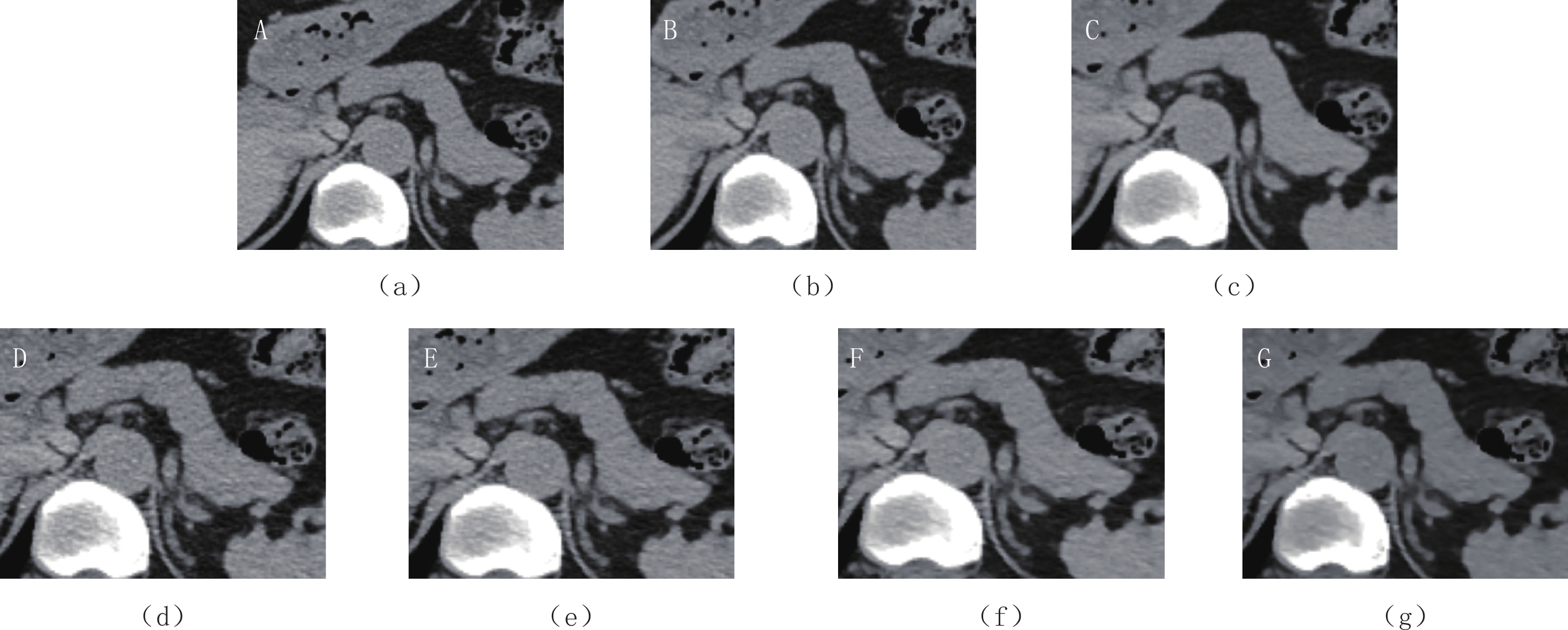
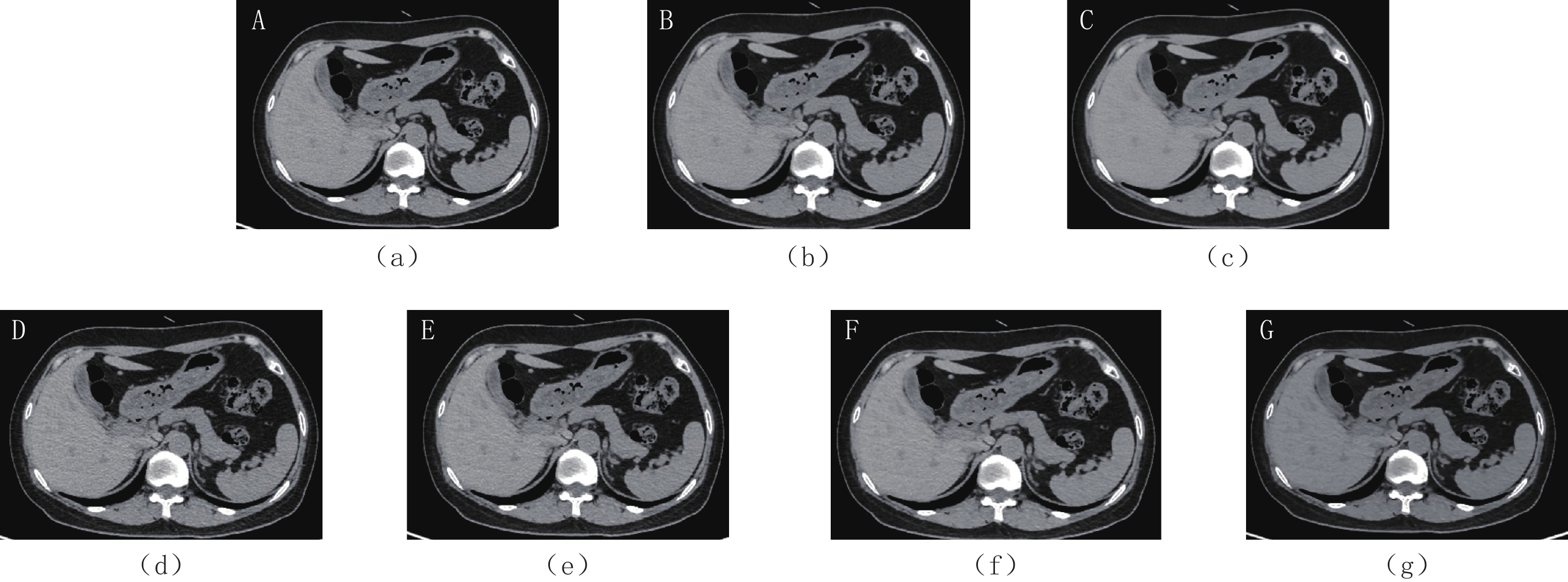
表 4 图像质量主观评分分析Table 4. Image quality subjective scoring analysis评分内容 评分者 DL-L DL-M DL-H ASIR 30% ASIR 50% ASIR 70% ASIR 90% 质量 Reader 1 4.41±0.59 4.79±0.41 4.97±0.16 3.57±0.68 3.89±0.68 4.42±0.57 4.71±0.45 Reader 2 4.30±0.56 4.84±0.36 4.96±0.19 3.53±0.57 3.87±0.61 4.36±0.48 4.75±0.43 噪声 Reader 1 2.21±0.57 1.18±0.39 1.01±0.11 3.79±0.41 3.37±0.56 2.66±0.55 1.83±0.50 Reader 2 2.18±0.51 1.11±0.31 1.05±0.22 3.91±0.29 3.18±0.39 2.36±0.55 1.68±0.46 ![]() 图 1 上腹部7组重建图像(a)~(g)依次是算法DL-L、DL-M、DL-H、ASIR 30%、ASIR 50%、ASIR 70%、ASIR 90%。Figure 1. 7 groups of reconstructed images of upper abdomen
图 1 上腹部7组重建图像(a)~(g)依次是算法DL-L、DL-M、DL-H、ASIR 30%、ASIR 50%、ASIR 70%、ASIR 90%。Figure 1. 7 groups of reconstructed images of upper abdomen![]() 图 2 上腹部7组重建图像局部放大图(a)~(g)依次是算法DL-L、DL-M、DL-H、ASIR 30%、ASIR 50%、ASIR 70%、ASIR 90%。Figure 2. Partial enlarged view of 7 groups of reconstructed images of upper abdomen
图 2 上腹部7组重建图像局部放大图(a)~(g)依次是算法DL-L、DL-M、DL-H、ASIR 30%、ASIR 50%、ASIR 70%、ASIR 90%。Figure 2. Partial enlarged view of 7 groups of reconstructed images of upper abdomen3. 讨论
本研究结果表明7组重建算法的图像CT值无明显统计学差异,DL-H算法的图像噪声低于ASIR、图像质量高于ASIR,且DL-H算法在7组重建算法中图像噪声最低、SNR值最高、图像质量最佳,能够满足上腹部诊断需求。虽然客观评价指标显示DL-L与ASIR 50%、DL-M与ASIR 70%、DL-H与ASIR 90% 无差异,但DLIR主观评分明显优于ASIR,图像细腻程度更高。这可能与算法本身有关,ASIR利用滤波反投影得到的图像信息作为重建图像的初始输入,使用矩阵代数将每个像素的测量值转换为新估计值,再将该像素值与噪声模型预测的理想值进行比较,在连续的迭代步骤中重复该过程,直到最终的估计像素值和理想像素值最终收敛为止[11-12]。而DLIR直接采用原始数据生成图像,基于DL的TrueFidelityTM算法,是业界首个还原原始图像的深度学习CT影像重建算法,它通过持续学习接近理想条件(高剂量、完整的采样轨迹和高分辨率的图像矩阵)下获得的高清真实影像数据,不断对图像进行优化,重建输出与标准图像较小差异的图像[11]。
图像主观噪声随着ASIR权重的升高持续改善,虽然ASIR 90% 的图像信噪比高于其他3种权重的迭代重建,但较高权重的迭代重建图像具有蜡质样伪影而不利于病变诊断[13-14],有研究显示[15],权重在60%~70% 是其最佳状态;本研究中ASIR 90% 的主观评分较高,蜡质伪影不明显,可能是由于采用的常规剂量扫描。3种DLIR算法间,不存在类似情况,保持临床诊断性能的前提下,随着等级升高,噪声依次降低、SNR升高,图像质量提高[14]。
近年来,随着人工智能在医疗领域的应用发展,涌现了大量基于深度学习的影像重建算法的研究[16-19],致力于突破医学影像“过度平滑”的技术瓶颈,还原真实影像数据,进一步优化、提升医学影像图像质量。随着CT在临床的广泛应用,放射医师一直致力于保持图像质量的同时降低辐射剂量,DLIR算法表现出了极大的剂量潜力[20-22]。Noda等[20]发现与混合迭代重建(hybrid iterative reconstruction)相比,用DLIR重建的低剂量CT(low-dose CT,LDCT)图像可使辐射剂量减少75% 以上,同时保持图像质量和病变检测率以及优越的SNR。
本研究所有的上腹部图像均采用常规管电压120 kV,未考虑DL算法在低于常规管电压条件下对图像质量的影响,以及在临床实践的应用,未来将进一步探讨DL算法对上腹部低剂量图像质量提高的优势应用。本研究中样本量较小,总共纳入病例数75例,有待进一步扩大样本量,使研究结果更具可靠性和说服力。
综上所述,通过DL算法重建图像能够有效降低上腹部CT图像的噪声,提高图像质量,且随DL等级升高,噪声依次降低,图像质量依次提高,DL-H图像噪声最低,图像质量最佳。
-
![]()
图 1 上腹部7组重建图像
(a)~(g)依次是算法DL-L、DL-M、DL-H、ASIR 30%、ASIR 50%、ASIR 70%、ASIR 90%。
Figure 1. 7 groups of reconstructed images of upper abdomen
![]()
图 2 上腹部7组重建图像局部放大图
(a)~(g)依次是算法DL-L、DL-M、DL-H、ASIR 30%、ASIR 50%、ASIR 70%、ASIR 90%。
Figure 2. Partial enlarged view of 7 groups of reconstructed images of upper abdomen
表 1 图像主观评价5级评分标准
Table 1 5-level scoring standard for subjective image evaluation
评分 评分细节 5分 肝脏、胰腺等上腹部组织结构显示非常清晰,图像细腻,能提供充分的诊断信息 4分 肝脏、胰腺等上腹部组织结构显示较为清晰,图像较细腻,能提供足够的诊断信息 3分 肝脏、胰腺等上腹部组织结构显示欠清晰,图像欠细腻,能提供一定的诊断信息 2分 肝脏、胰腺等上腹部组织结构显示模糊,图像粗糙,图像提供的诊断信息不足 1分 肝脏、胰腺等上腹部组织结构无法清晰显示,伪影重,图像不清,不能提供诊断信息  下载: 导出CSV
表 2 七组重建图像的CT值、SD值分析
Table 2 Analysis of CT value and SD value of seven groups of reconstructed images
算法 CT值/HU SD值/HU ROI1 ROI2 ROI3 ROI1 ROI2 ROI3 DL-L 60.65±8.64 44.63±7.45 49.43±8.70 14.05±4.21 15.38±4.73 15.25±5.57 DL-M 61.24±9.62 44.33±7.46 49.46±8.67 11.38±3.27 12.81±3.94 12.68±4.97 DL-H 60.64±8.43 44.33±7.27 49.48±8.66 8.41±2.15 9.53±3.16 9.90±4.54 ASIR 30% 60.58±8.80 44.50±7.68 49.46±8.55 17.70±5.25 19.34±5.54 18.79±6.26 ASIR 50% 60.65±8.81 44.29±7.48 49.45±8.60 14.69±4.33 16.14±4.95 15.77±5.46 ASIR 70% 60.59±8.96 44.32±7.28 49.51±8.66 11.86±4.43 13.07±4.24 12.85±4.92 ASIR 90% 60.75±8.70 44.29±7.53 49.55±8.88 8.74±2.83 10.1±3.57 10.27±4.70 P >0.05 >0.05 >0.05 <0.05 <0.05 <0.05
下载: 导出CSV
表 3 七组重建图像的SNR值、CNR值分析
Table 3 Analysis of SNR and CNR values of seven groups of reconstructed images
算法 SNR CNR ROI1 ROI2 ROI3 ROI1 ROI2 DL-L 4.67±1.53 3.18±1.05 3.67±1.30 0.71±0.70 -0.41±0.52 DL-M 5.80±1.91 3.80±1.28 4.47±1.64 0.90±0.89 -0.53±0.64 DL-H 7.66±2.30 5.11±1.64 5.85±2.17 1.07±1.03 -0.70±0.80 ASIR 30% 3.71±1.28 2.51±0.86 2.94±1.03 0.58±0.58 -0.33±0.42 ASIR 50% 4.49±1.58 3.02±1.04 3.54±1.29 0.70±0.67 -0.42±0.51 ASIR 70% 5.67±2.09 4.47±1.76 4.42±1.67 0.84±0.86 -0.22±0.63 ASIR 90% 7.70±2.94 4.96±1.83 5.79±2.48 1.09±1.05 -0.72±0.84 P <0.05 <0.05 <0.05 <0.05 >0.05
下载: 导出CSV
表 4 图像质量主观评分分析
Table 4 Image quality subjective scoring analysis
评分内容 评分者 DL-L DL-M DL-H ASIR 30% ASIR 50% ASIR 70% ASIR 90% 质量 Reader 1 4.41±0.59 4.79±0.41 4.97±0.16 3.57±0.68 3.89±0.68 4.42±0.57 4.71±0.45 Reader 2 4.30±0.56 4.84±0.36 4.96±0.19 3.53±0.57 3.87±0.61 4.36±0.48 4.75±0.43 噪声 Reader 1 2.21±0.57 1.18±0.39 1.01±0.11 3.79±0.41 3.37±0.56 2.66±0.55 1.83±0.50 Reader 2 2.18±0.51 1.11±0.31 1.05±0.22 3.91±0.29 3.18±0.39 2.36±0.55 1.68±0.46
下载: 导出CSV
-
[1] 张卓璐, 王征, 刘卓, 等. 迭代重建算法对冠状动脉Agatston钙化积分的影响[J]. 临床放射学杂志, 2020,39(10): 2093−2097. ZHANG Z L, WANG Z, LIU Z, et al. Influence of iterative reconstruction algorithm on coronary artery agatston calcium score[J]. Journal of Clinical Radiology, 2020, 39(10): 2093−2097. (in Chinese).
[2] 张喜荣, 贺太平, 贾永军, 等. 低辐射剂量下FBP、ASIR和ASIR-V 3种不同重建算法对上腹部CT图像质量的影响[J]. 中国中西医结合影像学杂志, 2020,18(3): 305−308. doi: 10.3969/j.issn.1672-0512.2020.03.027 ZHANG X R, HE T P, JIA Y J, et al. Effect of three different reconstruction algorithms (FBP, ASIR and ASIR-V) on the upper abdominal CT image quality with low radiation dose[J]. Chinese Imaging Journal of Integrated Traditional and Western Medicine, 2020, 18(3): 305−308. (in Chinese). doi: 10.3969/j.issn.1672-0512.2020.03.027
[3] 贾永军, 于勇, 贺太平, 等. 新一代基于模型的迭代重建在低剂量上腹部CT中的应用[J]. 中国医学影像技术, 2017,33(12): 1882−1887. JIA Y J, YU Y, HE T P, et al. Application of new model-based iterative reconstruction in low-dose upper abdominal CT[J]. Chinese Journal of Medical Imaging Technology, 2017, 33(12): 1882−1887. (in Chinese).
[4] 陈其锋, 林梓朗, 杨宇凌. 低剂量扫描联合迭代重建CTA在颈部疾病患者中的应用效果及价值研究[J]. 医学理论与实践, 2021,34(7): 1205−1207. [5] 姜一, 秦立新, 李宝学, 等. 80kV结合低剂量对比剂和迭代重建在胸部CT增强检查中的运用[J]. 中国医疗设备, 2021,36(3): 102−105. doi: 10.3969/j.issn.1674-1633.2021.03.022 JIANG Y, QIN L X, LI B X, et al. Application of 80kV Combined with low-dose contrast medium and iterative reconstruction in enhanced chest CT examination[J]. China Medical Devices, 2021, 36(3): 102−105. (in Chinese). doi: 10.3969/j.issn.1674-1633.2021.03.022
[6] FRANCK C, ZHANG G, DEAK P, et al. Preserving image texture while reducing radiation dose with a deep learning image reconstruction algorithm in chest CT: A phantom study[J]. Physica Medica, 2021, 81: 86−93. doi: 10.1016/j.ejmp.2020.12.005
[7] BENZ D C, BENETOS G, RAMPIDIS G, et al. Validation of deep-learning image reconstruction for coronary computed tomography angiography: Impact on noise, image quality and diagnostic accuracy[J]. Journal of Cardiovascular Computed Tomography, 2020, 14(5): 444−451. doi: 10.1016/j.jcct.2020.01.002
[8] PARAKH A, CAO J, PIERCE T T, et al. Sinogram-based deep learning image reconstruction technique in abdominal CT: Image quality considerations[J]. European Radiology, 2021: 1−12.
[9] NJØLSTAD T, SCHULZ A, GODT J C, et al. Improved image quality in abdominal computed tomography reconstructed with a novel deep learning image reconstruction technique: Initial clinical experience[J]. Acta Radiologica Open, 2021, 10(4): 20584601211008391.
[10] 贾秀川, 陈英敏, 暴云锋, 等. 双源CT小肠造影双能量虚拟平扫与常规平扫对比研究[J]. 中国医疗设备, 2021,36(1): 87−89, 93. doi: 10.3969/j.issn.1674-1633.2021.01.018 JIA X C, CHEN Y M, BAO Y F, et al. Comparative study of virtual and conventional non-contrast CT enterography using dual-source and energy CT[J]. China Medical Devices, 2021, 36(1): 87−89, 93. (in Chinese). doi: 10.3969/j.issn.1674-1633.2021.01.018
[11] 曾文, 曾令明, 徐旭, 等. 基于深度学习的图像重建算法在胸部薄层CT中的降噪效果评估[J]. 四川大学学报(医学版), 2021,52(2): 286−292. ZENG W, ZENG L M, XU X, et al. Noise reduction effect of deep-learning-based image reconstruction algorithms in thin-section chest CT[J]. Journal of Sichuan University (Medical Sciences), 2021, 52(2): 286−292. (in Chinese).
[12] SILVA A C, LAWDER H J, HARA A, et al. Innovations in CT dose reduction strategy: Application of the adaptive statistical iterative reconstruction algorithm[J]. American Journal of Roentgenology, 2010, 194(1): 191−199. doi: 10.2214/AJR.09.2953
[13] JENSEN C T, WAGNER-BARTAK N A, VU L N, et al. Detection of colorectal hepatic metastases is superior at standard radiation dose CT versus reduced dose CT[J]. Radiology, 2019, 290(2): 400−409. doi: 10.1148/radiol.2018181657
[14] LI L L, WANG H, SONG J, et al. A feasibility study of realizing low-dose abdominal CT using deep learning image reconstruction algorithm[J]. Journal of X-ray Science and Technology, 2021, 29(2): 361−372. doi: 10.3233/XST-200826
[15] 孙记航, 王帆宁, 段晓岷, 等. 自适应迭代重建技术结合高分辨算法提高儿童低剂量胸部CT肺脏病变显示的能力[J]. 中国医学影像技术, 2017,33(5): 773−777. SUN J H, WANG F N, DUAN X M, et al. Improve image resolution in low-dose pediatric chest CT scans with combination of adaptive statistical iterative reconstruction and sharp recon kernel[J]. Chinese Journal of Medical Imaging Technology, 2017, 33(5): 773−777. (in Chinese).
[16] HATA A, YANAGAWA M, YOSHIDA Y, et al. The image quality of deep-learning image reconstruction of chest CT images on a mediastinal window setting[J]. Clinical Radiology, 2021, 76(2): 155.e15−155.e23. doi: 10.1016/j.crad.2020.10.011
[17] SINGH R, DIGUMARTHY S R, MUSE V V, et al. Image quality and lesion detection on deep learning reconstruction and iterative reconstruction of submillisievert chest and abdominal CT[J]. American Journal of Roentgenology, 2020, 214(3): 566−573. doi: 10.2214/AJR.19.21809
[18] HIGAKI T, NAKAMURA Y, ZHOU J, et al. Deep learning reconstruction at CT: Phantom study of the image characteristics[J]. Academic radiology, 2020, 27(1): 82−87. doi: 10.1016/j.acra.2019.09.008
[19] KIM J H, YOON H J, LEE E, et al. Validation of deep-learning image reconstruction for low-dose chest computed tomography scan: Emphasis on image quality and noise[J]. Korean Journal of Radiology, 2021, 22(1): 131. doi: 10.3348/kjr.2020.0116
[20] NODA Y, KAGA T, KAWAI N, et al. Low-dose whole-body CT using deep learning image reconstruction: Image quality and lesion detection[J]. The British Journal of Radiology, 2021, 94: 20201329. doi: 10.1259/bjr.20201329
[21] NAM J G, HONG J H, KIM D S, et al. Deep learning reconstruction for contrast-enhanced CT of the upper abdomen: Similar image quality with lower radiation dose in direct comparison with iterative reconstruction[J]. European Radiology, 2021, 31(8): 5533−5543. doi: 10.1007/s00330-021-07712-4
[22] CHENG Y, HAN Y, LI J, et al. Low-dose CT urography using deep learning image reconstruction: A prospective study for comparison with conventional CT urography[J]. The British Journal of Radiology, 2021, 94(1120): 20201291. doi: 10.1259/bjr.20201291
-
期刊类型引用(1)
1. 刘方韬,刘隺是,陈勇,姜江,王凌云,董海鹏,张勇,张璇,孔德艳,常蕊. 深度学习重建算法的图像质量体模研究. CT理论与应用研究. 2022(03): 351-356 .  本站查看
本站查看
其他类型引用(2)
计量
- 文章访问数: 474
- HTML全文浏览量: 322
- PDF下载量: 79
- 被引次数: 3


