
A Preliminary Study on the Value of Texture Analysis in Predicting Bleeding Complications of CT-guided Percutaneous Lung Biopsy
-
摘要: 目的:探讨基于CT图像的纹理分析对CT引导下经皮肺穿刺出血并发症的预测价值。方法:回顾性分析130例行CT引导下经皮肺穿刺活检患者的术前平扫图像,术区有大于或等于2级肺出血者为有出血组,0级或1级肺出血者为无/少量出血组。首先随机选取100例作为训练组,采用MaZda 软件,分别手动勾画出平扫肺窗图像上预穿刺路径周边的肺野作为感兴趣区(ROI),分别通过Fisher系数、分类错误概率联合平均相关系数(POE+ACC)、交互信息(MI)法筛选出区分出血组及无/少量出血组最具有价值的纹理特征,然后分别采用原始数据分析(RDA)、主要成分分析(PCA)、线性分类分析(LDA)和非线性分类分析(NDA)四种特征分类统计方法进行判断,结果以错判率形式表示;最后再根据得到的最优纹理参数及特征分类方法分别对另外30例图像加以验证。结果:以穿刺路径周边的肺野作为ROI时,最低错判率为11.00%(11/100),该结果出现在特征选择方法采用POE+ACC或MI,特征分类统计方法采用NDA时,以此结果进行验证的错判率分别为13.33%(4/30)和16.67%(5/30),两者差异无统计学意义。结论:分析预穿刺路径周边肺野的纹理特征有助于预测CT引导下肺穿刺并发出血的风险,为选择合适的穿刺路径以减少肺出血并发症提供依据。Abstract: Objective: In this paper we intends to explore the value of texture analysis based on CT image in predicting the complications of percutaneous pulmonary puncture hemorrhage under the guidance of CT. Methods: The preoperative plain scan images of 130 patients who underwent CT-guided percutaneous lung biopsy were analyzed retrospectively. The patients with pulmonary hemorrhage greater than or equal to grade 2 in the operative area were assined into the bleeding group while the patients with grade 0 or grade 1 pulmonary hemorrhage were assined into the no / small bleeding group. 100 cases were randomly selected as the training group, and the lung field around the pre-puncture path on the plain scan lung window image was manually drawn as the region of interest (ROI) by using MaZda software. The most valuable texture features were selected by methods of Fisher coefficient, classification error probability joint average correlation coefficient (POE+ACC) and interactive information (MI) to distinguish between bleeding group and no / small amount of bleeding group. Then, the four following feature classification statistical methods; raw data analysis (RDA), principal component analysis (PCA), linear classification analysis (LDA) and nonlinear classification analysis (NDA), were used for judgement, and the results were shown by way of error rate. Finally, the other 30 cases were verified according to the optimal texture parameters and feature classification method. Results: The lowest error rate was 11.00% (11/100) when the lung field around the puncture path was used as ROI. The error rates were respectively 13.33% (4/30) and 16.67% (5/30), when the feature selection method was POE+ACC or MI, and the feature classification statistical method was NDA, there was no significant difference between the two groups. Conclusion: The analysis of the texture characteristics of the lung field around the puncture path is helpful in predicting the risk of pulmonary puncture complicated with hemorrhage under the guidance of CT, and can provide certain basis for selecting a suitable puncture path to reduce the complications of pulmonary hemorrhage.
-
-
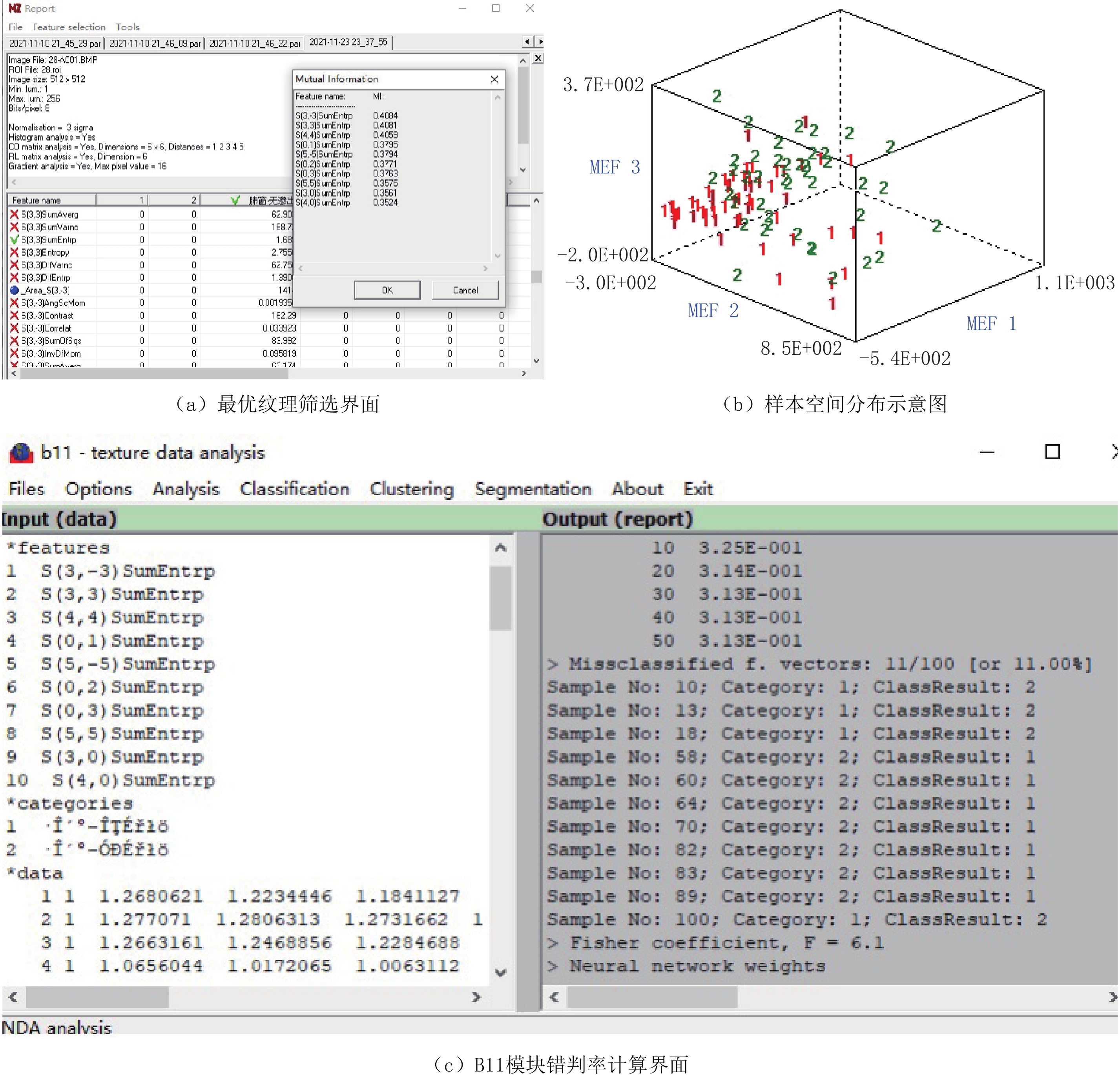
表 1 三种纹理特征提取法分别筛选出的的10个最优特征
Table 1 The 10 optimal features screened out respectively by the three texture feature extraction methods
方法 纹理特征 Fisher S(0,3)SumEntrp、S(4,0)SumEntrp、S(3,3)SumEntrp、S(0,2)SumEntrpS、(3,0)SumEntrp、S(0,4)SumEntrp、S(4,4)SumEntrp、S(5,0)SumEntrp、S(0,5)SumEntrp、S(2,-2)SumEntrp POE+ACC Skewness、Kurtosis、Teta3、S(5,5)SumVarnc、S(0,2)DifVarnc、Vertl_RLNonUni、WavEnLH_s-2、WavEnHH_s-4、
WavEnHL_s-4、WavEnHL_s-3MI S(3,3)SumEntrp、S(4,4)SumEntrp、S(0,2)SumEntrp、S(0,3)SumEntrp、S(3,0)SumEntrp、S(5,-5)SumEntrp、S(5,0)SumEntrp、S(4,0)SumEntrp、S(0,1)SumEntrp、S(5,5)SumEntrp  下载: 导出CSV
表 2 纹理分析预测穿刺并发出血的错判率[%(例)]
Table 2 Misjudgment rate of puncture complicated with bleeding predicted by texture analysis [% (cases)]
纹理特征 鉴别方法 RDA PCA LDA NDA 预穿刺路径
周边的肺野Fisher 27.00
(27/100)33.00
(33/100)24.00
(24/100)12.00
(12/100)POE+AsCC 41.00
(41/100)40.00
(40/100)33.00
(33/100)11.00
(11/100)MI 30.00
(30/100)31.00
(31/100)33.00
(33/100)11.00
(11/100)注:括号内为错判数/所有例数。
下载: 导出CSV
-
[1] POULOU L S, TSAGOULI P, ZIAKAS P D, et al. Computed tomography-guided needle aspiration and biopsy of pulmomary lesions: A single-center experience in 1000 patients[J]. Acta Radiology, 2013, 54: 640−645. doi: 10.1177/0284185113481595
[2] TAVARE A N, PATEL A, SAINI A, et al. Systemic air embolism as a complication of percutaneous lung biopsy[J]. British Journal of Hospital Medicine, 2018, 79(2): 106−107. doi: 10.12968/hmed.2018.79.2.106
[3] IALONGO P, CIARPAGLINI L, TINTI M D, et al. Systemic air embolism as a complication of percutaneous computed tomography guided transthoracic lung biopsy[J]. Annals of the Royal College of Surgeons of England, 2017, 99(6): 174−176. doi: 10.1308/rcsann.2017.0091
[4] SMIT D R, KLEIJN S A, DEVOOGT W G. Coronary and cerebral air embolism: A rare complication of computed tomography-guided transthoracic lung biopsy[J]. Netherlands Heart Journal, 2013, 21(10): 464−466. doi: 10.1007/s12471-013-0411-1
[5] 董鑫哲, 邢立刚, 于金明. 肿瘤异质性的医学影像学分析及临床应用[J]. 中华肿瘤杂志, 2013,(2): 81−84. DOI: 10.3760/cma.j.issn.0253-3766.2013.02.001. DONG X Z, XING L G, YU J M. Medical imaging analysis and clinical application of tumor heterogeneity[J]. Chinese Journal of Oncology, 2013, (2): 81−84. DOI: 10.3760/cma.j.issn.0253-3766.2013.02.001. (in Chinese).
[6] GILLIES R J, KINAHAN P E, HRICAK H. Radiomics: Images are more than pictures, they are data[J]. Radiology, 2016, 278(2): 563−577. doi: 10.1148/radiol.2015151169
[7] 中国抗癌协会肿瘤介入学专业委员会, 中国抗癌协会肿瘤介入学专业委员会青委会. 胸部肿瘤经皮穿剌活检中国专家共识[J]. 中华介入放射学电子杂志, 2018,6(3): 188−194. DOI: 10.3877/cma.j.issn.2095-5782.2018.03.002. The Professional Committee of Tumor Intervention of China Anti Cancer Association, the Youth Committee of the Professional Committee of Tumor Intervention of China Anti Cancer Association. Consensus of Chinese experts on percutaneous biopsy of thoracic tumors[J]. Chinese Journal of Interventional Radiology (Electronic Edition), 2018, 6(3): 188−194. DOI: 10.3877/cma.j.issn.2095-5782.2018.03.002. (in Chinese).
[8] TAI R, DUNNE R M, TROTMAN-DICKENSON B, et al. Frequency and severity of pulmonary hemorrhage in patients undergoing percutaneous CT guided transthoracic lung biopsy: Singleinstitution experience of 1175 cases[J]. Radiology, 2016, 279(1): 287−296. doi: 10.1148/radiol.2015150381
[9] 刘丹, 耿左军, 朱青峰, 等. CT引导下经皮肺穿刺活检的临床应用[J]. 介入放射学杂志, 2018,27(6): 539−543. DOI: 10.3969/j.issn.1008-794X.2018.06.010. LIU D, GENG Z J, ZHU Q F, et al. Clinical application of CT-guided percutaneous needle biopsy of lung[J]. Journal of Interventional Radiology, 2018, 27(6): 539−543. DOI: 10.3969/j.issn.1008-794X.2018.06.010. (in Chinese).
[10] WANG Y, JIANG F M, TAN X B, et al. CT-guided percutaneous transthoracic needle biopsy for paramediastinal and nonparamediastinal lung lesions: Diagnostic yield and complications in 1484 patients[J]. Medicine, 2016, 95(31): e4460. doi: 10.1097/MD.0000000000004460
[11] 张皓, 李琳, 吕发金. 基于Fisher判别构建CT引导下肺穿刺活检并发症的预测模型[J]. 介入放射学杂志, 2020,29(1): 45−50. DOI: 10.3969/j.issn.1008-794X.2020.01.009. ZHANG H, LI L, LV F J. The establishment of a model for predicting complications of CT-guided percutaneous transthoracic needle biopsy based on Fisher discriminant[J]. Journal of Interventional Radiology, 2020, 29(1): 45−50. DOI: 10.3969/j.issn.1008-794X.2020.01.009. (in Chinese).
[12] WIENER R S, WIENER D C, GOULD M K. Risks of transthoracic needle biopsy: How high?[J]. Clinical Pulmonary Medicine, 2013, 20: 29−35. doi: 10.1097/CPM.0b013e31827a30c1
[13] 杨肖华, 黄新宇, 汪国祥. CT引导下经皮肺穿刺活检术并发症的影响因素分析[J]. 介入放射学杂志, 2013,22(8): 658−662. DOI: 10.3969/j.issn.1008-794X.2013.08.011. YANG X H, HUANG X Y, WANG G X. Complications of CT-guided percutaneous lung puncture biopsy: an analysis of influencing factors[J]. Journal of Interventional Radiology, 2013, 22(8): 658−662. DOI: 10.3969/j.issn.1008-794X.2013.08.011. (in Chinese).
[14] ZHU H, ZHANG L, WANG Y, et al. Improved image quality and diagnostic potential using ultra-high-resolution computed tomography of the lung with small scan FOV: A prospective study[J]. PLoS One, 2017, 12(2): e0172688. doi: 10.1371/journal.pone.0172688
[15] KIM G R, HUR J, LEE S M, et al. CT fluoroscopy-guided lung biopsy versus conventional CT-guided lung biopsy: A prospective controlled study to assess radiation doses and diagnostic performance[J]. European Radiology, 2011, 21(2): 232−239. doi: 10.1007/s00330-010-1936-y
[16] SHESHADRI A, RODRIGUEZ A, CHEN R, et al. Effect of reducing field of view on multi-detector quantitative computed tomography parameters of airway wall thickness in asthma[J]. Journal of Computer Assisted Tomography, 2015, 39(4): 584−590. doi: 10.1097/RCT.0000000000000238
[17] 刘玉良, 陈麦林, 朱海滨, 等. 小视野扫描方法在肺小结节CT引导下经皮穿刺活检中的应用[J]. 中国介入影像与治疗学, 2019,16(9): 517−521. DOI: 10.13929/j.1672-8475.201905002. LIU Y L, CHEN M L, ZHU H B, et al. Application of small field of view scan in CT-guided transthoracic needle lung biopsy for pulmonary small nodule[J]. Chinese Journal of Interventional Imaging and Therapy, 2019, 16(9): 517−521. DOI: 10.13929/j.1672-8475.201905002. (in Chinese).
[18] DAVNALL F, YIP C S, LJUNGQVIST G, et al. Assessment of tumour heterogeneity: An emerging imaging tool for clinical practice[J]. Insights Imaging, 2012, 3(6): 573−589.
[19] 周林丽, 冯峰. CT纹理分析对吉非替尼治疗肺腺癌疗效评估的应用[J]. CT理论与应用研究, 2020,29(4): 473−480. DOI: 10.15953/j.1004-4140.2020.29.04.10. ZHOU L L, FENG F. CT texture analysis on the response evaluation of lung adenocarcinoma treated by gifitinib[J]. CT Theory and Applications, 2020, 29(4): 473−480. DOI: 10.15953/j.1004-4140.2020.29.04.10. (in Chinese).
[20] 凡健, 高斌, 夏春华. CT平扫与动脉期图像纹理分析在鉴别膀胱乳头状瘤和膀胱癌中的应用价值[J]. CT理论与应用研究, 2020,29(6): 742−750. DOI: 10.15953/j.1004-4140.2020.29.06.13. FAN J, GAO B, XIA C H. The value of CT non-enhanced and enhanced image texture analysis in differentiating bladder papilloma from bladder cancer[J]. CT Theory and Applications, 2020, 29(6): 742−750. DOI: 10.15953/j.1004-4140.2020.29.06.13. (in Chinese).
-
期刊类型引用(2)
1. 孟凡军. 基于CT平扫纹理特征诊断良恶性肺结节的应用价值. 影像研究与医学应用. 2024(11): 92-94 .  百度学术
百度学术
2. 涂永辉,王巍颋,李璠婷,熊景良. CT多平面重组技术在肺结节穿刺路径选择中的应用. 实用中西医结合临床. 2022(13): 6-10 . 百度学术
其他类型引用(1)
计量
- 文章访问数: 447
- HTML全文浏览量: 129
- PDF下载量: 31
- 被引次数: 3


