
Correlation between Arterial Enhancement Fraction on CT and Efficacy of Transcatheter Arterial Chemoembolization for Hepatocellular Carcinoma
-
摘要: 目的:探讨CT动脉增强分数(AEF)与肝癌经导管动脉化疗栓塞(TACE)治疗效果的相关性。方法:对我院2019年1月至2020年1月43例原发性肝细胞癌(HCC)患者临床资料进行回顾性分析,所有患者均接受TACE治疗,术前及术后1个月均行CT动态增强扫描检查,根据治疗效果分为治疗有效组及无效组,采用电化学发光法检测各组甲胎蛋白(AFP)水平,比较两组治疗前后AEF、AFP、肿瘤大小变化;采用Pearson系数探究AEF、AFP与肿瘤大小变化的相关性,采用受试者工作特征曲线(ROC)及曲线下面积(AUC)分析肝脏动态增强扫描联合AEF彩图对TACE疗效的评估价值。结果:有效组治疗后AEF、AFP、肿瘤大小均小于治疗前、无效组;Pearson相关性分析结果显示,TACE治疗后AEF、AFP均与肿瘤大小呈正相关;ROC曲线分析结果显示,肝脏动态增强扫描联合AEF评估TACE疗效的AUC为0.902,明显高于肝脏动态增强扫描及AEF单独评估的0.793和0.771。结论:AEF对TACE治疗效果具有良好的评估价值,联合肝脏动态增强扫描能进一步提高准确度,可作为TACE治疗HCC疗效评估的辅助手段,值得临床推广。Abstract: Objective: To explore the correlation between arterial enhancement fraction (AEF) on Computed Tomography (CT) and the curative effect of Transcatheter arterial chemoembolization (TACE) for hepatocellular carcinoma (HCC). Methods: We enrolled 43 patients who presented with HCC to our hospital between January 2019 and January 2020 and extracted their clinical data. Three-phase contrast-enhanced CT was performed 1 month before and after TACE; based on TACE efficacy, the enrolled patients were divided into the “effective” and “ineffective” groups. The alpha-fetoprotein (AFP) level was determined using electrochemical luminescence. Changes in the AEF, AFP level, and tumor size before and after treatment were compared between the two groups. The correlation among AEF, AFP level, and tumor-size changes was explored using Pearson’s correlation coefficient. Receiver operating characteristic (ROC) curves and the area under these curves (AUCs) were used to determine the evaluation value of dynamic enhanced scanning combined with AEF for TACE efficacy. Results: After TACE, the AEF, AFP level, and tumor size decreased in the effective group and were lower than the corresponding values in the ineffective group. Pearson’s correlation analysis revealed that AEF and the AFP level were positively correlated with the tumor size after TACE. ROC analysis revealed that the AUC for dynamic enhanced scanning combined with AEF for TACE efficacy evaluation was 0.902; this was significantly greater than the AUC of dynamic enhanced scanning (0.793) and AEF (0.771) alone. Conclusion: AEF is a reliable parameter for evaluating the therapeutic effect of TACE. The evaluation of TACE efficacy can be further improved by combining AEF with dynamic enhanced scanning; this approach should be used as an auxiliary method for evaluating TACE efficacy for HCC.
-
肝细胞癌(hepatocellular carcinoma,HCC)是具有较高发病率及死亡率的肝脏恶性肿瘤,具有起病隐匿、病情进展迅速及生存率低等特点,其致死率较高,居所有恶性肿瘤的第3位[1-2]。现阶段经导管动脉化疗栓塞(transcatheter arterial chemoembolization,TACE)是临床治疗HCC的重要手段,可以阻断HCC的供血动脉,引起癌组织缩小、坏死,最终发挥消除肿瘤的作用[3-4]。
有研究报道,部分患者在TACE治疗后病灶未完全坏死,加之肿瘤血管再形成,存在较高的肿瘤复发风险[5]。因此,采取安全、快速的方法准确评估HCC患者TACE的治疗效果对指导临床后续治疗具有重要的价值。血清肿瘤标志物监测、计算机断层扫描(computed tomography,CT)及磁共振是临床评估TACE治疗效果的常用方法,但均存在一定不足[6]。肝脏CT动脉增强分数(arterial enhancement fraction,AEF)彩图是近年来逐渐被了解的一种新型方法,有研究发现,AEF在评估肝脏疾病患者治疗效果等方面具有较大优势[7]。基于此,本研究探讨AEF与肝癌TACE治疗效果的相关性,为临床诊疗提供参考。
1. 资料与方法
1.1 一般资料
对我院2019年1月至2020年1月43例HCC患者临床资料进行回顾性分析。纳入标准:①满足《原发性肝癌诊疗规范》[8]中诊断标准,并经病理学检查确诊;②首次确诊,并接受TACE治疗;③术前及术后 1个月均行CT扫描检查;④肿瘤为巨块型或者结节型;⑤肝功能 Child-Pugh分级为A-B级;⑥无肝外转移病灶;⑦患者签署知情同意书。
排除标准:①肝癌复发者;②近期接受放疗或肝脏手术切除者;③存在其他恶性肿瘤者;④合并凝血功能障碍、感染性疾病及免疫功能异常者;⑤心、肺、肾等脏器严重损伤者;⑥临床资料不全者。
43例HCC患者中男性31例,女性12例;年龄36~76岁,平均(53.98±7.69)岁;肿瘤大小3~12 cm,平均(6.16±1.46)cm;肝功能Child-Pugh分级:A级19例,B级24例。
1.2 方法
1.2.1 TACE治疗
患者取平卧位,常规消毒双侧腹股沟区皮肤后铺巾,采用5 mL 2% 利多卡因进行局部麻醉,随后作一5 mm切口,对右侧股动脉进行穿刺,置入动脉鞘,并将导管置于肝总动脉、腹腔干、肝左/右动脉,行DSA,显示肿瘤部位及大小,按方案化疗栓塞,结束后拨出动脉鞘及导管,加压包扎。药物:超液化碘油、羟基喜数碱、表阿霉素。
1.2.2 CT扫描
仪器选择西门子128层螺旋CT扫描仪(西门子双源Definition Flash CT)。检查前,指导患者进行呼吸训练,嘱患者扫描时按口令,在吸气末屏住呼吸。CT平扫及增强扫描动脉期、门脉期呼吸幅度保持一致,在吸气末屏住气扫描。动脉期、门脉期数据采集时间分别为注药后30 s、注药后60 s。入选患者扫描:仰卧位,双手抱头,头先进。在吸气末从膈顶至肝底行全肝扫描(一定把肝组织包全)。
扫描参数:螺旋采集模式,2×128×0.6 mm。吸气相:管电压100 kV,管电流50 mA;呼气相:管电压100 kV,管电流50 mA。机架旋转时间0.33 s/周,矩阵512×512,重建层厚1 mm,重建间隔0.6 mm。
1.2.3 CT数据处理
分别选取TACE治疗前、治疗后1月CT平扫、动脉期和门静脉期图像导入西门子Syngo.VIA后处理工作站,选取MM肿瘤学卡片,生成AEF彩图。
由两名具有10年以上腹部影像诊断经验的放射科医师取得一致意见后选取多个感兴趣区(region of interest,ROI),选取肝肿瘤实质部分,尽量避开囊变、坏死及周围血管区,取病变区域所在相同层面作为ROI进而计算ROI内AEF分布,尽量保证TACE治疗前后病灶的ROI保持一致。
1.2.4 甲胎蛋白水平检测
在治疗前及治疗后1个月所有患者于清晨空腹采集肘静脉血3 mL置于干燥试管中,室温下静置1 h后离心处理,离心时间为5 min,离心速率为3000 r/min。取上清液保存在 -20℃冰备用,采用电化学发光法检测甲胎蛋白(alpha-fetoprotein,AFP)水平。
1.3 评价标准
根据术后CT影像结果并结合临床指标按照mRECIST标准[9]进行疗效评价:①完全缓解(complete remission,CR):所有目标病灶动脉期增强显影均消失;②部分缓解(partial remission,PR):目标病灶(动脉期增强显影)的直径总和缩小≥30%;③病情进展(progressive Disease,PD):目标病灶(动脉期增强显影)的直径总和增加≥20% 或出现新病灶;④病情稳定(stable disease,SD):缩小未达PR或增加未达PD。本研究根据疗效标准,将CR及PR视为有效组(n=28),将PD及SD视为无效组(n=15)。
1.4 观察指标
①记录并比较两组 AEF、AFP、肿瘤大小;②探究 AEF、AFP与肿瘤大小变化的相关性;③分析肝脏动态增强扫描联合 AEF彩图对TACE疗效的评估价值。
1.5 统计学方法
采用统计学软件SPSS 21.0处理分析数据。计数资料以例(%)表示,行χ2检验;计量资料以均数±标准差
$ (\bar x \pm s) $ 表示,两组间比较采用t检验;采用Pearson相关性系数探究AEF、AFP与肿瘤大小变化的相关性;采用受试者工作特征曲线(receiver operating characteristic curve,ROC)及曲线下面积(area under curve,AUC)分析肝脏动态增强扫描联合AEF彩图对TACE疗效的评估价值。以P<0.05为差异具有统计学意义。2. 结果
2.1 两组治疗AEF、AFP、肿瘤大小比较
治疗前,有效组与无效组相比,AEF、AFP、肿瘤大小三者差异均无统计学意义。治疗后,有效组AEF小于无效组,有效组AFP小于无效组、有效组肿瘤大小小于无效组(表1)。
表 1 两组治疗AEF、AFP、肿瘤大小比较($ \bar x \pm s $ )Table 1. Comparison of the AEF, AFP level and tumor size between the two groups ($ \bar x \pm s $ )组别 AEF AFP/(ng/L) 肿瘤大小/cm 治疗前 治疗后 治疗前 治疗后 治疗前 治疗后 有效组 0.51±0.13 0.34±0.05* 385.12±10.29 84.89±8.39* 6.28±1.05 3.01±0.41* 无效组 0.52±0.09 0.48±0.07 382.87±21.54 373.25±17.82 6.05±1.13 5.96±1.05 t 0.265 7.594 0.420 72.436 0.667 13.209 P 0.792 0.000 0.676 0.000 0.509 0.000 注:与本组治疗前,*-P<0.05。 2.2 AEF、AFP与肿瘤大小的相关性
Pearson相关性分析结果显示(表2),经TACE治疗后AEF与肿瘤大小呈正相关(r=0.537),AFP与肿瘤大小呈正相关(r=0.649)。
表 2 AEF、AFP与肿瘤大小的相关性Table 2. Correlation between the AEF, AFP level and tumor size指标 肿瘤大小 P AEF 0.537 0.002 AFP 0.649 0.000 2.3 肝脏动态增强扫描联合AEF对TACE疗效的评估价值
ROC曲线分析结果显示,肝脏动态增强扫描联合AEF评估TACE疗效的AUC为0.902,明显高于肝脏动态增强扫描及AEF单独评估的0.793和0.771(图1和表3)。
![]() 图 1 肝脏动态增强扫描联合AEF评估TACE疗效的ROC曲线Figure 1. ROC curve of dynamic enhanced liver scanning combined with AEF for evaluating TACE efficacy表 3 肝脏动态增强扫描联合AEF对TACE疗效的评估价值Table 3. Evaluation value of dynamic enhanced liver scanning combined with AEF for TACE efficacy
图 1 肝脏动态增强扫描联合AEF评估TACE疗效的ROC曲线Figure 1. ROC curve of dynamic enhanced liver scanning combined with AEF for evaluating TACE efficacy表 3 肝脏动态增强扫描联合AEF对TACE疗效的评估价值Table 3. Evaluation value of dynamic enhanced liver scanning combined with AEF for TACE efficacy项目 AUC P 95% CI 肝脏动态增强扫描 0.793 <0.05 0.642~0.901 AEF 0.771 <0.05 0.618~0.885 肝脏动态增强扫描联合AEF 0.902 <0.05 0.773~0.972 2.4 典型病例
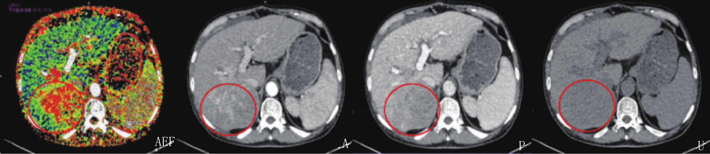
男性,58岁,肝硬化30余年,发现肝占位3天,AFP 487.52 ng/mL。肝右叶巨块型肝癌,A为动脉期图像,呈明显高强化,P为门静脉期图像,原动脉期高强化区域强化退出,与肝实质相比呈低密度影,U为CT平扫图像,与肝实质相比为稍低密度区,境界模糊,AEF彩图肿块大部分为红色区域,能够直观显示肿块、并提示肿块为丰富肝动脉供血(图2)。
![]() 图 2 HCC患者AEF伪彩图,CT增强扫描动脉期、门脉期及CT平扫图像Figure 2. CT image with an AEF, CT image taken during the arterial phase, CT image taken during the portal venous phase, and plain CT of a patient with HCC
图 2 HCC患者AEF伪彩图,CT增强扫描动脉期、门脉期及CT平扫图像Figure 2. CT image with an AEF, CT image taken during the arterial phase, CT image taken during the portal venous phase, and plain CT of a patient with HCC3. 讨论
HCC是原发性肝癌最为常见的类型,病死率居于我国恶性肿瘤的第3位[1]。TACE是一种有效的非外科手术姑息性治疗方法,将不同药物颗粒经动脉注入肿瘤血管,使肿瘤缺氧缺血而坏死[3]。但TACE治疗后若病灶并未完全坏死,会导致恶性肿瘤再次复发[10-11]。现阶段临床主要通过CT、磁共振等影像学检查评估TACE治疗后肝脏肿瘤的存活情况,但磁共振具有检查用时较长、费用较高等缺点,且对患者呼吸配合要求较高。因此,临床仍以CT为主要手段评估TACE治疗效果。
临床研究发现,HCC患者接受TACE治疗后血供明显减少,可使AEF明显降低[12]。本研究结果显示,TACE治疗后有效组AEF低于TACE治疗前及无效组,与既往研究结果一致,提示AEF可作为临床评估TACE治疗效果的参考指标。肝脏是门静脉及肝动脉的双重供血脏器,在肝脏功能正常情况下,主要以门静脉供血为主,故正常肝实质在门静脉期密度达最高。但HCC患者肿瘤组织主要以肝动脉供血为主,因此在HCC患者肝实质在门静脉期呈低密度。
AEF=(动脉期CT值-平扫期CT值)/(门静脉期CT值 - 平扫期CT值) (1)
AEF是感兴趣区域在肝动脉期内绝对强化增量与门静脉期内绝对强化增量的比值。
HCC是富血供病变,且由肝动脉供血,在动态增强CT扫描中动脉期迅速强化呈稍高密度,门静脉期和平衡期快速退出呈稍低密度,可见,TACE治疗前HCC患者的AEF相对较高。HCC患者在TACE治疗后肿瘤内部和周围动脉发生狭窄,甚至闭塞,使得肝脏固有动脉流速明显降低,对TACE治疗效果良好的肿瘤而言,肿瘤内部血流减少与肝脏固有动脉流有关,从而导致AEF较TACE治疗前明显减少。
蒋长斌等[13]研究发现,AFP在多数HCC患者血清中呈高水平状态,是诊断HCC的常用肿瘤标志物。本研究中有效组治疗后血清AFP水平明显低于治疗前、无效组,与其研究结果一致,表明AFP可作为临床评估HCC患者TACE治疗效果的参考指标。本研究采用Pearson系数分析AEF、AFP与肿瘤大小的相关性发现,HCC患者TACE治疗后AEF、AFP均与肿瘤大小呈正相关,进一步证实了AEF、AFP可用于评估HCC患者TACE的治疗效果。
王曦等[14]认为,相比于常规CT扫描,肝脏动态增强扫描的速度较快、处理技术强大,可以清楚反映肿瘤部位、大小及内部碘油沉积情况,有利于临床判断TACE治疗后肿瘤残留情况。有报道显示,肝脏动态增强扫描评估TACE治疗后肿瘤残留情况的准确度为85.30%[15]。本研究结果显示,肝脏动态增强扫评估TACE疗效的准确度仅为79.30%,低于上述研究结果,这可能是碘油影响对肿瘤血管的遮挡所致。
本研究中AEF评估TACE治疗效果的AUC为0.771,分析原因为TACE治疗后部分患者病灶大小不超过1 cm,此时AEF彩图可能出现失真,易与小血管混淆,造成漏诊;此外,扫描时患者的呼吸、心脏运动也会产生运动伪影,即使经软件矫正后仍会出现剪影,引发误诊[16],故扫描开始前需指导患者进行呼吸训练。本研究结果显示,肝脏动态增强扫描联合AEF评估TACE疗效的AUC为0.902,明显高于肝脏动态增强扫描及AEF单独评估的0.793和0.771,提示肝脏动态增强扫描联合AEF能提高TACE疗效评估的准确度,可用于指导临床疗效评估。
在临床工作中发现,患者在吸气末屏住气扫描与呼气末屏住气扫描相比能够较好的保持呼吸幅度一致,从而在后处理中不同扫描时期的图像能都较好的进行匹配,得到良好的后处理图像,目前尚无统计学支持。
综上所述,AEF对TACE治疗效果具有良好的评估价值,联合肝脏动态增强扫描能进一步提高准确度,可作为评估TACE疗效的辅助手段。本研究尚有不足之处,研究样本较少且为回顾性分析,后期需开展多中心、大样本量、前瞻性研究,以及较长时间的随访,以期得到更为准确的AEF与HCC患者预后的关系,进而指导临床诊疗。
-
![]()
图 1 肝脏动态增强扫描联合AEF评估TACE疗效的ROC曲线
Figure 1. ROC curve of dynamic enhanced liver scanning combined with AEF for evaluating TACE efficacy
![]()
图 2 HCC患者AEF伪彩图,CT增强扫描动脉期、门脉期及CT平扫图像
Figure 2. CT image with an AEF, CT image taken during the arterial phase, CT image taken during the portal venous phase, and plain CT of a patient with HCC
表 1 两组治疗AEF、AFP、肿瘤大小比较(
$ \bar x \pm s $ )Table 1 Comparison of the AEF, AFP level and tumor size between the two groups (
$ \bar x \pm s $ )组别 AEF AFP/(ng/L) 肿瘤大小/cm 治疗前 治疗后 治疗前 治疗后 治疗前 治疗后 有效组 0.51±0.13 0.34±0.05* 385.12±10.29 84.89±8.39* 6.28±1.05 3.01±0.41* 无效组 0.52±0.09 0.48±0.07 382.87±21.54 373.25±17.82 6.05±1.13 5.96±1.05 t 0.265 7.594 0.420 72.436 0.667 13.209 P 0.792 0.000 0.676 0.000 0.509 0.000 注:与本组治疗前,*-P<0.05。  下载: 导出CSV
表 2 AEF、AFP与肿瘤大小的相关性
Table 2 Correlation between the AEF, AFP level and tumor size
指标 肿瘤大小 P AEF 0.537 0.002 AFP 0.649 0.000
下载: 导出CSV
表 3 肝脏动态增强扫描联合AEF对TACE疗效的评估价值
Table 3 Evaluation value of dynamic enhanced liver scanning combined with AEF for TACE efficacy
项目 AUC P 95% CI 肝脏动态增强扫描 0.793 <0.05 0.642~0.901 AEF 0.771 <0.05 0.618~0.885 肝脏动态增强扫描联合AEF 0.902 <0.05 0.773~0.972
下载: 导出CSV
-
[1] BOSETTI C, TURATI F, LA VECCHIA C. Hepatocellular carcinoma epidemiology[J]. Best Practice & Research. Clinical gastroenterology Gastroenterol, 2014, 28(5): 753−770.
[2] 张文伟, 翁乐逸, 王建华. MRI对原发性肝细胞癌TACE术疗效评价及其复发的预测价值[J]. 中国CT和MRI杂志, 2020,18(3): 7−10. doi: 10.3969/j.issn.1672-5131.2020.03.003 ZHANG W W, WENG L Y, WANG J H. Curative effect evaluation on tace for primary hepatocellular carcinoma and its predictive value of recurrence by MRI[J]. Chinese Journal of CT and MRI, 2020, 18(3): 7−10. (in Chinese). doi: 10.3969/j.issn.1672-5131.2020.03.003
[3] 黄勇慧. 进化中的肿瘤经导管动脉化疗栓塞术[J]. 中山大学学报 (医学科学版), 2020,41(6): 825−833. HUANG Y H. Evolution of transcatheter arterial chemoembolization[J]. Journal of Sun Yat-sen University (Medical Sciences), 2020, 41(6): 825−833. (in Chinese).
[4] 张少平. MRI对原发性肝癌介入术的疗效评估[J]. 中国医学影像学杂志, 2019,27(5): 397−400. doi: 10.3969/j.issn.1005-5185.2019.05.019 [5] 周彬彬, 孙姚晨, 黄海帆, 等. TACE术后肝细胞癌患者功能磁共振成像参数变化研究[J]. 实用肝脏病杂志, 2020,23(5): 723−726. doi: 10.3969/j.issn.1672-5069.2020.05.030 ZHOU B B, SUN Y C, HUANG H F, et al. Evaluation of functional magnetic resonance imaging in patients with hepatocellular carcinoma after TACE[J]. Journal of Practical Hepatology, 2020, 23(5): 723−726. (in Chinese). doi: 10.3969/j.issn.1672-5069.2020.05.030
[6] 张智坚, 吴孟超, 刘崎, 等. 不同影像方法对射频消融治疗肝癌疗效的评价[J]. 中华肿瘤杂志, 2005,27(10): 616−619. doi: 10.3760/j.issn:0253-3766.2005.10.011 ZHANG Z J, WU M C, LIU Q, et al. Imaging evaluation of efficacy of radiofrequency ablation treatment for hepatic cancer[J]. Chinese Jouenal of Oncology, 2005, 27(10): 616−619. (in Chinese). doi: 10.3760/j.issn:0253-3766.2005.10.011
[7] 崔毛毛, 翟亚楠, 高玉岭, 等. AEF值评估肝硬化门静脉高压TIPS术疗效的应用价值[J]. 临床放射学杂志, 2020,39(1): 77−80. CUI M M, ZHAI Y N, GAO Y L, et al. AEF value in evaluating the efficacy of TIPS for portal hypertension in cirrhosis[J]. Journal of Clinical Radiology, 2020, 39(1): 77−80. (in Chinese).
[8] 中华人民共和国卫生和计划生育委员会医政医管局. 原发性肝癌诊疗规范(2017年版)[J]. 中华消化外科杂志, 2017,16(7): 635−647. doi: 10.3760/cma.j.issn.1673-9752.2017.07.001 Bureau of Medical Administration, National Health. Standardization of diagnosis and treatment for hepatocellular carcinoma (2017 edition)[J]. Chinese Journal of Digestive Surgery, 2017, 16(7): 635−647. (in Chinese). doi: 10.3760/cma.j.issn.1673-9752.2017.07.001
[9] 翁炜, 吕秀玲, 张倩倩, 等. 基于磁共振影像组学技术对肝癌经肝动脉化疗栓塞术后短期疗效的预后价值分析[J]. 中华医学杂志, 2020,100(11): 828−832. doi: 10.3760/cma.j.cn112137-20190705-01502 WENG W, LV X L, ZHANG Q Q, et al. Prediction of short-term prognosis of hepatocellular carcinoma after TACE surgery based on MRI texture analysis technology[J]. National Medical Journal of China, 2020, 100(11): 828−832. (in Chinese). doi: 10.3760/cma.j.cn112137-20190705-01502
[10] JEONG S O, KIM E B, JEONG S W, et al. Predictive factors for complete response and recurrence after transarterial chemoembolization in hepatocellular carcinoma[J]. Gut and liver, 2017, 11(3): 409−416. doi: 10.5009/gnl16001
[11] 张燕军, 蒋强, 张倩, 等. 增强CT与MRI在原发性肝癌介入治疗后疗效评估中的价值对比分析[J]. 实用癌症杂志, 2020,35(9): 1520−1523. doi: 10.3969/j.issn.1001-5930.2020.09.034 ZHANG Y J, JIANG Q, ZHANG Q, et al. CT combined with MRI in the diagnosis of primary liver cancer and evaluation of tace effect[J]. Journal of Practical Cancer, 2020, 35(9): 1520−1523. (in Chinese). doi: 10.3969/j.issn.1001-5930.2020.09.034
[12] 刘璐璐, 章浙伟, 杨永波, 等. CT灌注参数动脉增强分数值在评估肝癌TACE术后疗效中的初步研究[J]. 介入放射学杂志, 2017,26(11): 988−992. doi: 10.3969/j.issn.1008-794X.2017.11.006 LIU L L, ZHANG Z W, YANG Y B, et al. Application of quantitative arterial enhancement fraction of multiphase perfusion CT imaging in evaluating the curative effect of transcatheter arterial chemoembolization for hepatocellular carcinoma[J]. Journal of Interventional Radiology, 2017, 26(11): 988−992. (in Chinese). doi: 10.3969/j.issn.1008-794X.2017.11.006
[13] 蒋长斌, 崔邦平, 代文莉, 等. 超检测限AFP、CA19-9稀释后的检测结果对监测原发性肝癌栓塞治疗疗效的临床意义[J]. 标记免疫分析与临床, 2019,26(6): 1020−1022. JIANG C B, CUI B P, DAI W L, et al. The clinical value of the dilution values of AFP and CA19-9 exceeding the upper detection limit in the monitoring of the hepatocellular carcinoma embolization treatment effect[J]. Labeled Immunoassays and Clinical Medicine, 2019, 26(6): 1020−1022. (in Chinese).
[14] 王曦, 李东, 何芬, 等. 超声造影与增强CT评估TACE治疗原发性肝癌疗效的价值对比[J]. 河北医学, 2018,24(7): 1113−1116. doi: 10.3969/j.issn.1006-6233.2018.07.014 WANG X, LI D, HE F, et al. Contrastive analysis of contrast-enhanced ultrasound and enhanced CT in evaluating the efficacy of TACE in the treatment of primary liver cancer[J]. Hebei Medicine, 2018, 24(7): 1113−1116. (in Chinese). doi: 10.3969/j.issn.1006-6233.2018.07.014
[15] 曾春. DWI联合CT增强扫描在评估TACE治疗肝癌患者的临床价值[J]. 中国CT和MRI杂志, 2016,14(4): 80−83. doi: 10.3969/j.issn.1672-5131.2016.04.025 ZENG C. Clinical value of DWI combined with CT enhanced scan in the evaluation of transcatheter arterial chemoembolization in the treatment of patients with liver cancer[J]. Chinese Journal of CT and MRI, 2016, 14(4): 80−83. (in Chinese). doi: 10.3969/j.issn.1672-5131.2016.04.025
[16] 姜梅, 高德宏, 张凯, 等. CT平均动脉增强分数彩图在肝细胞型肝癌中的诊断价值[J]. 实用放射学杂志, 2020,36(4): 583−587. doi: 10.3969/j.issn.1002-1671.2020.04.019 JIANG M, GAO D H, ZHANG K, et al. The value of CT mean arterial enhancement fraction color maps in the diagnosis of hepatocellular carcinoma[J]. Journal of Practical Radiology, 2020, 36(4): 583−587. (in Chinese). doi: 10.3969/j.issn.1002-1671.2020.04.019
-
期刊类型引用(4)
1. 席晓旭,陈志晔. 光谱CT定量增强参数对肾细胞癌与肾血管平滑肌脂肪瘤的诊断价值. 分子影像学杂志. 2025(02): 218-222 .  百度学术
百度学术
2. 刘静垚,樊文萍,刘梦琦,葛文浩,姚慧,刘明波,陈志晔. 基于碘图的光谱CT多模态参数成像在甲状腺乳头状癌中的诊断价值. 分子影像学杂志. 2024(02): 138-142 . 百度学术
3. 王兴龙,冯坤鹏,袁牧,孟令武. CT动脉增强分数、甲胎蛋白异质体3、中性粒细胞与淋巴细胞比值联合检测对肝癌患者介入化疗疗效的评估价值. 癌症进展. 2024(10): 1146-1150 . 百度学术
4. 徐石. MSCT增强扫描对乙型肝炎肝硬化背景下原发性肝癌的诊断价值. 影像研究与医学应用. 2023(13): 132-134 . 百度学术
其他类型引用(0)


计量
- 文章访问数: 344
- HTML全文浏览量: 109
- PDF下载量: 39
- 被引次数: 4


