
Study on the Optimal Dosage of Gadolinium Contrast Agent for Lower Extremity Artery CE-MRA Angiography
-
摘要: 目的:探讨三维动态增强磁共振血管成像(3D CE-MRA)在双下肢动脉病变成像中对比剂最佳用量。方法:45例我院进行双下肢动脉3D CE-MRA血管成像的患者,根据就诊顺序随机分为A、B和C三组,三组钆对比剂用量分别为0.1、0.2和0.3 mmol/kg,分别与盐水进行1∶1配比,分别对不同对比剂用量的后处理图像进行主观及客观评分,并对小腿及足部血管进行整体静脉重叠评分,最后采用Wilcoxon检验比较3种扫描方案差异。客观评分是对3组的原始增强图像进行信号强度(SI)、信噪比(SNR)、对比噪声比(CNR)的测量及计算,比较股动脉、腘动脉及小腿动脉水平信号强度差异,进行t检验比较 3种扫描方案差异。结果:对比剂用量为0.1 mmol/kg时股动脉、腘动脉及小腿动脉的图像质量评分分别为(3.35±0.25)、(2.97±0.25)、(2.35±1.15);对比剂用量为0.2 mmol/kg时,上述部位的评分分别为(3.75±0.35)、(3.55±0.32)、(2.97±0.70);对比剂用量为0.3 mmol/kg时,上述部位的评分分别为(3.90±0.41)、(3.83±0.52)、(3.10±0.75)。A组有33.33%图像不满足诊断需要,B组和C组图像全部满足诊断需要,A组与B组差异有统计学意义,B组与C组差异无统计学意义。B和C组的股动脉、腘动脉的SI、SNR、CNR明显高于A组,且B组和C组差异无统计学意义。B组和C组图像优于A组。结论:适当提高对比剂用量有助于提高患者双下肢动脉全程的3D CE-MRA成像质量,0.2 mmol/kg的对比剂用量对血管的评估是可靠准确的,能够为临床外周动脉疾患的治疗方案提供准确可靠的影像依据。Abstract: Objective: To investigate the optimal dosage of contrast agent in three-dimensional dynamic enhanced magnetic resonance angiography (3D CE-MRA) in the imaging of arterial lesions of both lower limbs. Methods: 45 patients who underwent 3D CE-MRA angiography of lower extremity arteries in our hospital were randomly divided into three groups A, B and C. The gadolinium dosages of the three groups were respectively 0.1 mmol/kg, 0.2 mmol/kg and 0.3 mmol/kg. They were injected intravenously with saline at the ratio of 1:1. The images of different dose scanning schemes were scored subjectively and objectively. Subjective scoring: the lower limb arteries were divided into femoral artery, popliteal artery and calf artery (posterior tibial artery, anterior tibial artery and common peroneal artery). The quality of MIP reconstruction images was evaluated, and the overall venous overlap scoring of calf and foot vessels was also carried out. Finally, the differences of the three scanning schemes were compared by Wilcoxon test.The objective scoring is to measure and calculate the signal intensity Si, signal-to-noise ratio SNR and contrast-to-noise ratio of the original enhanced images of the three groups, compare the horizontal signal intensity differences of femoral artery, popliteal artery and calf artery, and compare the differences of the three scanning schemes by t-test. Results: When the contrast medium dosage was 0.1 mmol/kg, the image quality scores of femoral artery, popliteal artery and calf artery were (3.35±0.25), (2.97±0.25), (2.35±1.15) respectively; When the dosage of contrast agent was 0.2 mmol/kg, the scores of the above parts were (3.75±0.35), (3.55±0.32), (2.97±0.70) respectively; When the dosage of contrast agent was 0.3 mmol/kg, the scores of the above parts were (3.90±0.41), (3.83±0.52), (3.10±0.75) respectively. 33.33% of the images in group A did not meet the diagnostic needs, while all the images in group B and C met the diagnostic needs. There was significant difference between group A and group B, and there was no significant difference between group B and group C. B The Si, SNR and CNR of femoral artery and popliteal artery in group C were significantly higher than those in group A, and there was no significant difference between group B and C. The images of group B and C were better than those of group A. Conclusion: Appropriate increase of the contrast medium dosage is helpful to improve the 3D CE-MRA imaging quality of both lower limb arteries. The contrast medium dosage of 0.2 mmol/kg is reliable and accurate for the evaluation of blood vessels, and can provide accurate and reliable imaging basis for the formulation of treatment plan for patients with peripheral artery diseases.
-
Keywords:
- angiography /
- magnetic resonance /
- lower extremity
-
-
![]()
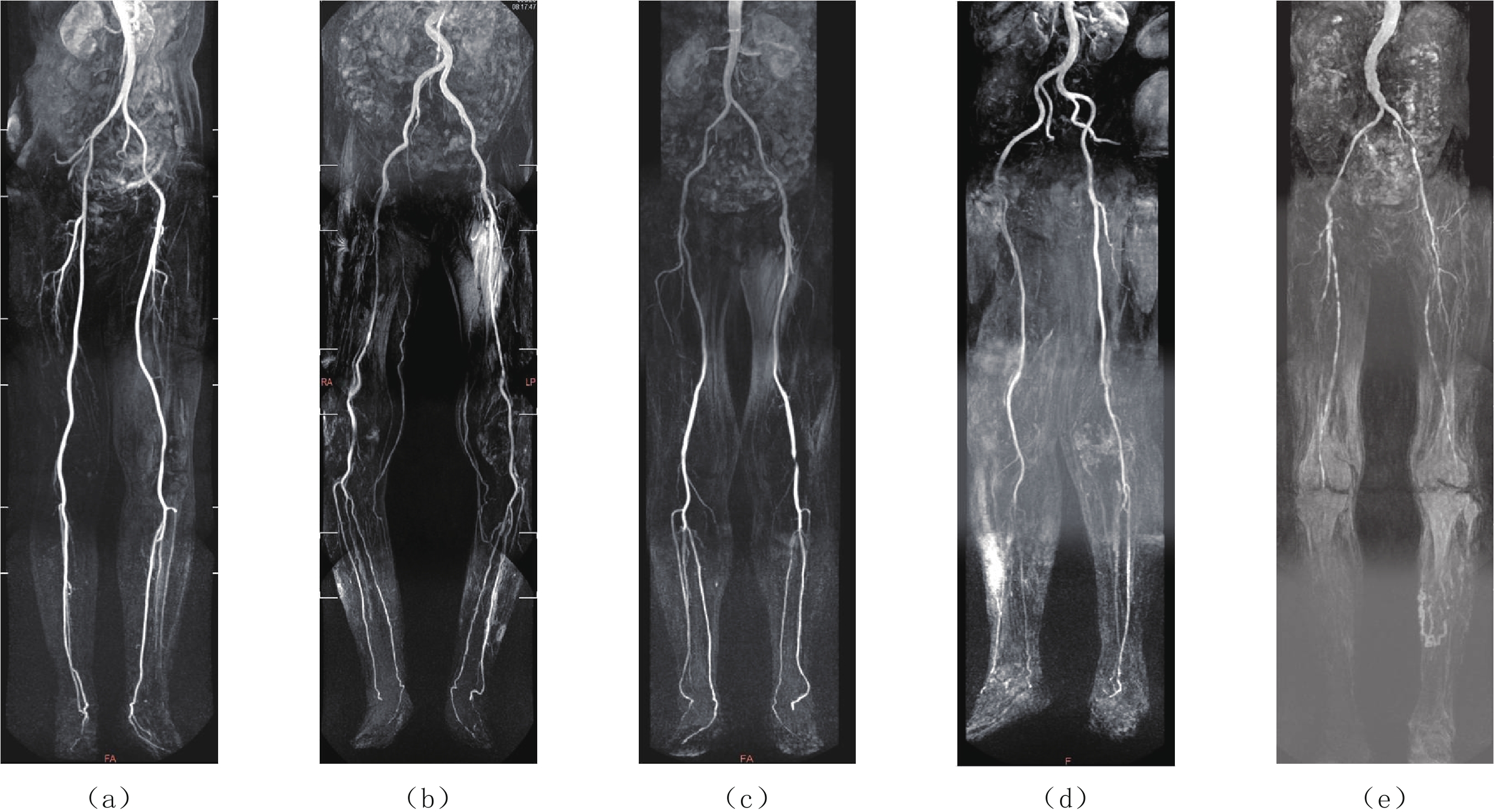
图 1 不同对比剂用量条件下双下肢动脉成像显示
Figure 1. Arterial imaging of both lower limbs under different dosage of contrast medium
![]()
图 2 下肢动脉血管信号强度的测量
Figure 2. Measurement of arterial signal intensity of lower extremity study on the optimal dosage of gadolinium CE-MRA contrast
表 1 45例患者不同对比剂用量条件下MRA图像质量结果比较(分,
$\bar x \pm s $ )Table 1 Comparison of MRA image quality results of 45 patients with different dosage of contrast agents (points,
$\bar x \pm s $ )扫描方案 图像质量评分 股动脉 腘动脉 小腿动脉 静脉污染 A组 3.35±0.25 2.97±0.25 2.35±1.15 0.98±0.75 B组 3.75±0.35 3.55±0.32 2.97±0.70 1.16±0.65 C组 3.90±0.41 3.83±0.52 3.10±0.75 1.42±0.35 A组与B组$z $值 0.63 1.86 3.21 0.32 $P $ >0.05 <0.05 <0.05 <0.05 B组与C组$z $值 1.11 3.06 2.71 0.56 $P $ >0.05 >0.05 >0.05 >0.05  下载: 导出CSV
表 2 45例患者下肢动脉在不同造影剂用量条件下客观评价结果(
$\bar x \pm s $ )Table 2 Objective evaluation results of lower limb arteries of 45 patients under different dosage of contrast media (
$\bar x \pm s $ )扫描方案 A组 B组 C组 A组与
B组t值P值 B组与
C组t值P值 股动脉 SI 364.38±21.43 619.81±36.33 766.72±47.30 3.74 <0.05 2.65 >0.05 SNR 181.09±16.34 303.43±19.45 409.33±23.12 4.89 <0.05 1.07 >0.05 CNR 152.73±11.22 271.70±15.53 340.56±15.98 2.77 <0.05 1.98 >0.05 腘动脉 SI 336.19±30.73 607.43±28.12 760.31±57.92 3.28 <0.05 1.01 >0.05 SNR 179.67±14.59 302.20±17.42 374.53±27.27 5.69 <0.05 2.18 >0.05 CNR 149.29±16.38 272.35±12.76 319.21±24.74 3.96 >0.05 3.04 >0.05 胫前/
后动脉SI 91.67±9.40 214.40±22.17 416.44±38.33 1.78 <0.05 1.05 >0.05 SNR 47.74±4.76 107.73±12.31 223.65±16.74 3.72 <0.05 2.67 <0.05 CNR 35.20±4.79 86.43±6.93 199.46±7.47 2.99 >0.05 2.04 <0.05
下载: 导出CSV
-
[1] 张磊, 畅坚, 吴云, 等. 对比增强MR血管成像诊断糖尿病下肢动脉闭塞症膝以下动脉狭窄的价值[J]. 中华放射学杂志, 2014,(8): 664−669. doi: 10.3760/cma.j.issn.1005-1201.2014.08.011 ZHANG L, CHANG J, WU Y, et al. Value of contrast-enhanced MR angiography in diagnosis of lower extremity arterial stenosis in diabetic patients with lower extremity arterial occlusive disease[J]. Chinese Journal of Radiology, 2014, (8): 664−669. (in Chinese). doi: 10.3760/cma.j.issn.1005-1201.2014.08.011
[2] 吴戈, 张藜莉, 霍健伟, 等. 64排螺旋CT血管造影在下肢动脉病变人工血管旁路移植术后随访的价值[J]. 实用放射学杂志, 2007,(5): 639−642. doi: 10.3969/j.issn.1002-1671.2007.05.018 WU G, ZHANG L L, HUO J W, et al. The application of 64-detector helical CT angiography in the evaluation of artery of lower extremity in follow-up after artificial vascular graft[J]. Journal of Practical Radiology, 2007, (5): 639−642. (in Chinese). doi: 10.3969/j.issn.1002-1671.2007.05.018
[3] 吴戈, 张藜莉, 邓刚, 等. 下肢动脉硬化闭塞症的多层螺旋(HD750) CTA和3.0T磁共振3DCE-MRA的对比研究[J]. 中国CT和MRI杂志, 2016,(5): 112−113. doi: 10.3969/j.issn.1672-5131.2016.05.035 WU G, ZHANG L L, DENG G, et al. Compariative Study of multi-sliceshelical (HD750) CT angiography and 3.0T Three dimensional dynamic contrast-enhanced MRA inLower extremities arteriosclerotic occlusive disease[J]. Chinese Journal of CT and MRI, 2016, (5): 112−113. (in Chinese). doi: 10.3969/j.issn.1672-5131.2016.05.035
[4] 孙凤, 舒政. 非对比增强MR下肢动脉成像技术研究进展[J]. 医学综述, 2018,(17): 3487−3491. doi: 10.3969/j.issn.1006-2084.2018.17.031 SUN F, SHU Z. Technological development of non-contrast enhanced MR angiography in lower extremity[J]. Medical Recapitulate, 2018, (17): 3487−3491. (in Chinese). doi: 10.3969/j.issn.1006-2084.2018.17.031
[5] 张玲, 李大鹏. 低剂量对比剂在下肢动脉疾病3D CE-MRA中的应用[J]. 医疗卫生装备, 2017,(4): 74−77. ZHANG L, LI D P. Application of low-dose contrast agent in 3D dynamic contrast enhanced MR angiography of lower extremity arterial disease[J]. Chinese Medical Equipment Journal, 2017, (4): 74−77. (in Chinese).
[6] HIATT W R. Medical treatment of peripheral arterial disease and claudication[J]. New England Journal of Medicine, 2001, 344(21): 1608−1621. doi: 10.1056/NEJM200105243442108
[7] HABIBI R, KRISHNAM M S, LOHAN D G, et al. High-spatial-resolution lower extremity MR angiography at 3.0T: Contrast agent dose comparison study[J]. Radiology, 2008, 248(2): 680−692. doi: 10.1148/radiol.2482071505
[8] 唐辉. 多模态磁共振成像在下肢缺血性疾病中的应用研究[D]. 上海: 上海交通大学, 2019. TANG H. Multi-modal medical imaging methods in assessment and prediction of lower limb ischemic disease[D]. Shanghai: Shanghai Jiaotong University, 2019. (in Chinese).
[9] 王洁, 李小鹰, 何耀, 等. 北京市万寿路地区老年人群周围动脉硬化闭塞病横断面调查[J]. 中华流行病学杂志, 2004,25(3): 221−224. doi: 10.3760/j.issn:0254-6450.2004.03.010 WANG J, LI X Y, HE Y, et al. A cross-sectional study of peripheral arterial occlusive disease in Wanshoulu area[J]. Chinese Journal of Epidemiology, 2004, 25(3): 221−224. (in Chinese). doi: 10.3760/j.issn:0254-6450.2004.03.010
[10] GUAN H, LI Y J, XU Z R, et al. Prevalence and risk factors of peripheral arterial disease in diabetic patients over 50 years old in China[J]. China Medical Sciences Journal, 2007, 22(2): 83−88.
[11] KOELEMAY M J, LIJMER J G, STOKER J, et al. Magnetic resonance angiography for the evaluation of lower extremity arterial disease: A meta-analysis[J]. Journal of the American Medical Association, 2001, 285(10): 1338−1345. doi: 10.1001/jama.285.10.1338
[12] 杨敏星, 李选, 金斌, 等. 3.0T磁共振下肢动脉3D CE-MRA对比剂用量[J]. 中国介入影像与治疗学, 2010,7(4): 358−362. YANG M X, LI X, JIN B, et al. Dose of contrast agent in 3D CE-MRA of lower extremity arteries at 3.0T MR system[J]. Chinese Journal of Interventional Imaging and Therapy, 2010, 7(4): 358−362. (in Chinese).
[13] GROBNER T. Gadolinium A—Specific trigger for the development of nrphrogenic fibrosing dermopathy and nephrogenic systemic fibrosis?[J]. Nephrology Dialysis Transplantion, 2006, 21(6): 1104−1108. DOI: 10.1093/ndt/gfk062.
[14] BROOME D R, GIRGUIS M S, BARON P W, et al. Gadodiamide associated nephrogenic systemivfibrosis: Why radiologists should be concerned?[J]. American Journal of Roentgenology, 2007, 188(2): 586−592. doi: 10.2214/AJR.06.1094
[15] PRINCE M R, ZHANG H, MORRIS M, et al. Incidence of nephrogenic systemic fibrosis at two large medical centers[J]. Radiology, 2008, 248(3): 807−816. doi: 10.1148/radiol.2483071863
[16] 张春秋. 三维增强磁共振血管成像在下肢动脉硬化病变中临床应用[J]. 当代医学, 2012,18(30): 48−49. [17] 孟祥水, 侯金文, 曾庆师, 等. 3.0T磁共振CE-MRA在腹部至下肢动脉疾病中的应用价值[J]. 实用放射学杂志, 2007,(5): 633−638. doi: 10.3969/j.issn.1002-1671.2007.05.017 MENG X S, HOU J W, ZENG Q S, et al. Diagnostic value of three dimensional dynamic contrast-enhanced MRA in arterial lesions of abdomen and lower extremities with 3.0 MR[J]. Journal of Practical Radiology, 2007, (5): 633−638. (in Chinese). doi: 10.3969/j.issn.1002-1671.2007.05.017
[18] XU Y, WANG L, HE J, et al. Prevalence and control of diabetes in Chinese adults[J]. Journal of the American Medical Association, 2013, 310(9): 948−959. doi: 10.1001/jama.2013.168118
[19] 张艺, 邓力刚, 靳二虎, 等. 全下肢动脉磁共振血管成像技术的临床应用[J]. CT理论与应用研究, 2010,(3): 75−81. ZHANG Y, DENG L G, JIN E H, et al. Clinical application of MR angiography of whole lower extremity artery[J]. CT Theory and Applications, 2010, (3): 75−81. (in Chinese).
-
期刊类型引用(3)
1. 郭虎. 多层螺旋CT在腮腺肿瘤性质鉴别诊断中的应用价值分析. 临床研究. 2025(03): 28-32 .  百度学术
百度学术
2. 施久刚,茅枭骁,唐银,马树声,张磊,卢亮. 基于CT平扫纹理分析预测腮腺多形性腺瘤包膜浸润的初步研究. 影像研究与医学应用. 2024(03): 67-69 . 百度学术
3. 孟宪鑫,王栋,庞华军,徐瑞辰. CT纹理分析对腮腺肿瘤良恶性的鉴别价值. 影像研究与医学应用. 2024(15): 68-70 . 百度学术
其他类型引用(1)


计量
- 文章访问数: 277
- HTML全文浏览量: 159
- PDF下载量: 17
- 被引次数: 4


