
Analysis of the Manifestations of ENT in Low-dose Oral CBCT Examination
-
摘要: 目的:探讨锥形束CT在口腔疾病检查中对耳鼻喉相关组织的显示情况及其影像学表现。方法:回顾性分析2019年7月至2020年5月期间医院300例口腔CBCT检查患者资料,统计其牙齿以外周围组织结构的显示情况,解剖结构主要包括鼻窦、鼻腔、咽隐窝、乳突及周围软组织。用双盲法独立评判成像图像中以上组织的解剖显示率(上颌窦、蝶窦、额窦、筛窦),并对组织结构显示情况及组织结构间成像对比度进行评分。所有数据进行统计学分析。结果:口腔CBCT检查中在低剂量X线下能够很好显示鼻窦中的上颌窦及蝶窦,而额窦及筛窦无法显示;鼻腔及咽隐窝显示优良,双侧乳突显示良好;组织间气体、骨质显示好,与软组织、脂肪及液体间评分有显著差异,软组织、脂肪及液体显示不佳,三者间评分无显著差异。结论:低剂量口腔CBCT检查能够很好显示上颌窦、蝶窦、鼻腔及咽隐窝这些耳鼻喉专科相关解剖,在影像结果分析中不要忽视这些组织结构的疾病检出,但是软组织结构间对比度差,这些组织结构疾病的定性诊断还需进一步检查。Abstract: Objective: To investigate the manifestations of ear, nose and throat (ENT) related tissues and imaging manifestations by cone beam computed tomography (CBCT) examination in oral diseases. Methods: The data of 300 patients who underwent oral CBCT examinations in our hospital from July 2019 to May 2020 were retrospectively analyzed, and the display of tissue structures outside their teeth was recorded. Anatomical structures mainly include sinuses, nasal cavity, pharyngeal recesses, mastoid and surrounding soft tissues. The anatomical display rate of the above tissues (maxillary sinus, sphenoid sinus, frontal sinus and ethmoid sinus) in the imaging image was independently evaluated by double-blind method, and the display of tissue structure and the imaging contrast between tissue structures were scored. All data were analyzed statistically, and the results were statistically significant. Results: In oral CBCT, under the low-dose X-ray, the maxillary and sphenoid sinuses in the paranasal sinuses can be well displayed on CBCT, but the frontal and ethmoid sinuses cannot be displayed; nasal cavity and ethmoid sinus pharyngeal crypts got excellent results, and bilateral mastoid got good results. The gas and bone quality between the tissues were displayed well, and the scores between the soft tissue, fat and fluid were significantly different, while the soft tissue, fat and fluid were not displayed well, and there was no significant difference in the scores among the three. Conclusion: Low-dose CBCT in oral examination can well show the relevant anatomy of the maxillary sinus, sphenoid sinus, nasal cavity, and pharyngeal recess. We shouldn’t ignore the disease detection of these tissue structures in the analysis of imaging results, but the contrast between soft tissue structures is poor, the qualitative diagnosis of these tissue structure diseases needs further examination.
-
-
![]()
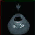
图 1 扫描在最上层图像中出现环形伪影
信噪比评分2分,上颌窦及蝶窦显示清楚评分4分,鼻道及鼻前庭评分2分。
Figure 1. The scan shows a ring artifact in the topside image
![]()
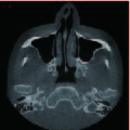
图 2 扫描在最下层图像中出现环形伪影
信噪比评分2分,口咽结构可见,评分2分。
Figure 2. The scan shows a ring artifact in the lowest-side level image
![]()
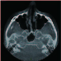
图 3 CBCT扫描图片
图片信噪比评分4分,双层上颌窦显示优良评分为4分,鼻腔、前庭、鼻咽部显示清楚评分为4分;双侧乳突部分结构显示,显示情况为良好评分3分,气体及骨质显示及对比度评分3分;软组织、脂肪及液体显示及对比度评分1分。
Figure 3. The scanning images of CBCT
表 1 图像SNR及牙齿周围组织结构解剖显示评估
Table 1 Image SNR and evaluation of anatomical display of tissue structure around teeth
项目 A医师 B医师 平均 ≥3分/% ≤2分/% Kappa系数 感知信噪比 3.83±0.41 3.79±0.43 4# 98.7~99.0 1.0~1.3 0.754 上颌窦、蝶窦 4 4 4# 100 0 1.000 额窦、筛窦 1 1 1* 0 100 1.000 咽隐窝 3.96±0.27 3.96±0.28 3.96±0.28# 98.7~99.0 1.0~1.3 0.864 鼻腔、前庭 3.99±0.10 3.99±0.12 3.99±0.12# 99.7 0.3 1.000 乳突 2.43±0.69 2.36±0.73 2.40±0.85${}^\nabla $ 46.3~49.3 50.7~53.7 0.820 注:# vs $\nabla $、# vs *、* vs $\nabla $,P<0.05差异有统计学意义;# vs #,P>0.05差异没有统计学意义。  下载: 导出CSV
表 2 耳鼻喉相关不同密度组织显示及对比度评估
Table 2 Contrast evaluation and display of different density tissues related to ear, nose and throat
项目 A医师 B医师 平均 ≥3分(%) ≤2分(%) Kappa系数 气体 4 4 4# 100 0 1.000 骨质 4 4 4# 100 0 1.000 软组织 0.85±0.36 0.77±0.42 0.81±0.38${}^{\nabla} $ 0 100 0.535 脂肪 1 1 1${}^{\nabla} $ 0 100 1.000 液体 0.81±0.39 0.71±0.46 0.75±0.45${}^{\nabla} $ 0 100 0.543 注:# vs $\nabla $,P<0.05差异有统计学意义;# vs #、$\nabla $ vs $\nabla $,P>0.05差异没有统计学意义。
下载: 导出CSV
-
[1] 徐家刚. CBCT在口腔临床诊断治疗中的应用研究[J]. 系统医学, 2018,3(16): 146−148. XU J G. Application research on CBCT in the clinical diagnosis and treatment in the department of stomatology[J]. Systems Medicine, 2018, 3(16): 146−148. (in Chinese).
[2] MCGUIGAN M B, DUNCAN H F, HORNER K. An analysis of effective dose optimization and its impact on image quality and diagnostic efficacy relating to dental cone beam computed tomography (CBCT)[J]. Swiss Dental Journal, 2018, 128(4): 297−316.
[3] EZELDEEN M, WYATT J, AL-RIMAWI A, et al. Use of CBCT guidance for tooth auto-transplantation in children[J]. Journal of Dental Research, 2019, 98(4): 406−413. doi: 10.1177/0022034519828701
[4] DELPHINE M, JEAN-NOEL V, OVE A P, et al. Recent advances in cone-beam CT in oral medicine[J]. Current Medical Imaging, 2020, 16(5): 553−564(12). doi: 10.2174/1573405615666190114152003
[5] 徐菁玲, 李伟力. 锥型束CT在牙周手术中的应用进展[J]. 中国介入影像与治疗学, 2013, 10(12): 768-771. XU J L, LI W L. Applications of cone beam CT in periodontal surgery[J]. Chinese Journal of Interventional Imaging and Therapy, 2013, 10(12): 768-771. (in Chinese).
[6] SAFADI A, KLEINMAN S, GIGI D, et al. Surgical management of odontogenic cysts involving the maxillary sinus: A retrospective study[J]. Journal of Craniomaxillofac Surgery, 2020, 48(8): 800−807. doi: 10.1016/j.jcms.2020.06.011
[7] ROH A T, XIAO Z, CHENG J Y, et al. Conical ultrashort echo time (UTE) MRI in the evaluation of pediatric acute appendicitis[J]. Abdominal Radiology, 2019, 44(1): 22−30. doi: 10.1007/s00261-018-1705-y
[8] 李一新, 梁健东, 余敏祥. CBCT三维成像技术在口腔种植术中的应用[J]. 临床医学工程, 2014,21(4): 413−414. doi: 10.3969/j.issn.1674-4659.2014.04.0413 LI Y X, LIANG J D, YU M X. Discussion on CBCT three-dimensional imaging technology in dental implantation[J]. Clinical Medicine & Engineering, 2014, 21(4): 413−414. (in Chinese). doi: 10.3969/j.issn.1674-4659.2014.04.0413
[9] 王嘉艺, 王珊, 王林. CBCT在口腔正畸学头影测量中的应用与发展[J]. 口腔医学, 2016,36(11): 1047−1050. WANG J Y, WANG S, WANG L. Application and development of CBCT in the orthodontics cephalometrics[J]. Stomatology, 2016, 36(11): 1047−1050. (in Chinese).
[10] 中华医学会放射学分会头颈学组, 中华医学会影像技术分会辐射防护学组. 头颈部CT检查和辐射剂量管理专家共识[J]. 中华放射学杂志, 2020, 54(9): 827-838. Head and Neck Group of Chinese Society of Radiology Chinese Medical Association, Radiation Protection Group of Chinese Society of Imaging Technology Chinese Medical Association. Expert consensus on head and neck CT examination and radiation dose management[J]. Chinese Journal of Radiology, 2020, 54(9): 827-838. (in Chinese).
[11] 滕跃辉, 洪渊, 林梓桐, 等. 上颌窦底壁与上颌后牙根尖位置关系的临床与锥形束CT研究[J]. 口腔医学研究, 2015,31(12): 1216−1219. TENG Y H, HONG Y, LIN Z T, et al. Assessment of topographic relationship between maxillary sinus floors and upper molar root apices by cone-beam computed tomography[J]. Journal of Oral Science Research, 2015, 31(12): 1216−1219. (in Chinese).
[12] 李功臣, 陆永健, 徐泽淼, 等. 应用CBCT回顾分析牙源性上颌窦病变[J]. 口腔医学, 2015,35(3): 192−196. LI G C, LU Y J, XU Z M, et al. The evaluation of maxillary sinusitis using cone beam computed tomography[J]. Stomatology, 2015, 35(3): 192−196. (in Chinese).
[13] 潘继. 副鼻窦炎并发症的临床治疗分析与进展性研究[J]. 中国医药指南, 2016,14(16): 93−94. PAN J. Clinical analysis and progress of complications of paranasal sinusitis[J]. Guide of China Medicine, 2016, 14(16): 93−94. (in Chinese).
[14] GULDNER C, DIOGO I, WINDFUHR J, et al. Analysis of the fossa olfactoria using cone beam tomography (CBT)[J]. Acta Oto-laryngologica, 2011, 131(1): 72−78. doi: 10.3109/00016489.2010.506653
[15] BREMKE M, SESTERHENN A M, MURTHUM T, et al. Digital volume tomography (DVT) as a diagnostic modality of the anterior skull base[J]. Acta Oto-laryngologica, 2009, 129(10): 1106−1114. doi: 10.1080/00016480802620621
[16] DEMESLAY J, VERGEZ S, SERRANO E, et al. Morphological concordance between CBCT and MDCT: A paranasal sinus-imaging anatomical study[J]. Surgical & Radiologic Anatomy, 2016, 38(1): 71−78.
[17] 秦力军, 樊明成, 郭景涛, 等. 低剂量CT扫描在儿童鼻窦检查中的应用及防护价值[J]. 河北医药, 2011,33(21): 3241−3242. QIN L J, FAN M C, GUO J T, et al. Application and protective value of low-dose CT scan in children's sinus examination[J]. Hebei Medical Journal, 2011, 33(21): 3241−3242. (in Chinese).
[18] 陈静, 雷薇薇, 华清泉. 慢性鼻窦炎患者睡眠障碍的影响因素及护理对策[J]. 中国医药导报, 2018,15(5): 140−143. CHEN J, LEI W W, HUA Q Q. Influencing factors of sleep disorders and nursing strategies for patients with chronic sinusitis[J]. China Medical Herald, 2018, 15(5): 140−143. (in Chinese).
-
期刊类型引用(0)
其他类型引用(2)
计量
- 文章访问数: 273
- HTML全文浏览量: 99
- PDF下载量: 22
- 被引次数: 2


