
Evaluation of CT Imaging in Secondary Lower Extremity Lymphedema: A Prospective Study
-
摘要: 目的:探讨CT双下肢体积测量法在继发性下肢淋巴水肿分级评价中的应用价值。材料及方法:收集2019年6月至2020年1月因继发性下肢淋巴水肿入院并行CT双下肢扫描的患者,临床医师于CT扫描一周内完成双下肢多径线实体测量,分别记录CT双下肢体积测量法及临床多径线测量法所得双侧全下肢、大腿及小腿的体积。基于患侧比健侧体积增大的百分比,分别得到CT和临床测量结果的全下肢、大腿及小腿的淋巴水肿分级,采用Kappa软件分析CT与临床全下肢淋巴水肿分级之间的一致性。结果:最终入组患者38例。CT双下肢体积测量法和临床多径线测量法所得患侧的全下肢体积分别为(9984±2217)cm3和(11308±2373)cm3,健侧分别为(7154±1417)cm3和(8265±1704)cm3。全下肢淋巴水肿的CT分级为隐匿期1例,轻度6例,中度14例,重度17例;临床分级为隐匿期1例,轻度8例,中度11例,重度18例。CT与临床分级的一致性非常好(Kappa=0.878)。结论:CT双下肢体积测量可以作为继发性下肢淋巴水肿分级诊断的影像检查工具。Abstract: Objective: To explore the application value of CT bilateral lower extremity volume measurement in the grading of secondary lower extremity lymphedema. Materials and methods: Patients with secondary lower extremity lymphedema who were admitted to our hospital and underwent CT of both lower extremities were collected from June 2019 to January 2020. The clinician completed the multiple-circumference measurement of lower extremity within one week of CT scanning, and recorded the volumes of both lower extremities, thighs and calfs obtained by CT bilateral lower extremity volume measurement and clinical multiple-circumference method, respectively. Based on the percentage enlargement in the volume of the affected side compared with the healthy side, the grading of the whole lower extremity, thigh and calf was obtained by CT and clinical measurement results,respectively. Kappa analysis was used to compare the consistency between CT and clinical in the grading of whole lower extremity. Results: Thirty-eight patients were finally enrolled. The volumes of lower extremities measured respectively by CT bilateral lower extremity volume measurement and clinical multiple-circumference method were (cm3): the affected side (9984±2217 and 11308±2373), the healthy side (7154±1417 and 8265±1704). The CT grading of whole lower extremity was 1 case in preclinical stage, 6 cases of mild, 14 cases of moderate, and 17 cases of severe. The clinical grading of whole lower extremity was 1 case in preclinical stage, 8 cases of mild, 11 cases of moderate, and 18 cases of severe. The consistency between CT and clinical in grading of whole lower extremity is excellent (Kappa=0.878). Conclusion: CT bilateral lower extremity volume measurement can be used as an imaging tool in the grading of secondary lower extremity lymphedema.
-
Keywords:
- CT /
- lower extremity lymphedema /
- grade
-
继发性下肢淋巴水肿是指后天性淋巴回流受阻,淋巴液淤滞在组织间隙中导致下肢淋巴源性肿胀,多继发于腹、盆腔恶性肿瘤根治性切除和/或放化疗后,在癌症幸存者中发生率高达22%[1]。患者的生活质量下降,频繁发作蜂窝组织炎、淋巴管炎、静脉炎等并发症,罕见情况下会诱发血管内皮瘤或淋巴管瘤等恶性肿瘤[2]。本病治疗方式的选择与疾病的严重程度相关,主要有保守治疗、吸脂术、淋巴静脉吻合术等[3]。根据2016版国际淋巴学会共识[4],评价下肢淋巴水肿严重程度依据患肢体积增大的百分比。
目前临床上广泛采用多径线测量法,该法简便易行,但受患者体型及测量者主观因素影响较大[5]。因此,寻找一种客观且重复性好的量化工具是临床应用的一项基本需求。
CT可量化分析脏器体积及脂肪含量,已广泛用于神经肌肉病、骨科创伤等肢体的体积测量[6-9],但目前尚无将CT应用于淋巴水肿的报道。因此,本文旨在探讨CT在继发性下肢淋巴水肿的应用价值。
1. 材料与方法
1.1 一般资料
本研究通过首都医科大学附属北京世纪坛医院医院伦理委员会批准,分析2019年6月至2020年1月因继发性下肢淋巴水肿入我院的患者。
入组标准:①继发性下肢淋巴水肿诊断明确,如继发于直肠癌、宫颈癌、子宫内膜癌等恶性肿瘤,治疗后患者出现单侧下肢淋巴水肿;②下肢 CT成像资料完整;③签署知情同意书。排除标准:①其它原因导致的下肢淋巴水肿,如血栓、心衰、肝肾功能不全,代谢性下肢水肿;②肿瘤复发和/或转移者。
1.2 CT成像检查及测量法
所有检查均在GE Revolution CT完成,扫描范围自盆腔至双足。扫描参数:管电压80/140 kVp切换,自动管电流调节模式100~200 mA,噪声指数为12,Asir比例设置为40%,球管旋转时间0.5 s,螺距0.992︰1,探测器宽8 cm,矩阵512×512,SFOV 50 cm,层厚与层间隔设置为5 mm。
所得原始数据均以2.5 mm层厚、2.5 mm层间隔进行重建,所得CT图像均导入GE AW 4.7工作站,使用感兴趣体积工具,设定CT阈值范围为 -200至1000。
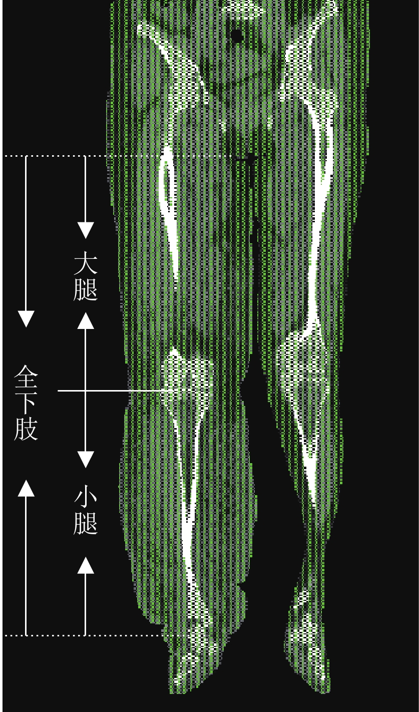
大腿测量范围上缘起自会阴水平,下缘达膝关节间隙水平,小腿测量范围以膝关节间隙水平以下至脚踝水平为准,全下肢测量范围以会阴水平以下至脚踝水平为准(图1)。分别记录双侧大腿、小腿及全下肢体积数据,计算(患侧大腿体积 - 健侧大腿体积)/健侧大腿体积、(患侧小腿体积 - 健侧小腿体积)/健侧小腿体积、(患侧全下肢体积 - 健侧全下肢体积)/健侧全下肢体积的百分比结果,并参照2016版国际淋巴协会共识[5]将所得结果进行分级。
![]() 图 1 CT双下肢冠状面图像,大腿测量范围由会阴水平至膝关节间隙水平,小腿测量范围由膝关节间隙水平至脚踝水平,全下肢测量范围由会阴水平至脚踝水平Figure 1. CT imaging of both lower extre- mities. The measurement range of thigh is from perineum to knee, the measurement range of calf is from knee to ankle, and the measurement range of whole lower extremity is from perineum to ankle
图 1 CT双下肢冠状面图像,大腿测量范围由会阴水平至膝关节间隙水平,小腿测量范围由膝关节间隙水平至脚踝水平,全下肢测量范围由会阴水平至脚踝水平Figure 1. CT imaging of both lower extre- mities. The measurement range of thigh is from perineum to knee, the measurement range of calf is from knee to ankle, and the measurement range of whole lower extremity is from perineum to ankle1.3 临床多径线测量法
由具有5年以上肢体测量经验的临床医师测量入组患者双侧下肢,分别记录患侧与健侧踝、小腿下1/3、小腿中1/2、小腿上1/3、膝、大腿下1/3、大腿中1/2、大腿上1/3、大腿根部的周径,以及相邻周径层面之间的长度距离(图2),采用圆锥体计算公式得出分段体积,将相应分段的体积相加计算为总体积。分别计算(患侧大腿体积 - 健侧大腿体积)/健侧大腿体积、(患侧小腿体积 - 健侧小腿体积)/健侧小腿体积、(患侧全下肢体积 - 健侧全下肢体积)/健侧全下肢体积的百分比结果,并参照2016版国际淋巴协会共识[5]将所得结果进行分级。
![]() 图 2 临床多径线测量法的双下肢病例展示图,黑线为临床医师选取的下肢截面,从下到上依次为踝、小腿下1/3、小腿中1/2、小腿上1/3、膝、大腿下1/3、大腿中1/2、大腿上1/3及大腿根部Figure 2. The image of clinical multiple-circumference for both lower extre-mities. The black line is the cross section selected by the clinician. From bottom to top, it is ankle, lower 1/3 of calf, 1/2 of calf, and upper 1/3 of calf, Knee, lower 1/3 of thigh, 1/2 of thigh, upper 1/3 of thigh, and the root of thigh
图 2 临床多径线测量法的双下肢病例展示图,黑线为临床医师选取的下肢截面,从下到上依次为踝、小腿下1/3、小腿中1/2、小腿上1/3、膝、大腿下1/3、大腿中1/2、大腿上1/3及大腿根部Figure 2. The image of clinical multiple-circumference for both lower extre-mities. The black line is the cross section selected by the clinician. From bottom to top, it is ankle, lower 1/3 of calf, 1/2 of calf, and upper 1/3 of calf, Knee, lower 1/3 of thigh, 1/2 of thigh, upper 1/3 of thigh, and the root of thigh1.4 统计学分析
所有数据采用SPSS 25.0版软件进行统计分析,分别计算临床双下肢多径线测量法及CT双下肢体积测量法所得双侧大腿、小腿及全下肢体积的平均数及标准差。
采用配对t检验分别比较两种方法测量同一部位体积之间的差异,采用Kappa方法分析CT全下肢淋巴水肿分级与临床全下肢淋巴水肿分级之间的一致性。
2. 结果
2.1 患者一般资料
最终入组患者38例,女/男,34/4例,年龄范围32~71岁,中位年龄(51±10)岁,病程中位年限(3±7)年。主要临床表现为单侧下肢肿胀、皮肤粗糙,其中继发于子宫内膜癌7例,宫颈癌22例,卵巢癌4例,腹股沟肿瘤3例,阴茎癌1例,下肢皮肤病变1例。
2.2 测量结果
临床双下肢多径线测量法与CT双下肢体积测量法所得双侧大腿、小腿及全下肢体积的结果详见表1和表2。基于2016版国际淋巴协会共识,依临床与CT所得患侧比健侧全下肢、大腿及小腿体积增大的百分比值分别进行分级诊断,结果如表3。
表 1 临床多径线与CT测量患侧肢体体积值Table 1. The volume of the affected side by clinical multiple-circumference and CT部位 不同方法患侧肢体体积/cm3 统计检验 临床多径线 CT t P 全下肢 11308±2373 9984±2217 5.320 0.000 大腿 7122±1847 6515±1455 3.182 0.003 小腿 3973±1162 3462±1078 8.180 0.000 表 2 临床多径线与CT测量健侧肢体体积值Table 2. The volume of the healthy side by clinical multiple-circumference and CT部位 不同方法健侧肢体体积/cm3 统计检验 临床多径线 CT t P 全下肢 8265±1704 7154±1417 5.788 0.000 大腿 5763±1618 4917±1004 3.211 0.003 小腿 2714±562 2174±575 12.056 0.000 表 3 基于临床多径线与CT测量的下肢淋巴水肿分级Table 3. The grading of lower extremity lymphedema by clinical multiple-circumference and CT方法 下肢淋巴水肿分级分布/例 隐匿期 轻度 中度 重度 临床多径线全下肢分级 1 8 11 18 CT全下肢分级 1 6 14 17 临床多径线大腿分级 0 10 19 9 CT大腿分级 0 10 18 10 临床多径线小腿分级 1 10 8 19 CT小腿分级 1 8 10 19 统计学分析显示,CT双下肢体积测量法与临床双下肢多径线测量法对全下肢淋巴水肿分级的一致性非常好(Kappa=0.878),CT全下肢分级与CT大腿、小腿分级的一致性中等(Kappa=0.486/0.511)。
3. 讨论
继发性下肢淋巴水肿作为一种严重影响癌症幸存患者生活质量的慢性疾病,临床治疗目标主要包括减轻患肢沉重与疼痛感、预防患肢感染、改善患肢外观及功能等,但仅从患者主观感受不足以决定治疗方式的选择及评估疗效。本研究引用2016版国际淋巴学会共识制定的客观分级标准,首次将CT双下肢体积测量法与当前临床应用最广泛的多径线测量法进行了对照分析,两种方法分级的一致性非常好。
继发性下肢淋巴水肿的准确分级诊断可有效帮助临床评估与治疗。目前该类疾病公认的分级指标为患侧肢体体积增大百分比,临床常用体积测量方法如排水测量法、多径线测量法等[10-13]均存在不同程度的局限性,不但容易受到测量者主观因素影响,甚至会增加患者感染的风险。
影像学检查是一种无创的可精准量化的评价工具,研究表明,CT可用于液体、脂肪、肌肉等不同密度组织的定量分析中,并可评价靶组织及脏器体积,如肺结节、肝脏等,特别是对下肢肌肉萎缩和脂肪化程度的定量分析,在神经肌肉病、骨科创伤后等疾病的评价中已得到广泛认可[6-9]。因此本研究将CT应用于继发性下肢淋巴水肿患者肢体体积的测量中,根据患肢体积增大百分比进行分级,与临床多径线测量法分级一致的例数共有35/38例,高达92%。
研究结果表明,临床多径线测量法所得全下肢的体积值均大于CT双下肢体积测量法所得全下肢的体积值,这种差异也存在于大腿和小腿的体积测量中。出现这种差异的原因在于CT体积值是基于体素测量得出的精确数据,不受影像医师主观影响,而临床基于圆锥体计算公式的多径线测量法是估值数据,且圆锥体计算公式对不规则形态的淋巴水肿肢体存在一定的计算误差。
本研究中分别以患侧大腿或小腿体积增大百分比作为分级标准时,与全下肢分级结果的一致性中等,尤其在中度和重度组,大腿和小腿分别出现了过低分级和过高分级的情况(表3),这符合继发性下肢淋巴水肿多从远端向近端蔓延的发病趋势,小腿肿胀程度常较大腿为重,因此单独计算大腿或小腿体积变化不能代表全下肢,更不能决定治疗方式。
此外,本研究中CT剂量指数为4.88 mGy,剂量长度乘积范围约421~596 mGy·cm,有效剂量约0.0842~0.1192 mSv,但本研究主要针对恶性肿瘤患者,且辐射剂量远远低于传统的多时相腹腔及盆腔成像,这是因为下肢的转换系数(k=0.0002~0.0110)比腹盆腔低(k=0.0180)[14-16]。
本研究的不足:①本研究对于继发性下肢淋巴水肿患者的分级是基于体积增大百分比,然而,CT图像表明,处于同一分级的患肢组织成分(脂肪、肌肉)比例可能存在明显差异,即患肢体积增大百分比一致,但其内纤维化程度明显不同[17],因此手术方式和难度存在很大差异,疗效和预后也大相径庭,所以基于体积的分级诊断有一定的局限性,下一步的研究将基于淋巴水肿肢体体积增大的不同组织成分进行精确量化分析与分级评价;②本研究样本量较小,下一步的研究将继续增加淋巴水肿病例数目及类型,不断寻求肢体淋巴水肿分级评价的最佳方法;③本研究未纳入MRI、核医学等影像学研究,期待进一步完善比较影像学研究。
4. 结论
CT双下肢体积测量法可以准确测量淋巴水肿肢体体积并进行分级,指导临床选择最佳治疗方案与评估疗效,对提高癌症幸存者生存质量意义深远。
-
![]()
图 1 CT双下肢冠状面图像,大腿测量范围由会阴水平至膝关节间隙水平,小腿测量范围由膝关节间隙水平至脚踝水平,全下肢测量范围由会阴水平至脚踝水平
Figure 1. CT imaging of both lower extre- mities. The measurement range of thigh is from perineum to knee, the measurement range of calf is from knee to ankle, and the measurement range of whole lower extremity is from perineum to ankle
![]()
图 2 临床多径线测量法的双下肢病例展示图,黑线为临床医师选取的下肢截面,从下到上依次为踝、小腿下1/3、小腿中1/2、小腿上1/3、膝、大腿下1/3、大腿中1/2、大腿上1/3及大腿根部
Figure 2. The image of clinical multiple-circumference for both lower extre-mities. The black line is the cross section selected by the clinician. From bottom to top, it is ankle, lower 1/3 of calf, 1/2 of calf, and upper 1/3 of calf, Knee, lower 1/3 of thigh, 1/2 of thigh, upper 1/3 of thigh, and the root of thigh
表 1 临床多径线与CT测量患侧肢体体积值
Table 1 The volume of the affected side by clinical multiple-circumference and CT
部位 不同方法患侧肢体体积/cm3 统计检验 临床多径线 CT t P 全下肢 11308±2373 9984±2217 5.320 0.000 大腿 7122±1847 6515±1455 3.182 0.003 小腿 3973±1162 3462±1078 8.180 0.000  下载: 导出CSV
表 2 临床多径线与CT测量健侧肢体体积值
Table 2 The volume of the healthy side by clinical multiple-circumference and CT
部位 不同方法健侧肢体体积/cm3 统计检验 临床多径线 CT t P 全下肢 8265±1704 7154±1417 5.788 0.000 大腿 5763±1618 4917±1004 3.211 0.003 小腿 2714±562 2174±575 12.056 0.000
下载: 导出CSV
表 3 基于临床多径线与CT测量的下肢淋巴水肿分级
Table 3 The grading of lower extremity lymphedema by clinical multiple-circumference and CT
方法 下肢淋巴水肿分级分布/例 隐匿期 轻度 中度 重度 临床多径线全下肢分级 1 8 11 18 CT全下肢分级 1 6 14 17 临床多径线大腿分级 0 10 19 9 CT大腿分级 0 10 18 10 临床多径线小腿分级 1 10 8 19 CT小腿分级 1 8 10 19
下载: 导出CSV
-
[1] CORMIER J N, ASKEW R L, MUNGOVAN K S, et al. Lymphedema beyond breast cancer: A systematic review and meta-analysis of cancer-related secondary lymphedema[J]. Cancer, 2010, 116(22): 5138−5149. doi: 10.1002/cncr.25458
[2] 中国妇幼保健协会妇科肿瘤防治专业委员会. 妇科肿瘤治疗后下肢淋巴水肿专家共识[J]. 中国临床医生杂志, 2021,49(2): 149−155. DOI: 10.3969/j.issn.2095-8552.2021.02.006. Professional Committee of Gynecological Cancer Prevention and Treatment of China Maternal and Child Health Association. Expert consensus on lower limb lymphedema after treatment of gynecological tumors[J]. Chinese Journal for Clinicians, 2021, 49(2): 149−155. DOI: 10.3969/j.issn.2095-8552.2021.02.006. (in Chinese).
[3] CIUDAD P, SABBAGH M D, AGKO M, et al. Surgical management of lower extremity lymphedema: A comprehensive review[J]. Indian Journal of Plastic Surgery, 2019, 52(1): 81−92. DOI: 10.1055/s-0039-1688537.
[4] Executive Committee. The diagnosis and treatment of peripheral lymphedema: 2016 consensus document of the International Society of Lymphology[J]. Lymphology. 2016, 49(4): 170-184.
[5] TIDHAR D, ARMER J M, DEUTSCHER D, et al. Measurement issues in anthropometric measures of limb volume change in persons at risk for and living with lymphedema: A reliability study[J]. Journal of Personalized Medicine, 2015, 5(4): 341−353. DOI: 10.3390/jpm5040341.
[6] TSUKADA A, UCHIDA K, AIKAWA J, et al. Unilateral-dominant reduction in muscle volume in female knee osteoarthritis patients: Computed tomography-based analysis of bilateral sides[J]. Journal of Orthopaedic Surgery and Research, 2020, 15(1): 543−549. doi: 10.1186/s13018-020-02074-x
[7] GUO Z J, LIN Q, LIU H T, et al. The preliminary exploration of 64-slice volume computed tomography in the accurate measurement of pleural effusion[J]. Acta Radiologica, 2013, 54(7): 765−769. DOI: 10.1177/0284185113483675.
[8] 杨献峰, 杨尚文, 胡安宁, 等. 基于CT值阈值法测量骨骼肌体积[J]. 中国医学计算机成像杂志, 2017,23(3): 287−291. DOI: 10.3969/j.issn.1006-5741.2017.03.019. YANG X F, YANG S W, HU A N, et al. Measurement of skeletal muscle volume based on CT value threshold method[J]. Chinese Journal of Medical Computer Imaging, 2017, 23(3): 287−291. DOI: 10.3969/j.issn.1006-5741.2017.03.019. (in Chinese).
[9] 张国来, 包强, 陈光辉, 等. CT阈值法测量乳突气房体积与面神经管垂直段位置的相关性[J]. 中国医学影像学杂志, 2016,24(3): 175−178. DOI: 10.3969/j.issn.1005-5185.2016.03.004. ZHANG G L, BAO Q, CHEN G H, et al. Correlation between the volume of the mastoid air chamber and the position of the vertical segment of the facial nerve canal measured by the CT threshold method[J]. Chinese Journal of Medical Imaging, 2016, 24(3): 175−178. DOI: 10.3969/j.issn.1005-5185.2016.03.004. (in Chinese).
[10] ASTRÖM K G, ABDSALEH S, BRENNING G C, et al. MR imaging of primary, secondary, and mixed forms of lymphedema[J]. Acta Radiologica, 2001, 42(4): 409−416. DOI: 10.1034/j.1600-0455.2001.420412.x.
[11] RABE E, STÜCKER M, OTTILLINGER B. Water displacement leg volumetry in clinical studies: A discussion of error sources[J]. BMC Medical Research Methodology, 2010, 10(5): 1−9. DOI: 10.1186/1471-2288-10-5.
[12] DELTOMBE T, JAMART J, RECLOUX S, et al. Reliability and limits of agreement of circumferential, water displacement, and optoelectronic volumetry in the measurement of upper limb lymphedema[J]. Lymphology, 2007, 40(1): 26−34.
[13] TAYLOR R, JAYASINGHE U W, KOELMEYER L, et al. Reliability and validity of arm volume measurements for assessment of lymphedema[J]. Physical Therapy, 2006, 86(2): 205−214. doi: 10.1093/ptj/86.2.205
[14] ERENDS M, TEIKE V D A, de GRZYMALA A P, et al. Validity and reliability of three-dimensional imaging for measuring the volume of the arm[J]. Lymphatic Research and Biology, 2014, 12(4): 275−281. DOI: 10.1089/lrb.2014.0007.
[15] SALTYBAEVA N, JAFARI M E, HUPFER M, et al. Estimates of effective dose for CT scans of the lower extremities[J]. Radiology, 2014, 273(1): 153−159. DOI: 10.1148/radiol.14132903.
[16] SMITH-BINDMAN R, WANG Y, YELLEN-NELSON T R, et al. Predictors of CT radiation dose and their effect on patient care: A comprehensive analysis using automated data[J]. Radiology, 2017, 282(1): 182−193. DOI: 10.1148/radiol.2016151391.
[17] CELLINA M, MARTINENGHI C, PANZERI M, et al. Noncontrast MR lymphography in secondary lower limb lymphedema[J]. Journal of Magnetic Resonance Imaging, 2021, 53(2): 458−466. DOI: 10.1002/jmri.27328.
-
期刊类型引用(1)
1. 郭佳,信建峰,张春燕,过丽芳,王梦君,王仁贵,董健. 能谱CT多物质解析评价继发性下肢淋巴水肿分级. CT理论与应用研究. 2022(03): 365-371 .  本站查看
本站查看
其他类型引用(0)

计量
- 文章访问数: 201
- HTML全文浏览量: 155
- PDF下载量: 21
- 被引次数: 1


