
Clinicopathological and CT Findings of Cystic Lung Cancer
-
摘要: 目的:囊腔型肺癌是一种少见的、具有特殊形态学表现的肺癌,在影像学上尚未被充分认识,本研究通过分析其CT特征及临床病理,以提高对该类特殊类型肺癌的认识及诊断能力。方法:回顾性总结分析本院2015年1月至2021年6月期间确诊的囊腔型肺癌患者的临床信息、病理类型及CT图像特征。结果:病理类型:腺癌44例(86.27%),其中微浸润性腺癌7例(13.73%),浸润性腺癌35例(68.63%)(组织学类型:贴壁为主型11例(21.57%)、乳头状为主型8例(15.69%)、腺泡为主型15例(29.41%)且其中2例(3.92%)含微乳头成分占比大于20%,1例(1.96%)未分型),1例(1.96%)化疗后手术病理提示腺癌,1例(1.96%)淋巴结穿刺活检提示肺转移性腺癌;鳞癌5例(9.80%),其中角化性鳞状细胞癌3例(5.89%),非角化性鳞状细胞癌2例(3.92%);大细胞癌1例(1.96%),高级别粘液表皮样癌1例(1.96%)。病灶以外周分布为主40例(80.30%);平均直径为(4.7±2.7)cm,主体成分:囊腔伴磨玻璃成分20例(39.22%)、囊腔伴实性成分22例(43.14%)、囊腔伴混合成分9例(17.65%);Mario分型Ⅰ型24例(47.06%)、Ⅱ型8例(15.69%)、Ⅲ 型9(17.65%)、Ⅳ型10例(19.61%);囊腔壁不均匀42例(82.35%);病灶边缘分叶状28例(54.90%)、毛刺征14例(27.45%);病灶内分隔33例(64.71%)、内壁光滑36例(70.59%);与胸膜关系:胸膜牵拉24例(47.06%)、紧贴胸膜11例(21.57%);周围血管情况:血管穿行于病灶内8例(15.69%)、血管紧贴/走向于病灶边缘16(31.37%);周围支气管情况:支气管穿行于病灶内8例(15.69%)、支气管紧贴/走向于病灶边缘21例(41.18%)。结论:囊腔型肺癌的CT表现有一定的特征性,掌握其特征表现可做出肺癌的提示性诊断。Abstract: Objective: Cystic lung cancer is a rare type of lung cancer with special morphological manifestations on imaging that have not been completely recognized. This study aimed to improve the understanding and diagnosis of this lung cancer by analyzing its computed tomography (CT) features and clinicopathological findings. Methods: The clinical data, pathological types, and CT features of patients with cystic lung cancer who were diagnosed at our hospital between January 2015 and June 2021 were analyzed retrospectively. Results: We identified 44 cases (86.27%) of adenocarcinomas; these included 7 (13.73%) and 35 (68.63%) cases of microinvasive adenocarcinomas and invasive adenocarcinomas, respectively. Regarding the histological type, 11 (21.57%), 8 (15.69%), and 15 (29.41%) cases were of the adjoint, papillary, and acinar types, respectively. Two cases (3.92%) contained micropapillary components that accounted for more than 20% . Furthermore, one case (1.96%) was not classified, and in another case (1.96%), pathological examination after chemotherapy suggested an adenocarcinoma. Lymph node biopsy in one case (1.96%) suggested a metastatic lung adenocarcinoma. Squamous cell carcinoma was observed in five cases (9.80%); these included three (5.89%) and two (3.92%) cases of keratinizing and non-keratinizing squamous cell carcinomas, respectively. A large cell carcinoma and a high-grade mucoepidermoid carcinoma were observed in one case each (1.96%). A peripheral distribution was observed in 40 cases (80.30%). The mean diameter was 4.7± 2.7 cm. A cystic cavity with ground glass, cystic cavity with solid components, and cystic cavity with mixed components were observed in 20 (39.22%), 22 (43.14%), and 9 (17.65%) cases, respectively. According to the Mario classification, 24 (47.06%), 8 (15.69%), 9 (17.65%), and 10 (19.61%) cases were of types I, II, III, and IV, respectively. The cyst wall was uneven in 42 cases (82.35%); furthermore, a lobular lesion edge and the burr sign were observed in 28 (54.90%) and 14 (27.45%) cases, respectively. Moreover, 33 cases (64.71%) showed a separation within the lesions, while 36 cases (70.59%) had a smooth inner lesion wall. Regarding the relationship with the pleura, pleural traction and closed pleura were observed in 24 (47.06%) and 11 (21.57%) cases, respectively. The peripheral blood vessels passed through the lesion in eight cases (15.69%) and adhered closely to the edge of the lesion in 16 cases (31.37%). The surrounding bronchi passed through the lesion in eight cases (15.69%) and moved toward the edge of the lesion in 21 cases (41.18%). Conclusion: Cystic lung cancer has certain features on CT that can allow its suggestive diagnosis.
-
Keywords:
- computed tomography /
- lung cancer /
- cystic cavity
-
骨质疏松症(osteoporosis,OP)是一种以骨量低下、骨组织微结构损坏,导致骨脆性增加,易发生骨折为特征的全身性骨病[1]。OP按病因可分为原发性和继发性两大类,常见于绝经后女性和老年男性。我国是全球老年人口最多的国家,目前我国骨质疏松症患者数约为9千万,其中女性约7千万[2]。骨质疏松症的严重后果是容易引发骨质疏松性骨折(或称脆性骨折),严重影响生活质量。骨密度(bone mineral density,BMD)可通过骨密度测量技术客观、如实的反映骨量在程度上的变化,对OP的临床诊断、疗效评估及防治具有重要作用。
本研究通过应用定量CT(quantitative CT,QCT)对佛山市健康成年居民椎体BMD进行检测,分析骨量变化与性别和年龄的关系,探讨佛山地区成年居民骨密度数据参考值范围。
1. 资料与方法
1.1 一般资料
2021年10月至2023年3月期间在佛山市中医院健康管理中心进行胸部CT/低剂量胸部CT平扫行肺结节筛查联合QCT骨密度测量检查者共3819例,根据健康管理中心数据库录入资料,所选符合条件的健康成年人1065例。纳入标准:①20周岁以上的健康体检者;②佛山市常住人口,居住时间至少10年以上(含10年)。排除标准:①患有系统性代谢性疾病,椎体骨肿瘤或陈旧性骨折的病患;②上腹部扫描范围内有金属植入物的病患。其中男性648例,女性417例,年龄20~89岁。
按性别、年龄分组,每10岁为1个年龄段:20~29岁62例(男41例,女21例);30~39岁157例(男93例,女64例);40~49岁265例(男168例,女97例);50~59岁338例(男211例,女127例);60~69岁146例(男79例,女67例);70~79岁65例(男40例,女25例);80~89岁32例(男16例,女16例)。
1.2 检测方法
(1)质量控制:在数据采集前和项目进行中采用校准体模(Model 4)对CT机进行质控分析校准。体模扫描范围包括体模上端1 cm处至下端1 cm处;图像重建的参数是120 kV,1 mm层厚;标准重建算法,50 cm SFOV。
(2)QCT扫描规范:按常规/低剂量胸部CT扫描摆位,严格执行辐射防护要求做好体检者辐射防护。扫描范围包含肺尖至第1腰椎下缘,患者仰面躺卧,双手交叉上举,于吸气末单次屏气扫描。
(3)QCT结果分析与测量:图像上传QCT工作站,通过Mindways QCT Pro 6.1v软件行椎体骨密度测量,测量时选择第12胸椎和第1腰椎椎体的正中层面的骨松质区域,注意避开骨皮质、骨岛和椎体后静脉区(图1),测量结果保存在工作站数据库。如果发现第12胸椎或第1腰椎椎体有明显骨折或其他病理改变(如骨肿瘤、骨水泥等),则不宜进行骨密度测量,需要做好标记。
![]() 图 1 椎体骨密度QCT测量方法QCT测量的是椎体中央骨松质的体积骨密度,兴趣区避开骨皮质、骨岛和椎体后静脉区;其测量结果不受身高体重、脊柱侧弯、退变和血管钙化等因素的影响。Figure 1. Measurement of vertebral bone mineral density by QCT
图 1 椎体骨密度QCT测量方法QCT测量的是椎体中央骨松质的体积骨密度,兴趣区避开骨皮质、骨岛和椎体后静脉区;其测量结果不受身高体重、脊柱侧弯、退变和血管钙化等因素的影响。Figure 1. Measurement of vertebral bone mineral density by QCT1.3 诊断依据
按《中国定量CT(QCT)骨质疏松症诊断指南(2018)》及《骨质疏松的影像学与骨密度诊断专家共识(2020)》所定标准进行诊断[3-4]。BMD>120 mg/cm3为骨密度正常,BMD=80~120 mg/cm3范围内为低骨量,BMD<80 mg/cm3为骨质疏松,BMD<80 mg/cm3且伴一处或多处的脆性骨折,为严重骨质疏松。
1.4 统计学方法
数据进行初步整理后,采用SPSS 22.0及Graph Pad 8.0.1软件,计数资料进行卡方检验,计量资料进行正态性检验,正态性分布的计量资料采用“平均值±标准差”
$(\bar x \pm s)$ 的形式表示,采用方差分析方法对多组间均值进行比较,采用LSD法进行组间两两比较,采用$\chi^2 $ 检验统计发病率的比较,采用Spearman法对等级资料进行相关分析,检验水准α=0.05。2. 结果
2.1 男女间各年龄段体检者的BMD比较
20~29岁、30~39岁、40~49岁3个年龄段男女之间BMD存在显著性差异,女性BMD显著高于男性,60~69岁;70~79岁两个年龄段男女之间BMD存在显著性差异,男性BMD显著高于女性。在40~49岁、50~59岁、60~69岁、70~79岁男性组及30~39岁、40~49岁、50~59岁、60~69岁女性组中,同一性别不同年龄段间的BMD比较差异也有统计学意义,且随年龄的增长呈下降趋势(表1)。
表 1 1065例健康成年人胸部CT体检者椎体BMD分析($\bar x \pm s $ ,mg/cm3)Table 1. Analysis of vertebral BMD in 1065 healthy adults undergoing chest CT examination年龄组/岁 男性 女性 t P 人数/例 BMD 人数/例 BMD 20~29 41 163.31±26.28 21 188.38±28.79 -3.442 0.001 30~39 93 160.62±26.86 64 172.42±29.21b -2.610 0.010 40~49 168 145.47±29.67a 97 158.05±32.50b -3.211 0.001 50~59 211 127.1±28.72a 127 127.95±38.05b -0.215 0.830 60~69 79 107.54±29.85a 67 92.36±25.64b 3.263 0.001 70~79 40 96.11±23.60a 25 84.43±18.11 2.114 0.038 80~89 16 91.51±26.78 16 77.38±17.21 1.775 0.086 F 56.893 74.37 P <0.001 <0.001 注:a表示在男性组中,该年龄段与前一年龄段比较P<0.05;b表示在女性组中,该年龄段与前一年龄段比较P<0.05。 2.2 各年龄段BMD正常、低骨量、骨质疏松及严重骨质疏松发病率比较
在男性组和女性组中,不同年龄段之间的OP发病率差异均有统计学意义,(表2)。采用Spearman法对等级资料进行相关分析,发现男女性年龄与OP发病率均呈正相关(r男=0.517,r女=0.636)。
表 2 1065例健康成年人胸部CT体检者骨质疏松筛查结果(例(%))Table 2. Screening results of osteoporosis in 1065 healthy adults undergoing chest CT examination年龄组/岁 男性 女性 正常 低骨量 OP 严重OP 正常 低骨量 OP 严重OP 20~29 38(92.68) 3(7.32) 0(0.00) 0(0.00) 21(100.00) 0(0.00) 0(0.00) 0(0.00) 30~39 90(96.77) 3(3.23) 0(0.00) 0(0.00) 61(95.31) 3(4.69) 0(0.00) 0(0.00) 40~49 137(81.55) 29(17.26) 1(0.60) 1(0.60) 85(87.63) 11(11.34) 0(0.00) 1(1.03) 50~59 125(59.24) 68(32.23) 13(6.16) 5(2.37) 68(53.54) 41(32.28) 12(9.45) 6(4.72) 60~69 25(31.65) 39(49.37) 13(16.46) 2(2.53) 8(11.94) 35(52.24) 22(32.84) 2(2.99) 70~79 4(10.00) 23(57.50) 11(27.50) 2(5.00) 1(4.00) 11(44.00) 10(40.00) 3(12.00) 80~89 2(12.50) 7(43.75) 4(25.00) 3(18.75) 0(0.00) 4(25.00) 10(62.50) 2(12.50) $\chi^2 $ 225.201 235.748 P <0.001 <0.001 3. 讨论
3.1 BMD测量的必要性及意义
随着我国社会人口老龄化加剧,OP作为一种中老年人最常见的全身性慢性骨骼疾病,越来越加重社会和医疗负担,已成为现阶段重要的公共卫生健康问题。OP患病率高危害大,但知晓率、诊断率、治疗率低(“一高三低”)是我国社会OP的防治现状[2,5];不同地区间及城乡间对OP诊疗水平更是存在着较大的差异[6-7]。原发性OP的基本病理改变是由于骨量不断减少而引起的一种全身慢性进行性骨结构退行性病变,包括绝经后OP(Ⅰ型)、老年OP(Ⅱ型)和特发性OP(包括青少年型),其发病机制复杂,近年来,国内外学者取得的研究新进展大大丰富了原发性OP发病机制[2,8]。
骨质疏松性骨折(又称脆性骨折),是指受到轻微外伤(接近站立高度或更低的高度跌倒)时,甚至在没有明显外伤的情况下即发生的骨折,是OP的严重临床后果。骨质疏松性骨折发生后,再骨折的风险显著增高,导致较高的致死率与致残率[9]。骨质疏松性骨折的好发部位包括脊柱椎体、腕部、股骨近端、肱骨近端等[10-11]。
骨密度(BMD)是指单位面积或单位体积所含的骨量。BMD测量技术是一种无创性定量测量人体骨矿含量、骨密度和体质成份的方法。椎体BMD测量能敏感、客观反映骨量变化,并作为诊断、防治OP的主要依据。当椎体局部BMD增高,或远超过该年龄段BMD或邻近正常椎体BMD,提示椎体骨挫伤、压缩骨折或成骨性病变;当椎体局部BMD降低,则提示骨质疏松或骨质破坏等椎体病变。椎体BMD对脊柱病变术前指导及术后疗效评估亦有重要的临床意义[12]。目前我国已经将骨密度检测项目纳入40岁以上人群常规体检内容[2]。
3.2 BMD的测量方法
双能X线(dual X-ray absorptiometry,DXA)BMD测量是目前认知度、认可度及临床应用最高的骨密度测量方法,DXA测量的是面积骨密度(area bone mineraldensity,aBMD),单位为g/cm2。目前国内外公认的OP诊断标准是以DXA测量的结果为依据[4]。DXA通常在椎体、股骨近端或桡骨远端等部位进行测量。DXA对骨密度水平的判断采用的是T值,须根据同种族同性别的正常参考值范围进行计算。
使用DXA诊断骨质疏松的标准由世界卫生组织(World Health Organization,WHO)专家组制定,T值≥-1.0 SD为正常,-2.5<T值<-1.0 SD为低骨量,T值≤-2.5 SD为骨质疏松,T值≤-2.5 SD且伴脆性骨折为严重骨质疏松;我国的骨质疏松学术组织基于2018年我国疾病控制中心组织的大范围多中心流行病学调查或大样本健康人群数据[13-15],也推荐该标准适用于中国绝经后女性和老年男性。对于儿童、青少年、绝经前女性以及50岁以下男性等人群,则采用中国人群数据库计算出的Z值对骨密度水平进行判断,Z值≤-2.0 SD(标准差)可诊断为“低于同年龄段预期范围”或低骨量[4,16-17]。由于受被检者的人种、年龄、性别、区域及检测方法等多种因素影响,以及采用不同的数据库,用DXA诊断骨质疏松症时,国内外报道OP发病率为14%~60%[18-19],会出现比较大的差距。而且,由于DXA是平面投影成像,其所测出的aBMD易受身高体重、脊柱侧弯、脊柱退变和血管钙化等因素影响,BMD测量的准确性降低,导致假阳性出现[20]。
定量CT(QCT)是一种经体模校准后,利用CT容积数据进行精准测量和定量分析的方法,可应用于骨密度、肝脏脂肪、体部脂肪及肌肉的测量,目前主要用于骨密度测量。QCT可以按需测量不同部位的骨密度,其中椎体和股骨上端最为常用。对于椎体,QCT测量的是椎体中央骨松质的体积骨密度,其测量的是真正的体积骨密度(volumetric bone mineraldensity,vBMD),单位为g/cm3,可敏感反映早期松质骨的丢失状况,且其测量结果不受身高体重、脊柱侧弯、退变和血管钙化等因素的影响[3]。近年来,随着QCT的技术优势和CT技术的快速发展,利用QCT开展骨密度领域研究和临床应用越来越受到关注。
目前的QCT诊断标准是国际临床骨密度学会(International Society for Clinical Densitometry,ISCD)和美国放射学院(American College of Radiology,ACR)分别于2007年和2018年提出,根据美国 Mindways QCT系统,采用120 kV管电压测量椎体骨密度结果数据制定的[21-22]。vBMD>120 mg/cm3为正常,80 mg/cm3≤vBMD≤120 mg/cm3为低骨量,vBMD<80 mg/cm3为骨质疏松,vBMD<80 mg/cm3且伴脆性骨折为严重骨质疏松。我国学者对QCT进行了积极探索、论证,建立了中国人群脊柱QCT正常参考值范围数据库,并认为此ISCD及ACR标准同样适用于中国人群骨质疏松症的诊断[23-24]。QCT如果单独使用,其辐射剂量高于DXA,研究证明低剂量胸部CT扫描与QCT相结合同样可以精准测量椎体骨密度[25-26],在健康管理中有较高的应用价值,因而建议在临床使用过程中,QCT扫描应尽量与临床CT扫描同步联合进行,而且推荐使用低剂量扫描技术[27-30]。
本研究通过对佛山市健康成年居民体检行常规/低剂量胸部CT平扫,应用QCT对胸12、腰1椎体BMD进行定量分析,探讨地区性BMD参考值范围。
3.3 BMD、OP与年龄、性别的关系
(1)表1显示,男女性BMD均随年龄的增长呈下降趋势,且女性下降总体幅度大于男性。在40~49岁、50~59岁、60~69岁、70~79岁男性组及30~39岁、40~49岁、50~59岁、60~69岁女性组中,同性别不同年龄段的BMD比较差异有统计学意义,说明女性骨量流失速度比男性提前10年,此与陈文清等[31]研究相符,考虑与女性怀孕哺乳期导致体内骨钙流失等有关。在70~79岁,女性骨量流失较男性有所减缓。在女性70~79岁年龄段及男性80~89岁年龄段,男女性BMD分别与各自前一年龄段比较均无明显区别,且均已接近OP临界值,亦说明女性BMD降低到病理临界值较男性提前10年。而80~89岁年龄段,男女性之间BMD及与各自前1年龄段比较均无明显区别。
(2)男女性BMD均在20~29岁达到峰值,在20~29岁、30~39岁、40~49岁年龄段男女性均随年龄增加BMD下降,女性的下降速度及幅度大于同年龄段男性,但女性的BMD仍高于同年龄组男性,考虑与此年龄段的女性为即将到来的生育、哺乳打下良好的基础作出充分准备有关。年龄低于40岁的男女性均未发现OP或严重OP。
(3)表1与表2显示,50~59岁年龄段男、女性BMD分别为127.1及127.95,此年龄段为女性绝经期前后,男、女性BMD数字相近,差异无统计学意义。在此年龄段后,男、女性BMD均出现下降,女性BMD下降更为明显,导致同年龄段的女性的BMD均低于男性。从40~49岁年龄段开始,随年龄增长,OP及严重OP在男、女性各年龄段均开始出现且发病率逐步上升。从50~59岁年龄段开始,女性OP及严重OP发病率均高于同年龄组男性。在60~69岁年龄段,男、女BMD分别为107.54及92.36,均达到低骨量诊断标准;此年龄段男、女OP发生率分别为16.46% 及32.84%,女性约是男性的两倍,且分别较同性别上一年龄段大幅增长。在80~89岁年龄段,女性BMD低于80,已全部达到OP;而男性的BMD仍在低骨量范围,但亦接近OP的诊断标准。
本研究采用的是Mindways公司的校准体模(Model 4)及QCT Pro 6.1v骨质密度测量软件,能够如实精准的反映BMD。研究显示,随年龄的增长,受试者的BMD逐渐下降,低骨量、OP及严重OP的发病率也逐渐上升,此结果与大多数文献报道的年龄与骨密度呈负相关、年龄与OP发病率呈正相关结论相符[32-33]。
4. 小结
本研究旨在初步探讨佛山地区成年居民骨密度数据参考值范围。研究发现本地区成年居民骨密度及骨质疏松发病率与全国水平相当。研究结果提示,对于40岁以上的人群,尤其是无症状人群,可以通过将QCT与胸部CT体检结合起来,进行骨密度测量,及时发现骨量减低的程度,以利于临床针对骨质疏松发展的不同阶段采取相应的预防和治疗措施,降低因骨质疏松引起的脆性骨折等潜在风险。
本研究不足之处:①未将20岁以下及90岁以上成年居民纳入样本,此年龄段人群参与胸部CT健康体检的样本量极少故未纳入;②未开展多中心研究,样本量相对局限。后续将继续针对不足之处进一步研究,为完善及建立本地区人群骨密度数据库提供更为可靠的理论支持。
-
![]()
图 1 囊腔型肺癌根据不同主体成分分型
Figure 1. Classification of cystic lung cancer according to the main components
![]()
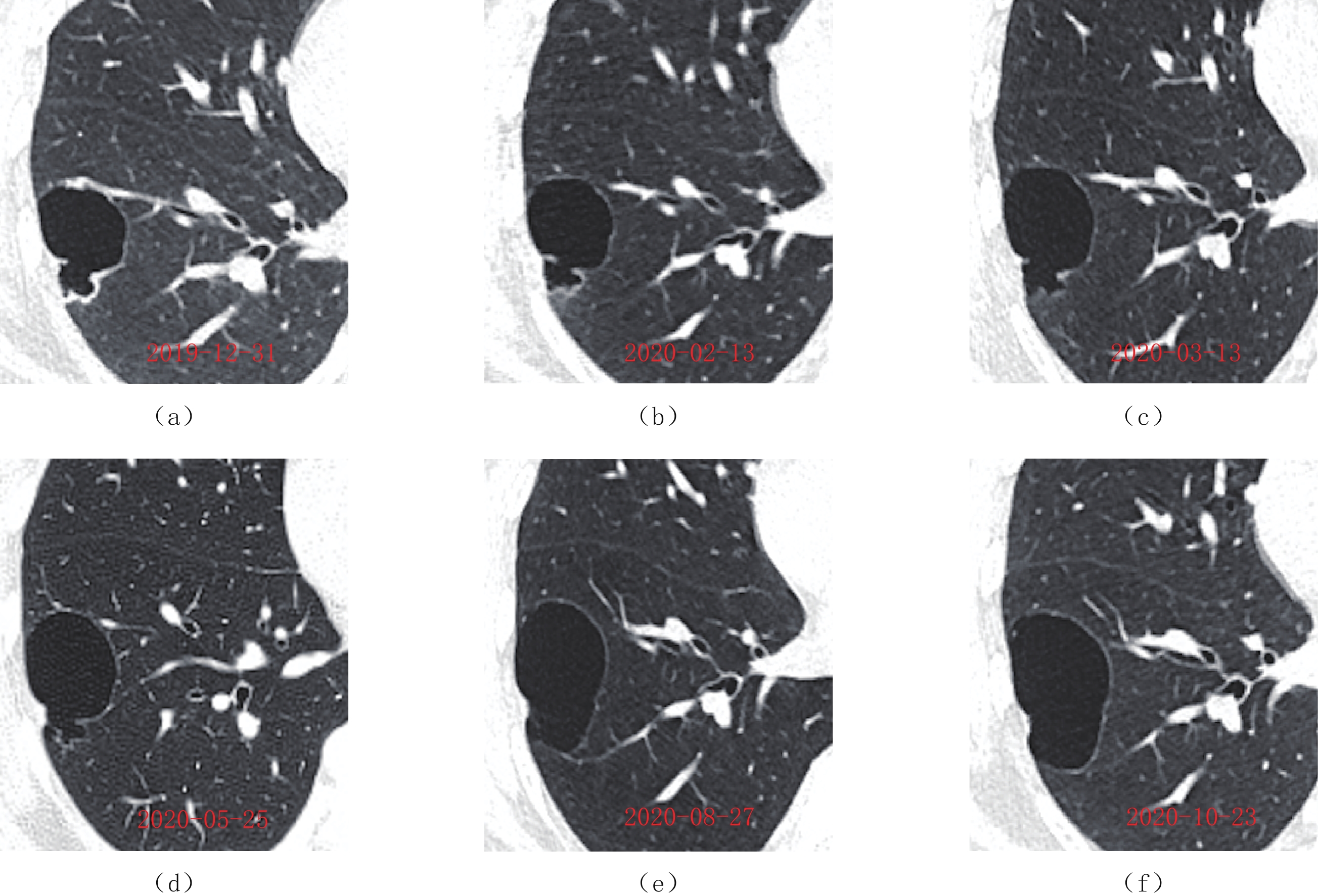
图 3 一例囊腔型肺癌接受化疗后的病灶改变
(a)~(f)分别于2020-01-17、02-01、03-13、04-08、05-06、05-26执行AP方案(力比泰+顺铂)第1~6疗程化疗。(a)患者化疗前的CT图像,可见右肺下叶前基底段一囊腔病变伴囊壁不均匀增厚;(b)~(e)分别为第2、3、5、6次化疗后CT图像,可见囊壁较前逐渐变薄,囊腔较前增大;(f)第6次化疗后的第2次CT图像,与第6次化疗后的初次图像相比,囊腔进一步增大且薄而均匀,边界清晰。
Figure 3. Changes in a lesion after chemotherapy in a case of cystic lung cancer
表 1 囊腔型肺癌多排螺旋CT征像分析
Table 1 Analysis of the signs of cystic lung cancer on multi-slice spiral CT
CT征像 类别 数量 构成比/% 形态 圆形/类圆形 29 56.86 不规则 22 43.14 病灶主体成分 囊腔伴磨玻璃成分 20 39.22 囊腔伴实性成分 22 43.14 囊腔伴混合成分 9 17.65 Mario分型 Ⅰ型(结节位于腔外) 24 47.06 Ⅱ型(结节位于腔内) 8 15.69 Ⅲ 型(环壁生长) 9 17.65 Ⅳ 型(多房囊腔与结节混合者) 10 19.61 囊腔壁厚度 均匀 9 17.65 不均匀 42 82.35 有无分隔 有分隔 33 64.71 无分隔 18 35.29 内壁 光整 36 70.59 毛糙 15 29.41 病灶边缘 分叶状 28 54.90 毛刺征 14 27.45 与胸膜关系 胸膜牵拉 24 47.06 紧贴胸膜 11 21.57 周围血管情况 血管穿行于病灶内 8 15.69 血管紧贴/走向于病灶边缘 16 31.37 周围支气管情况 支气管穿行于病灶内 8 15.69 支气管紧贴/走向于病灶边缘 21 41.18  下载: 导出CSV
-
[1] SUNG H, FERLAY J, SIEGEL R L, et al. Global cancer statistics 2020: GLOBOCAN estimates of incidence and mortality worldwide for 36 cancers in 185 countries[J]. CA: A Cancer Journal for Clinicians, 2021, 71(3): 209−249. doi: 10.3322/caac.21660
[2] RAMPINELLI C, CALLONI S F, MINOTTI M, et al. Spectrum of early lung cancer presentation in low-dose screening CT: A pictorial review[J]. Insights Imaging, 2016, 7(3): 449−59. doi: 10.1007/s13244-016-0487-4
[3] ANDERSON H J, PIERCE J W. Carcinoma of the bronchus presenting as thin-walled cysts[J]. Thorax, 1954, 9(2): 100−105. doi: 10.1136/thx.9.2.100
[4] 吴光耀, 伍建林. 肺部囊腔类肺癌的分型及其CT表现[J]. 放射学实践, 2016,31(10): 902−907. [5] SNOECKX A, REYNTIENS P, CARP L, et al. Diagnostic and clinical features of lung cancer associated with cystic airspaces[J]. Journal of Thoracic Disease, 2019, 11(3): 987−1004. doi: 10.21037/jtd.2019.02.91
[6] SHEARD S, MOSER J, SAYER C, et al. Lung cancers associated with cystic airspaces: Underrecognized features of early disease[J]. Radiographics, 2018, 38(3): 704−717. doi: 10.1148/rg.2018170099
[7] MASCALCHI M, ATTINÀ D, BERTELLI E, et al. Lung cancer associated with cystic airspaces[J]. Journal of Computer Assisted Tomography, 2015, 39(1): 102−108. doi: 10.1097/RCT.0000000000000154
[8] GUO J, LIANG C, SUN Y, et al. Lung cancer presenting as thin-walled cysts: An analysis of 15 cases and review of literature[J]. Asia-Pacific Journal of Clinical Oncology, 2016, 12(1): e105−112. doi: 10.1111/ajco.12126
[9] ARONBERG D J, SAGEL S S, LEFRAK S, et al. Lung carcinoma associated with bullous lung disease in young men[J]. American Journal of Roentgenology, 1980, 134(2): 249−252. doi: 10.2214/ajr.134.2.249
[10] 于晶, 王亮, 伍建林, 等. 周围型肺癌伴薄壁空腔的CT表现与征象分析[J]. 中华放射学杂志, 2015, (2): 99-102. YU J, WANG L, WU J L, et al. CT features in peripheral lung cancer with thin-walled cavity[J]. Chinese Journal of Radiology, 2015, (2): 99-102. (in Chinese).
[11] WANG X, TAO Y X, ZHANG M, et al. Solitary thin-walled cystic lung cancer with extensive extrapulmonary metastasis: A case report and review of the literature[J]. Medicine (Baltimore), 2018, 97(43): e12950. doi: 10.1097/MD.0000000000012950
[12] MENDOZA D P, HEEGER A, MINO-KENUDSON M, et al. Clinicopathologic and longitudinal imaging features of lung cancer associated with cystic airspaces: A systematic review and meta-analysis[J]. American Journal of Roentgenology, 2021, 216(2): 318−329. doi: 10.2214/AJR.20.23835
[13] 李媛, 谢惠康, 武春燕. WHO胸部肿瘤分类中肺肿瘤部分解读[J]. 中国癌症杂志, 2021,31(7): 574−580. LI Y, XIE H K, WU C Y, et al. Interpretation of lung tumours in the WHO classification of thoracic tumours[J]. China Oncology, 2021, 31(7): 574−580. (in Chinese).
[14] 王翅鹏, 程少容, 黄文才, 等. 囊腔型肺癌的CT表现及18F-FDG代谢特征分析[J]. 华南国防医学杂志, 2018, 32(7): 477-480, 485. WANG C P, CHENG S R, HUANG W C, et al. CT findings and analysis of 18F-FDG metabolism in cavitary lung cancer[J]. Military Medical Journal of South China, 2018, 32(7): 477-480, 485. (in Chinese).
[15] 俞霞, 金利, 顾梁瑞, 等. MSCT对含囊腔型肺癌的诊断价值[J]. 影像诊断与介入放学, 2019,28(4): 243−246. YU X, JIN L, GU L R, et al. Diagnostic value of multi-slice CT in cystic lung cancer[J]. Diagnostic Imaging & Interventional Radiology, 2019, 28(4): 243−246. (in Chinese).
[16] FINTELMANN F J, BRINKMANN J K, JECK W R, et al. Lung cancers associated with cystic airspaces: Natural history, pathologic correlation, and mutational analysis[J]. Journal of Thoracic Imaging, 2017, 32(3): 176−188. doi: 10.1097/RTI.0000000000000265
-
期刊类型引用(0)
其他类型引用(1)


计量
- 文章访问数: 453
- HTML全文浏览量: 249
- PDF下载量: 34
- 被引次数: 1


