
Correlation between Carotid Atherosclerotic Plaques and Acute Ischemic Stroke
-
摘要: 目的:运用颈动脉CTA分析颈动脉粥样硬化斑块,探讨斑块性质及部分因素与急性缺血性脑卒中的相关性,从而为急性缺血性脑卒中的预防和治疗提供参考和依据。方法:回顾性分析行颈动脉CTA检查、且在颈动脉CTA检查前或后一周内行磁共振DWI检查的患者,根据磁共振DWI判断有无前循环急性缺血性脑卒中,将患者分为急性缺血性脑卒中组(95例)和非急性缺血性脑卒中组(102例);对两组患者的临床资料、实验室资料和CTA显示的颈动脉斑块性质、斑块表面形态进行分析,比较两组之间有无统计学差异,对P<0.05的指标纳入急性缺血性脑卒中发生的多因素Logistic回归分析。结果:两组间在年龄、高血压病史、TC、TG、HDL、HCY、CysC、HbA1c指标中差异有统计学意义;两组间在斑块性质及斑块表面形态间差异有统计学意义,在颈动脉管腔狭窄程度间差异无统计学意义;多因素回归分析提示年龄≥65岁、高血压、TC、HDL、HCY及脂质斑块是急性缺血性脑卒中发生的危险因素。结论:颈动脉粥样硬化与急性缺血性脑卒中发生相关,脂质斑块是急性缺血性脑卒中发生的危险因素;颈动脉CTA可以对斑块性质进行判断,结合部分实验室指标,可以为急性缺血性脑卒中的防治提供参考和依据。Abstract: Objective: Carotid artery computed tomography angiography (CTA) was used to analyze carotid atherosclerotic plaques and explore the correlation of plaque properties and other factors with the occurrence of acute ischemic stroke. The aim was to provide a basis for the prevention and treatment of acute ischemic stroke. Methods: Patients who underwent carotid artery CTA and magnetic resonance diffusion-weighted imaging (DWI) within 2 weeks before or after carotid artery CTA were analyzed retrospectively. Based on magnetic resonance DWI data, these patients were divided into the acute ischemic stroke group (n=95) and non-acute ischemic stroke group (n=102). The clinical data, laboratory data, and nature and surface morphology of the carotid plaques on CTA were compared between the two groups. Variables with P<0.05 were included in a multivariate logistic regression analysis to determine the risk factors of acute ischemic stroke. Results: Age, hypertension history, and levels of total cholesterol (TC), triglyceride (TG), high-density lipoprotein (HDL), homocysteine (Hcy), cystatin C (Cys-C), and glycated hemoglobin (HbA1c) differed significantly between the two groups. Multivariate regression analysis revealed that age ≥65 years (odds ratio [OR]: 4.95), hypertension (OR: 9.91), high TC (OR: 2.78), high Hcy (OR: 3.07), high HbA1c (OR: 4.60), and lipid plaque (OR: 4.89) were the independent risk factors for acute ischemic stroke. A high HDL level (OR of 0.13) was identified as a protective factor for the development of acute ischemic stroke. Conclusion: Carotid atherosclerosis is related to acute ischemic stroke occurrence. Furthermore, the presence of lipid plaques is a risk factor for acute ischemic stroke. Combined with some laboratory indicators, carotid artery CTA can judge the nature of carotid plaques and provide a basis for the prevention and treatment of acute ischemic stroke.
-
Keywords:
- carotid CTA /
- carotid plaque /
- acute ischemic stroke
-
计算机断层成像(computed tomography,CT)是应用最为广泛的医学成像模态,由于其过高的X射线辐射剂量可能存在诱发癌症的风险。因此,低剂量CT成为当前研究的热点。目前,低剂量CT有两种实现方法,一种是降低每个投影角度下的辐射剂量,另一种是在稀疏角度下减少投影个数。在稀疏投影角度下重建的图像就是稀疏重建,其中以滤波反投影(filtered back projection,FBP)[1]算法为代表的解析法,是一类经典的CT图像重建算法。然而FBP算法稀疏重建的图像产生严重的条状伪影,可能导致出现错误的疾病判读,基于此,研究者们开始设计新的高精度稀疏重建算法,以压制条状伪影。
CT稀疏重建方法大致分为两类。一类方法是基于压缩感知(compressed sensing,CS)[2]的迭代法,2006年以来,Sidky等[3-4]提出了扇束和锥束CT的总变差(total variation,TV)最小算法,实现了高精度CT稀疏重建。随后,研究者们在TV算法的基础上,提出了自适应加权TV(adaptive-weighted total variation,awTV)[5]、保边TV(edge-preserving TV,EPTV)[6]、各向异性TV(anisotropic TV,aTV)[7]、高阶TV(high order TV,HOTV)[8]、非局部TV(non-local TV,NLTV)[9]及TpV(total p-variation,TpV)[10-11]等算法,进一步提高了重建精度,有力地推动了CT稀疏重建的发展。此外,其他基于压缩感知的重建算法也得到了深入研究,如基于字典学习的方法[12]和基于秩最小的方法[13]。
另一类方法是深度学习法。Chen等[14]提出的RED-CNN网络,结合了反卷积[15]和残差学习[16],在CT稀疏重建方面取得了不错的效果。Wolterink等[17]使用生成式对抗网络(generative adversarial networks,GAN)[18]在CT稀疏重建问题上也获得了很大的提升。Han等[19]证明学习条状伪影比学习原始信号更加简单,并提出了基于Unet网络[20-21]的深度残差学习方法来估计条状伪影,然后通过输入的含条状伪影的CT图像减去估计的条状伪影来获得高质量的CT图像。Jin等[22]提出的FBPConvNet网络将残差UNet网络和FBP算法结合起来解决稀疏重建中出现的条形伪影问题,并将该方法与传统的TV方法进行对比,取得了不错的效果。
Zhang等[23]提出的DD-Net网络结合密集连接[24]和反卷积,通过特征复用克服了梯度消失和梯度爆炸以及模型参数大小增加等问题,让网络结构进一步的压缩从而更容易训练,提高了网络训练的性能。Han等[25]揭示了Unet网络可能会产生模糊边缘的缺陷,提出了基于UNet的多分辨率深度学习网络结构:多级结构UNets和轻量级结构UNets,增强了图像的高频特征,并论证了该网络压制伪影的性能优于传统的TV算法。Steven等[26]提出的FD-UNet网络在UNet网络的收缩和扩展路径中均引入密集连接来抑制条状伪影,进一步实现高精度稀疏重建。
在这些利用深度学习的图像处理中,ResNet网络中残差连接可以在训练更深网络的同时,又能保证良好的性能。一定程度上解决了深度神经网络存在的退化问题和梯度消失、梯度爆炸的问题[16]。密集连接增加了浅层网络的特征映射在深层网络中的复用,提升了网络的表达能力[24];注意力机制通过对空间或通道信息进行加权,强调有用信息,提升了网络性能[27];对抗机制通过两个不同网络的博弈达到纳什均衡,在计算机视觉方面取得了良好的效果[18]。
本文拟在经典UNet网络的基础上,引入残差连接、密集连接、注意力机制和对抗机制,提出一种对抗式残差密集深度神经网络,用于压制由解析法FBP稀疏重建产生的条状伪影,以期进一步提高CT稀疏重建的精度。
1. GAN模型
GAN[18]网络被广泛应用于图像处理的各个领域。GAN由生成网络G(generator,G)和判别网络D(discriminator,D)两个网络构成,生成网络G用来生成图像,判别网络D用来判别图像的真假。标准GAN模型的目标函数为:
$$ \underset{G}{{\rm{min}}}\;\underset{D}{{\rm{max}}}V(D,G)={E}_x\sim{P}_{{\rm{data}}}\left(x\right)\Big(\mathit{{\rm{log}}}D\left(x\right)\Big)+{E}_z\sim{P}_{z}\left(z\right)\bigg(\mathit{{\rm{log}}}\Big(1-D\big(G\left(z\right)\big)\Big)\bigg), $$ (1) 其中
$G$ 为生成器;$ D $ 为判别器;$ z $ 为噪声,服从高斯分布;${P}_{{\rm{data}}}\left(x\right)$ 为真实数据$ x $ 服从的概率分布;$ {P}_{z}\left(z\right) $ 为$ z $ 服从的概率分布;$ {E}_{x} $ 和$ {E}_{z} $ 为期望值。对抗式残差密集网络的框架图如图1所示。在训练过程中,对抗式残差密集网络分别迭代训练残差密集生成网络和判别网络,从而达到纳什均衡,以压制条状伪影。2. 网络结构设计
2.1 残差密集生成网络结构
残差密集生成网络原理是将FBP算法稀疏重建的含条状伪影的CT图像作为输入,经过残差密集生成网络训练后,得到高精度CT图像。残差密集生成网络基于经典UNet进行了改进,残差密集生成网络结构如图2所示,网络结构以大小为256×256的CT图像作为输入,在U型结构的左侧,第1个卷积单元(这里将每次下采样后到下一次下采样之前的卷积操作称作1个卷积单元)包含一个通道数为32的3×3卷积层、BN(batch normalization)层、ReLU (Rectified linear unit)激活层和RAD块(residual attention-based dense block)等操作,其余4个卷积单元只包含RAD块操作。
在U型结构的右侧,每一次上采样之后的输出与U型结构左侧相同大小的特征映射图进行拼接,之后进行1×1卷积层、BN层、ReLU激活层和RD块(residual dense block)等操作。1×1卷积将输入的特征图的通道数减少为原来的1/4倍,RD块操作将输入的特征图通道数转换为原来的2倍。其中,上采样过程中转置卷积操作卷积核大小为4×4,步长为2,padding为1。
最后一个卷积单元的最后一层是通道数为1的1×1卷积层,用来输出图像。在U型结构的输入和输出之间加入了残差连接。这样,整个深度网络实际上是在学习含条状伪影图像到条状伪影图像的映射。
一种输入为32通道输出为64通道的RAD块如图3所示,它是整个网络第1单元中的RAD块,其余单元中的RAD块有类似结构。在图3中,RAD块包含6个单元。前4个单元构成密集连接块,具体的特征映射图变化流程如图3所示。
第5个单元是一个通道注意力模块,如图4所示。它通过学习到一个64×1的通道注意力权重,并将之与输入的通道数为64的特征映射图相乘,获得特定通道被加强的特征映射图。第6个单元是1×1卷积层,并在第4个单元与第6个单元之间加入了残差学习。RD块与RAD块的区别是没有第5个单元即注意力模块。
2.2 判别器结构
判别网络结构图如图5所示。判别网络采用4个卷积层,将大小为256×256的高精度CT图像和残差密集生成网络重建的CT图像分别作为网络的输入,然后进行4个卷积操作,4个卷积操作使用3×3大小的卷积核,步长为2。最后一个卷积操作将通道数转换为1,进行输出,通过这样训练的时候可以获取到更多的图像细节。图中n32s2代表卷积层的卷积核的数量为32,步长为2。
2.3 损失函数
本文对抗式残差密集网络采用原始GAN中的交叉熵二分类作为对抗损失,本文的对抗式残差密集网络的损失函数采用内容损失和对抗损失的加权和,内容损失使用
$ {\mathcal{l}}_{2} $ 损失可以获取到更好的效果。内容损失表示为:$$ {L}_{{\rm{Con}}}=\frac{1}{N}\sum _{i=0}^{N}{({y}_{i}-{x}_{i})}^{2}, $$ (2) 其中,
$ i $ 表示图像中像素的索引,$ N $ 为图像的大小, x为残差密集生成网络生成的CT图像,$ y $ 为相对应的高精度CT图像。本文损失函数表示为:$$ L={L}_{{\rm{Con}}}+{L}_{{\rm{adv}}} 。$$ (3) 3. 实验结果与分析
本节主要介绍数据集,网络训练参数设定,以及实验对比分析。实验选取经典的RED-CNN网络、FBPConvNet和FD-UNet网络3种算法作为稀疏重建的算法,来与本文算法进行对比。
3.1 数据集的创建
本文选取了2015幅不同的CT图像进行训练,来自TCIA数据集(https://www.cancerimagingarchive.net/),其中50幅为测试集,50幅为验证集。图像大小为256×256像素。含条状伪影的CT图像通过对高精度CT图像进行Radon变换得到相对应的稀疏投影图像,其中每幅CT图像选择60个投影进行稀疏重建。
3.2 网络训练超参数设定
在实验中,RED-CNN网络、FBPConvNet网络和FD-UNet网络使用
$ {\mathcal{l}}_{2} $ 损失函数。本文提出的对抗式残差密集网络使用$ {\mathcal{l}}_{2} $ 损失与对抗损失加权,其他参数保持一致。其中,所有的网络都使用Adam优化器,epoch为80,学习率为0.0002,batch_size为8。所有的卷积和反卷积滤波器都以均值为0、标准差为0.01的随机高斯分布初始化。3.3 实验平台
实验配置CPU是Inter(R) Xeon(R) CPU E5-2620 v4 @ 2.10 GHz,GPU是NVIDIA Geforce GTX 1080 Ti,使用Keras库,在Python上进行训练,训练时间约为4 h。
3.4 实验结果及分析
3.4.1 稀疏重建算法对比分析
该实验中,输入是在[0,π]范围内等间隔采集60个角度下的投影数据进行稀疏重建的CT图像。在测试集中分别选取两幅CT图像不同算法测试结果如图6和图7所示,将本文提出的方法与经典的RED-CNN、FBPConvNet和FD-UNet三种算法进行实验对比,实验结果表明,FBP稀疏重建的CT图像具有明显的条状伪影,其他3种经典稀疏重建算法都有效地压制了条状伪影,并取得了较好的效果,但图像中仍然存在一些明显的条状伪影,而本文提出的对抗式残差密集网络重建图像较为清晰,在去掉条状伪影的同时保留了更多的图像细节。
对于定量分析,本文采用峰值信噪比(PSNR)、均方根误差(RMSE)和结构相似性(SSIM)作为CT图像稀疏重建效果的评价指标,结果如表1所示。本文方法与经典的RED-CNN、FBPConvNet和FD-UNet三种方法相比,第1幅CT图像PSNR 高出 FBPConvNet网络大概1 dB,RMSE降低了将近0.002,与FD-UNet网络相比,SSIM略微领先。第2幅CT图像中本文方法指标也优于其他的稀疏重建方法。实验结果表明,本文提出的方法与经典的RED-CNN、FBPConvNet和FD-UNet三种算法相比,该方法保留的细节信息更多,重建出的CT图像精度更高。
表 1 稀疏重建结果的RMSE、SSIM和PSNR分析Table 1. RMSE, SSIM and PSNR analysis of sparse-view reconstruction results重建方法 FBP RED-CNN FBPConvNet FD-UNet 本文方法 PSNR 22.230 28.800 34.440 35.180 35.240 RMSE 0.077 0.036 0.019 0.017 0.017 SSIM 0.645 0.923 0.967 0.969 0.970 PSNR 19.740 27.210 35.660 35.930 36.150 RMSE 0.103 0.044 0.016 0.016 0.016 SSIM 0.593 0.941 0.981 0.979 0.981 3.4.2 本文算法在不同稀疏情形下的性能分析
为了探索本文提出的对抗式残差密集网络在不同稀疏情形下的重建图像算法的性能,在[0,π]范围内等间隔分别采集15、30和90个角度下的投影数据进行稀疏重建的CT图像作为本文算法的输入进行训练,并进行定性和定量分析。
实验结果如图8所示,在稀疏度为15时,该对抗式残差密集网络稀疏重建的CT图像效果最差,稀疏度为30时重建的图像仍然存在明显的条状伪影,稀疏度为60时,保留的细节信息明显增加,从图8(i)可以看出,90个投影角度下保留的细节信息更多,纹理结构更加明显。对于定量分析,比较结果如表2所示,在90个稀疏角度下重建出的CT图像PSNR比60个稀疏度下重建的CT图像高出将近0.2 dB。实验结果表明,在深度学习去条状伪影过程中,稀疏角度越多的CT图像,在该网络中越容易训练出高精度CT图像。
![]() 图 8 不同稀疏情形下的重建图像对比图Figure 8. Comparison of reconstructed images under different sparsity表 2 不同稀疏度下重建图像的PSNR值、SSIM值、RMSE值Table 2. RMSE, SSIM and PSNR analysis of reconstruction results under different sparsity
图 8 不同稀疏情形下的重建图像对比图Figure 8. Comparison of reconstructed images under different sparsity表 2 不同稀疏度下重建图像的PSNR值、SSIM值、RMSE值Table 2. RMSE, SSIM and PSNR analysis of reconstruction results under different sparsity投影个数 15 30 60 90 PSNR 29.770 31.450 34.690 34.810 RMSE 0.032 0.027 0.018 0.018 SSIM 0.908 0.930 0.964 0.964 4. 结语
在本文提出的对抗式残差密集深度神经网络中,将残差密集生成网络基于经典UNet进行了改进,判别网络提高了网络的表达能力。与经典的RED-CNN、FBPConvNet和FD-UNet三种稀疏重建算法相比,该算法能够有效地恢复图像细节,且重建出的CT图像精度更高。与此同时,稀疏角度越多,该网络越容易训练出高精度CT图像,说明该网络在提高CT图像高精度稀疏重建方面具有很大的潜力。
未来的研究方向将基于此网络架构的压制条状伪影网络进行改进,进一步提高CT图像稀疏重建的精度。现在,我们正将此网络应用于电子顺磁共振稀疏重建中,评估其在此成像模态下的性能。
-
![]()
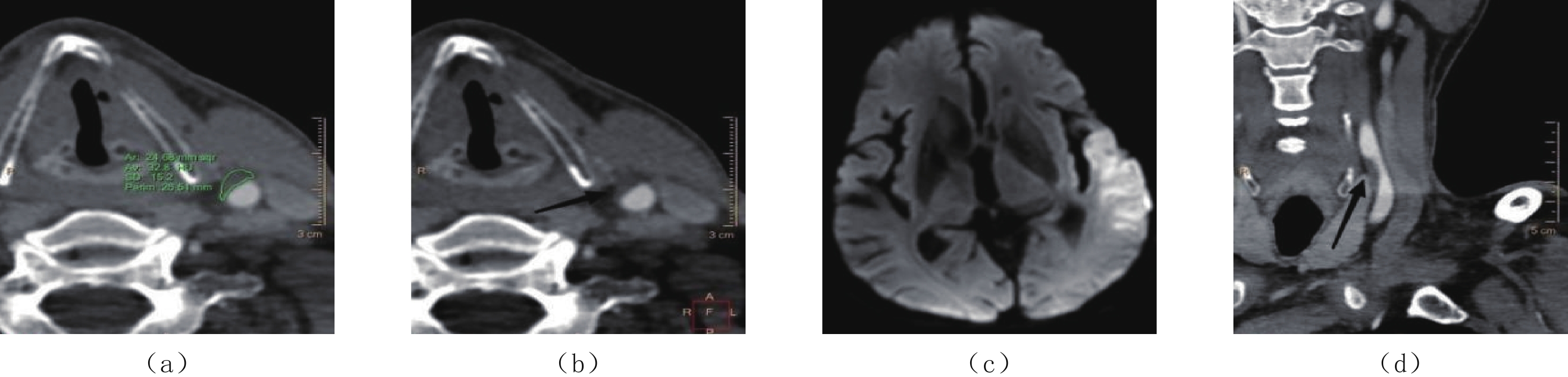
图 1 同一患者:(a)和(b)显示右侧颈总动脉脂质、溃疡斑块,(c)显示右侧基底节区及颞顶叶急性大面积脑梗塞,(d)显示右侧颈总动脉脂质斑块所致管腔中度狭窄(黑箭头所示)
Figure 1. In the same patient: (a) and (b) show lipid and ulcerative plaques in the right common carotid artery, respectively; (c) shows acute massive cerebral infarction of the right basal ganglia and temporo-parietal lobe; and (d) shows moderate stenosis of the right common carotid artery due to lipid plaques (black arrow)
![]()
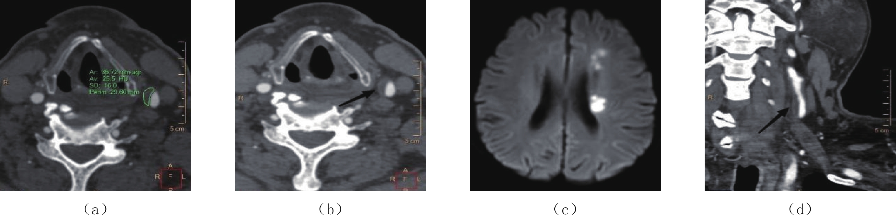
图 2 同一患者:(a)和(b)显示左侧颈总动脉脂质、不规则斑块,(c)显示左侧颞顶叶急性脑梗塞,(d)显示左侧颈总动脉脂质斑块所致管腔中度狭窄(黑箭头所示)
Figure 2. In the same patient: (a) and (b) show lipid and irregular plaques in the left common carotid artery, respectively; (c) shows acute cerebral infarction of the left temporo-parietal lobe; and (d) shows moderate stenosis of the left common carotid artery lumen due to lipid plaques (black arrow)
![]()
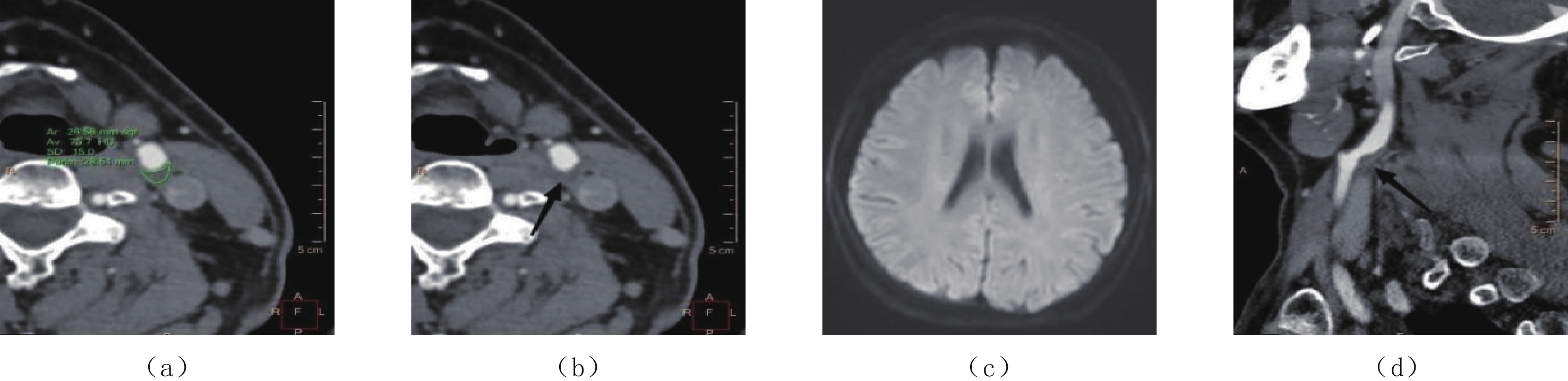
图 3 同一患者。(a)和(b)显示左侧颈总动脉脂质、光滑斑块,(c)显示左侧侧脑室旁小片状急性脑梗塞,(d)显示左侧颈总动脉脂质斑块所致管腔轻度狭窄(黑箭头所示)
Figure 3. In the same patient: (a) and (b) show lipid and smooth plaques in the left common carotid artery, respectively; (c) shows small patchy acute cerebral infarction near the left lateral ventricle; and (d) shows mild stenosis of the left common carotid artery lumen due to lipid plaques (black arrow)
![]()
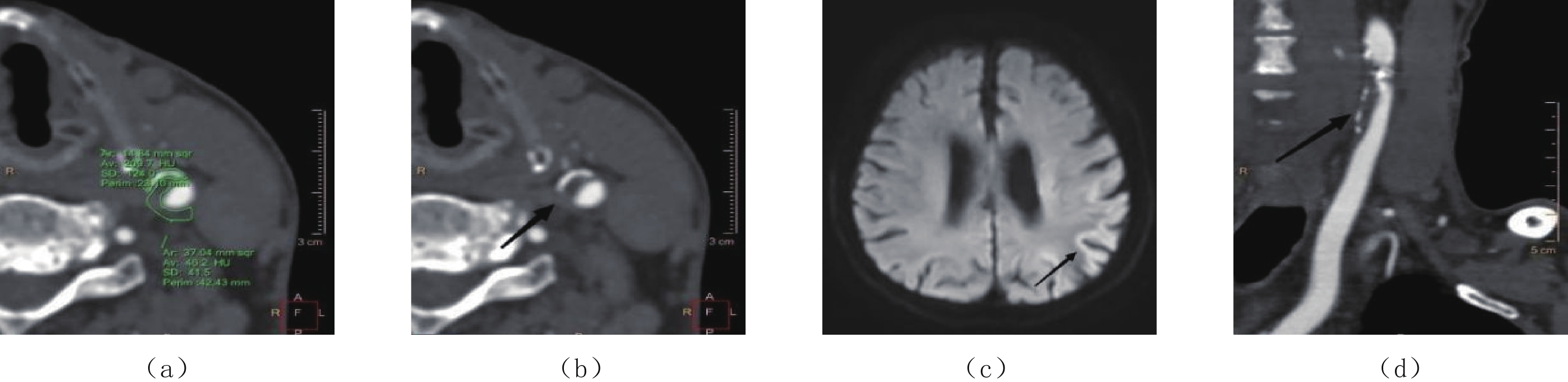
图 4 为同一患者:(a)和(b)显示左侧颈动脉分叉处纤维斑块,(c)显示该患者DWI未见明显急性脑梗塞,(d)显示左侧颈动脉分叉处纤维斑块所致管腔中度狭窄(黑箭头所示)
Figure 4. In the same patient: (a) and (b) show fibrous plaques at the left carotid bifurcation, (c) shows no significant acute cerebral infarction on DWI, and (d) shows moderate stenosis of the lumen at the left carotid bifurcation due to fibrous plaques (black arrow)
![]()
图 5 同一患者:(a)和(b)显示为左侧颈总动脉混合斑块、脂质成分为主,(c)显示左侧顶叶皮层下急性脑梗塞(黑箭头所示),(d)显示左侧颈总动脉混合斑块所致管腔中度狭窄(黑箭头所示)
Figure 5. In the same patient: (a) and (b) show mixed plaques in the left common carotid artery with a predominance of lipid components, (c) shows left parietal subcortical acute cerebral infarction (black arrow), and (d) shows moderate stenosis of the left common carotid artery lumen due to mixed plaques (black arrow)
表 1 病例组与对照组一般临床资料和实验室指标比较
Table 1 Comparison of general clinical data and laboratory indicators between the case and control groups
项目 参数 组别 统计检验 病例组(n=95)
例(%)或$\bar x \pm s$对照组(n=102)
例(%)或$\bar x \pm s$t/χ2 P 性别 男 71(81.4) 83(74.7) 1.269 0.260 女 24(18.6) 19(25.3) 年龄/岁 <50 5(5.3) 25(24.5) 32.672 <0.001 50~64 25(26.3) 25(24.5) ≥65 65(68.4) 52(51.0) 高血压* 是 66(69.5) 35(34.3) 24.339 <0.001 否 29(30.5) 67(65.7) 糖尿病 是 26(27.4) 17(16.7) 3.301 0.069 否 69(72.6) 85(83.3) 吸烟 是 14(14.7) 9(8.8) 1.668 0.197 否 81(85.3) 93(91.2) 饮酒 是 13(13.7) 7(6.9) 2.509 0.133 否 82(86.3) 95(93.1) TC/(mmol/L) 6.417±3.681 5.050±2.447 3.047 0.002 TG/(mmol/L) 2.235±1.849 1.742±1.504 2.043 0.042 HDL/(mmol/L) 1.241±0.344 1.086±0.302 3.337 0.001 LDL/(mmol/L) 2.609±0.837 2.385±0.918 1.789 0.075 HCY/(umol/L) 20.502±11.495 16.614±9.592 2.567 0.011 CysC/(mg/L) 1.143±0.251 1.073±0.245 1.978 0.049 HbA1c/% 正常 47(49.5) 81(79.4) 19.372 <0.001 不正常 48(50.5) 21(20.6)  下载: 导出CSV
表 2 病例组与对照组颈动脉斑块检出情况和颈动脉管腔狭窄程度比较
Table 2 Comparison of the features of carotid plaques and carotid artery stenosis
项目 参数 组别 统计检验 病例组(n=95)
例(%)或$\bar x \pm s$对照组(n=102)
例(%)或$\bar x \pm s$t/χ2 P 斑块类型* 无斑块 14a(14.7) 16a(15.7) 28.109 <0.001 钙化斑块 17a(17.9) 37a(36.3) 脂质斑块 46a(48.4) 15b(14.7) 纤维斑块 13a(13.7) 23a(22.5) 混合斑块 5a(5.3) 11a(10.8) 斑块表面形态* 无斑块 14a(14.7) 16a(15.7) 9.136 0.028 光滑斑块 34a(35.8) 56b(54.9) 不规则斑块 30a(31.6) 18b(17.6) 溃疡斑块 17a(17.9) 12a(11.8) 斑块数量/个* 4.37±3.252 3.00±2.325 3.415 0.001 管腔狭窄程度 无狭窄 15(15.8) 16(15.7) 1.153 0.679 轻度狭窄 47(49.5) 44(43.1) 中度狭窄 30(31.6) 40(39.2) 重度狭窄 3(3.2) 2(2.0) 注:* 表示P<0.05;a和b表示该因素在两组间经两两比较后存在统计学差异,字母相同则不存在统计学差异。
下载: 导出CSV
表 3 急性缺血性脑卒中发生的多因素相关Logistic回归分析
Table 3 Multivariate logistic regression analysis of the risk factors for acute ischemic stroke
因素 β $S_{\bar {\rm{x}}} $ χ2 P OR(95%CI) 年龄/岁 <50 1.000 50~65 1.294 0.946 1.871 0.171 3.647(0.571~23.281) ≥65* 1.892 0.799 5.600 0.018 6.632(1.384~31.777) 高血压* 否 1.000 是 2.341 0.507 21.309 <0.001 10.395(3.847~28.090) TC* 0.169 0.076 4.920 0.027 1.184(1.020~1.375) HDL* 1.803 0.797 5.144 0.024 6.067(1.272~28.940) HCY* 0.062 0.023 7.125 0.008 1.064(1.017~1.114) 脂质斑块* 否 1.000 是 1.328 0.494 7.217 0.007 3.773(1.432~9.938) 注:* 为P<0.05,差异有统计学意义。
下载: 导出CSV
-
[1] 彭斌, 吴波. 中国急性缺血性脑卒中诊治指南2018[J]. 中华神经科杂志, 2018,51(9): 666−682. doi: 10.3760/cma.j.issn.1006-7876.2018.09.004 PENG B, WU B. Chincese guidlines for dingnosis and treatment of acute ischemic stroke 2018[J]. Chinese Journal of Neurology, 2018, 51(9): 666−682. (in Chinese). doi: 10.3760/cma.j.issn.1006-7876.2018.09.004
[2] de WEERT T T, OUHLOUS M, MEIJERING E, et al. In vivo characterization and quantification of atherosclerotic carotid plaque components with multidetector computed tomography and histopathological correlation[J]. Arteriosclerosis, Thrombosis, and Vascular Biology, 2006, 26(10): 2366−2372. doi: 10.1161/01.ATV.0000240518.90124.57
[3] SABA L, ANZIDEI M, MARINCOLA B C, et al. Imaging of the carotid artery vulnerable plaque[J]. Cardiovasc Intervent Radiol, 2014, 37(3): 572−585. doi: 10.1007/s00270-013-0711-2
[4] RAFAILIDIS V, CHRYSSOGONIDIS I, TEGOS T, et al. Imaging of the ulcerated carotid atherosclerotic plaque: A review of the literature[J]. Insights Imaging, 2017, 8(2): 213−225. doi: 10.1007/s13244-017-0543-8
[5] WANG W, JIANG B, SUN H, et al. Prevalence, incidence, and mortality of stroke in China: Results from a nationwide population-based survey of 480687 adults[J]. Circulation, 2017, 135(8): 759−771. doi: 10.1161/CIRCULATIONAHA.116.025250
[6] CUI B, YANG D, ZHENG W, et al. Plaque enhancement in multi-cerebrovascular beds associates with acute cerebral infarction[J]. Acta Radiologica, 2021, 62(1): 102−112. doi: 10.1177/0284185120915604
[7] HYAFIL F, SCHINDLER A, SEPP D, et al. High-risk plaque features can be detected in non-stenotic carotid plaques of patients with ischaemic stroke classified as cryptogenic using combined 18F-FDG PET/MR imaging[J]. European Journal of Nuclear Medicine and Molecular Imaging, 2016, 43(2): 270−279. doi: 10.1007/s00259-015-3201-8
[8] DAI Y, QIAN Y, ZHANG M, et al. Associations between local haemodynamics and carotid intraplaque haemorrhage with different stenosis severities: A preliminary study based on MRI and CFD[J]. Journal of Clinical Neuroscience, 2019, 66: 220−225. doi: 10.1016/j.jocn.2019.05.041
[9] SAXENA A, NG E, LIM S T. Imaging modalities to diagnose carotid artery stenosis: Progress and prospect[J]. Biomedical Engineering Online, 2019, 18(1): 66. DOI: 10.1186/s12938-019-0685-7.
[10] ZHU G, LI Y, DING V, et al. Semiautomated characterization of carotid artery plaque features from computed tomography angiography to predict atherosclerotic cardiovascular disease risk score[J]. Journal of Computer Assisted Tomography, 2019, 43(3): 452−459. doi: 10.1097/RCT.0000000000000862
[11] LEE J, KIL J, KIM D W, et al. Usefulness of plaque magnetic resonance imaging in identifying high-risk carotid plaques irrespective of the degree of stenosis[J]. Journal of Cerebrovascular and Endovascular Neurosurgery, 2017, 19(4): 291−300. doi: 10.7461/jcen.2017.19.4.291
[12] COLLURA S, MORSIANI C, VACIRCA A, et al. The carotid plaque as paradigmatic case of site-specific acceleration of aging process: The microRNAs and the inflammaging contribution[J]. Ageing Research Reviews, 2020, 61: 101090. doi: 10.1016/j.arr.2020.101090
[13] PAN J, LIU J, WANG H, et al. Association of carotid atherosclerosis with lipid components in asymptomatic low-income Chinese: A population-based cross-sectional study[J]. Frontiers in Neurology, 2020, 11: 276. DOI: 10.3389/fneur.2020.00276.
[14] GARDENER H, DELLA M D, ELKIND M S, et al. Lipids and carotid plaque in the Northern Manhattan study (NOMAS)[J]. BMC Cardiovascular Disorders, 2009, 9: 55. DOI: 10.1186/1471-2261-9-55.
[15] 何爽, 江海强. 进展期脑卒中病人颈动脉斑块性质与生化指标的相关性分析[J]. 蚌埠医学院学报, 2017,42(12): 1630−1633. doi: 10.13898/j.cnki.issn.1000-2200.2017.12.018 HE S, JIANG H Q. The correlation analysis between carotid plaque nature and biochemical indicator in progressive stroke[J]. Journal Bengbu Medical College, 2017, 42(12): 1630−1633. (in Chinese). doi: 10.13898/j.cnki.issn.1000-2200.2017.12.018
[16] 刘杰, 吴东峰, 廖钦晨, 等. 颈动脉粥样硬化斑块性质与血清总同型半胱氨酸、血脂水平的相关性研究[J]. 内科, 2018,13(4): 544−546. doi: 10.16121/j.cnki.cn45-1347/r.2018.04.02 LIU J, WU D F, LIAO Q C, et al. Correlation between carotid atherosclerotic vulnerable plaque and serum total homocysteine and blood lipid levels[J]. Internal Medicine, 2018, 13(4): 544−546. (in Chinese). doi: 10.16121/j.cnki.cn45-1347/r.2018.04.02
-
期刊类型引用(0)
其他类型引用(2)


计量
- 文章访问数: 304
- HTML全文浏览量: 94
- PDF下载量: 25
- 被引次数: 2


