
CT Spectral Imaging Study in Patients with Carotid Body Tumors at High Altitudes
-
摘要: 目的:探讨CT能谱成像对颈动脉体瘤(CBT)的研究价值。方法:收集30例接受CT能谱检查并经手术确诊的CBT患者,通过GSI Viewer重建60 keV和40 keV单能量图像,对40 keV、60 keV单能量图像及120 kVp like三组图像的CBT供血动脉CT值、背景噪声(SD)、对比噪声比(CNR)、信噪比(SNR)及主观评分进行统计分析,分析CBT能谱参数及影像特征与手术结果的关系。结果:CBT动脉期及静脉期能谱参数与手术结果均无相关性;CBT横径、纵径、Shamblin分型与术中出血量呈强相关,供血动脉数量与术中出血量呈中度相关;横径、纵径、Shamblin分型与颅神经损伤呈中度相关,供血动脉数量与颅神经损伤呈强相关;40 keV是CBT供血动脉显示的最佳能级,CT值、SD、CNR及SNR均显著高于60 keV组及120 kVp like组,60 keV组CT值、噪声显著高于120 kVp like组,二者CNR及SNR差异无统计学意义;40 keV组主观评价分值最高,两位放射科医师主观评分一致性良好。结论:①CBT动脉期及静脉期能谱参数与GAPP评分无相关性;②CBT供血动脉数量是评估手术并发症重要参数之一,40 keV单能量图像可明显优化CBT供血动脉显示。Abstract: Objective: To investigate the value of CT spectral imaging in the study of carotid body tumors (CBT). Methods: Thirty patients with CBT who underwent CT energy spectrum examination and were confirmed by operation were included. Subsequently, 60 keV and 40 keV single energy images were reconstructed through the GSI viewer. The CT value, background noise (SD), contrast noise ratio (CNR), signal-to-noise ratio (SNR), and subjective score of CBT feeding arteries of the 40 keV, 60 keV single energy images and 120 kVp like images were statistically analyzed to evaluate the relationship between CBT energy spectrum parameters and imaging features with surgical results. Results: There was no correlation between energy spectrum parameters in the arterial and venous phases of CBT and the surgical results. However, CBT transverse diameter, longitudinal diameter, and Shamblin classification were strongly correlated with intraoperative bleeding, and the number of feeding arteries was moderately correlated with intraoperative bleeding. Additionally, the transverse diameter, longitudinal diameter, and Shamblin classification were moderately correlated with cranial nerve injury, and the number of feeding arteries was strongly correlated with cranial nerve injury. We also found that 40 keV was the best energy level for the CBT feeding artery display. Furthermore, the CT value, SD, CNR, and SNR of the 60 keV group were significantly higher than those of the 60 keV and 120 kVp like groups, and the CT value and noise of the 60 keV group were significantly higher than those of the 120 kVp like group. However, there was no significant difference in CNR and SNR between the two groups. The subjective evaluation score of the 40 keV group was the highest, and the subjective evaluation of the two radiologists had good consistency. Conclusion: (1) There was no correlation between the energy spectrum parameters in the arterial and venous phases of CBT and GAPP score. (2) The number of CBT feeding arteries is one of the important parameters for evaluating surgical complications and 40 keV single energy imaging can significantly optimize the display of CBT feeding arteries.
-
Keywords:
- CT spectral imaging /
- carotid body tumor /
- image quality /
- differentiation degree
-
颈动脉体瘤(carotid body tumor,CBT)是罕见的头颈部副神经节瘤,以中老年女性多见,高海拔地区发病率相对较高[1]。2017版WHO神经内分泌肿瘤分类标准中,CBT被定义为转移性肿瘤,转移率约为15%,转移的患者5年生存率低于50%[2-3]。根据肾上腺嗜铬细胞瘤和副神经节瘤病理评分系统(the grading system for adrenal pheochromocytoma and paraganglioma,GAPP),CBT分为高中低3种分化程度,分化程度越低转移风险越高[4],传统影像学检查的范围有限,尚未能评估CBT的分化程度。
早期外科手术切除是CBT首选治疗方法,但CBT位置特殊,手术易导致大量出血及颅神经损伤[5]。目前临床主要依据Shamblin分型来指导手术治疗,但仍有不足之处,有研究发现供血动脉数量较肿瘤大小、Shamblin分型等可以更好的预测CBT手术出血量,术前栓塞供血动脉可明显减少直径大于3 cm CBT的术中失血量及手术时间[6-8],但供血动脉多为细小血管,常规CTA难以清晰显示,评估较为困难。近年来随着CT能谱多参数成像技术的不断发展,极大地拓宽了CT的应用范围,为降低CBT术后并发症的发生率提供可能[9]。
本文旨在探讨CT能谱定量参数对CBT分化程度评估的价值及单能量图像对CBT供血动脉图像质量的优化价值,为临床治疗方案的制定提供参考。
1. 材料与方法
1.1 临床资料
回顾性收集2020年10月至2021年12月在青海大学附属医院医学影像中心行头颈部CT能谱扫描的患者。纳入标准:①患者均经手术确诊 CBT并签署知情同意书;②患者术前均行头颈部 CT能谱成像检查。排除标准:①有其他颈部肿瘤病史或颈部手术史者;②治疗前后临床及影像资料不完整者;③图像伪影严重。
1.2 仪器与方法
采用256排螺旋CT扫描仪(Revolution CT,GE Healthcare,Milwaukee,USA)进行头颈部CT能谱扫描。患者均取仰卧位,头先进,扫描时保持头颈部静止,避免吞咽动作。采用GSI扫描模式,扫描范围为主动脉弓至颅顶。扫描参数:管电压80 kVp/140 kVp瞬时切换,管电流320 mA,50% ASIR-V,螺距0.984︰1,转速0.5 s/rot,准直64×0.625 mm,DFOV 25 cm×25 cm,重建层厚0.625 mm。对比剂注射方案:使用高压注射器经右侧肘正中静脉依次注射18 mL生理盐水、1.5 mL/kg的优维显(370 mgI/mL)和30 mL生理盐水,生理盐水和优维显的注射速率分别为5 mL/s和4.5 mL/s。
采用对比剂智能追踪技术(Smart prep),监测点设置为主肺动脉窗层面的降主动脉,触发阈值80 HU,延迟3 s启动动脉期扫描,动脉期扫描结束后5 s行静脉期扫描。将带有datafile的120 kVp like图像传输至AW 4.7工作站进行后处理和分析。
1.3 图像分析
1.3.1 图像质量客观评价
通过GSI Viewer重建60 keV和40 keV单能量图像,并利用容积再现(volume rendering,VR)及最大密度投影(maximal intensity projection,MIP)后处理技术重组图像(图1)。在一级供血动脉开口端放置感兴趣区(region of interest,ROI),直径为1 mm,记录CT值。测量并记录同侧同一层面头夹肌CT值和皮下脂肪的标准差(standard deviation,SD),ROI直径为4~6 mm,以皮下脂肪的SD值作为图像背景噪声。保证同一患者在120 kVp like图像及60 keV、40 keV单能量图像的ROI大小和位置一致。分别计算供血动脉的信噪比(signal to noise ratio,SNR)和对比噪声比(contrast to noise ratio,CNR),公式如下:
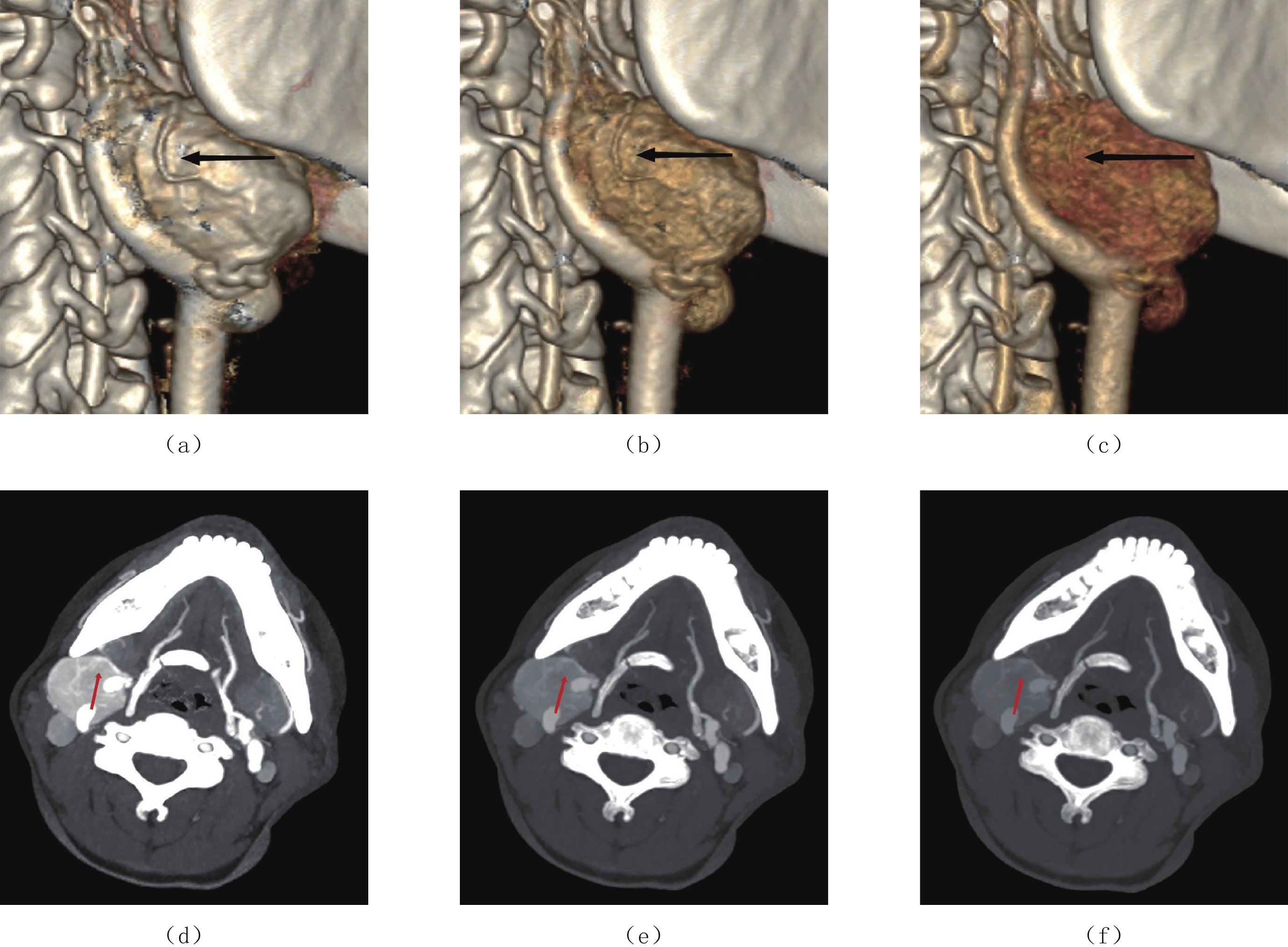
![]() 图 1 女,53岁,右侧颈动脉体瘤(a)~(c)分别为CBT 40 keV、60 keV及120 kVp like的VR图像,黑色箭头所指为供血动脉;(d)~(f)分别为CBT 40 keV、60 keV及120 kVp like的MIP图像,红色箭头所指为供血动脉。Figure 1. A right carotid body tumor of a 53-year-old female
图 1 女,53岁,右侧颈动脉体瘤(a)~(c)分别为CBT 40 keV、60 keV及120 kVp like的VR图像,黑色箭头所指为供血动脉;(d)~(f)分别为CBT 40 keV、60 keV及120 kVp like的MIP图像,红色箭头所指为供血动脉。Figure 1. A right carotid body tumor of a 53-year-old female$$ {\rm{SNR}}={\rm{CT}}_{供血动脉}/{\rm{SD}}_{脂肪}\text{,}{\rm{CNR}}= ({\rm{CT}}_{供血动脉}-{\rm{CT}}_{头夹肌}) /{\rm{SD}}_{脂肪}。 $$ 1.3.2 图像质量主观评价
两名高年资放射科医师采用盲法在相同窗宽、窗位下(窗宽1400 HU,窗位300 HU)评估3组图像的轴位图像、VR及MIP图像。
评估采用5分法。5分:供血动脉近端及远端分支显影清晰锐利;4分:供血动脉近端显影较清晰,远端分支显影完整;3分:供血动脉近端显影尚清晰,远端分支显影较淡;2分:供血动脉近端显影模糊,远端分支未显影;1分:供血动脉近端未显影,血管结构无法分辨。
1.3.3 CBT能谱参数测量及影像特征分析
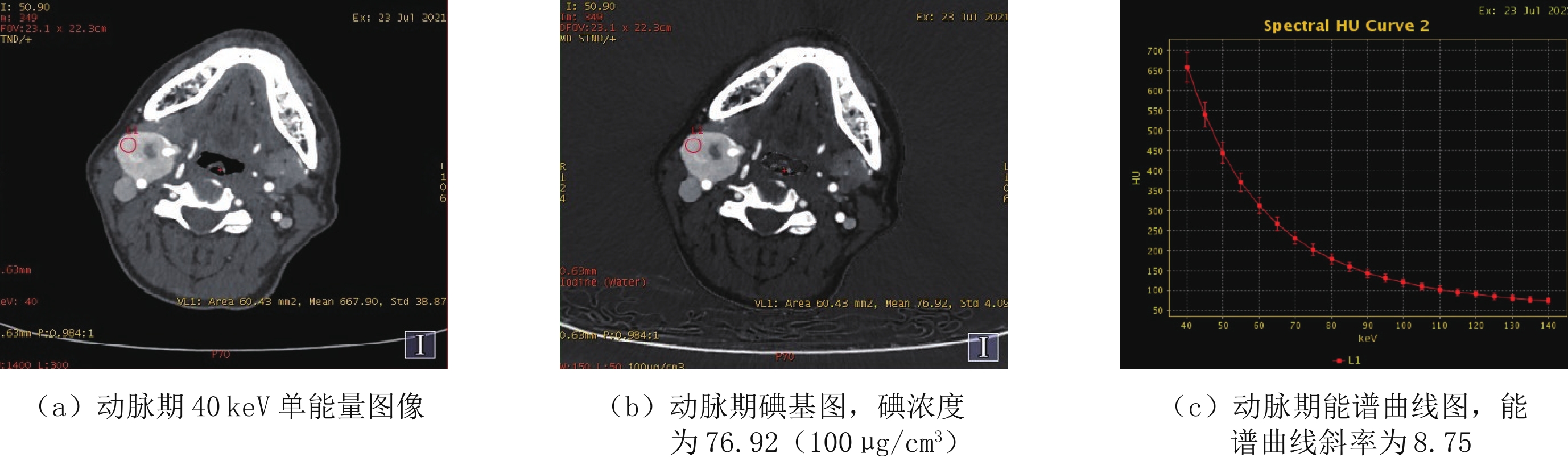
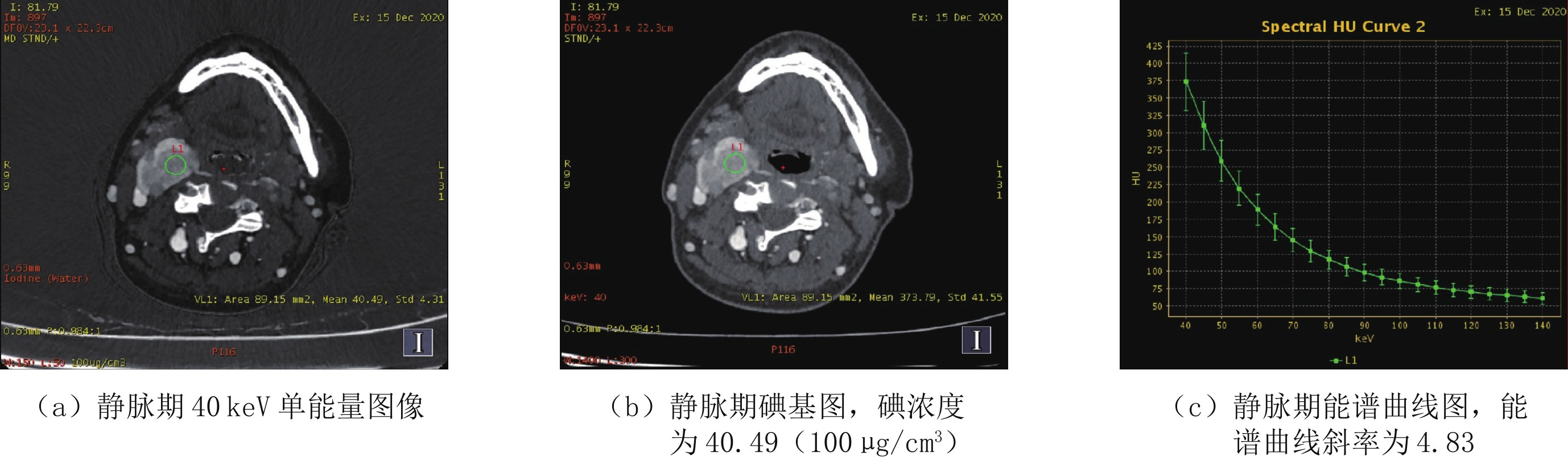
通过GSI Viewer在动脉期及静脉期CBT最大层面实质部分放置ROI,尽量避开液化、坏死、钙化及小血管,直径为4~8 mm。记录碘浓度、有效原子序数、40 keV及100 keV单能量图像的CT值,计算能谱曲线斜率=(CT40 keV - CT100 keV)/60。同一患者动脉期及静脉期ROI的大小和位置保持一致,测量值取ROI和上下两个连续层面的测量平均值。
CBT横径、纵径分别为瘤体横断面、纵切面径线的最大值,供血动脉数量在40 keV单能量图像下进行计数。对CBT进行Shamblin分型:Shamblin Ⅰ型为瘤体横径小于4 cm,与颈动脉及周围组织无粘黏;Shamblin Ⅱ 型为瘤体横径大于4 cm,与颈动脉及周围组织部分粘黏;Shamblin Ⅲ 型为瘤体横径大于4 cm,与颈动脉及周围组织紧密粘黏[10]。
1.3.4 手术结果分析
颅神经损伤定义:伸舌时舌尖偏向患侧为舌下神经损伤(Ⅻ),声音嘶哑、饮水呛咳为迷走神经损伤(X),吞咽困难为舌咽神经损伤(Ⅳ),口角歪斜为面神经损伤(Ⅶ)。
1.4 统计学方法
采用SPSS 23.0统计学软件行数据分析。符合正态分布的计量资料用均数±标准差(
$\bar x \pm s $ )表示;CBT的影像特征及能谱参数与手术结果分析采用Spearman相关性分析及多因素线性回归分析:|r|<0.4为弱相关或无相关;0.4≤|r|<0.6为中度相关;0.6≤|r|<0.8为强相关;0.8≤|r|<1为极强相关;供血动脉数量与影像特征及手术结果比较、各组图像的客观测量结果比较均使用单因素方差分析,两两比较采用最小显著性差异法。各组图像的主观评分比较采用非参数Kruskal-Wallis检验,两位医师主观评分及手动测量值的一致性均通过加权Kappa检验分析,Kappa值>0.75,一致性良好。P<0.05为差异有统计学意义。
2. 结果
2.1 患者一般资料
本研究共收集33例患者,其中2例因瘤体较大行保守治疗,1例最终病理诊断为神经鞘瘤,最终入组30例。其中男性3例,女性27例,平均年龄约(55.8±10.3)岁,居住地海拔为2200~5536 m,平均海拔3150 m。30例中有5例双侧CBT均行CBT切除术。患者术后出现颅神经损伤症状18例,共29支颅神经受损,其中舌下神经损伤15例,迷走神经损伤11例,舌咽神经损伤2例,面神经损伤1例。手动测量数据均一致性良好,Kappa值>0.75(表1)。
表 1 30例患者的一般资料($\bar x \pm s $ )Table 1. The general information of 30 patients$(\bar x \pm s )$ 变量 参数 数值(%) 变量 参数 数值(%) 年龄/岁 55.8±10.3 供血动脉数量/例 1支 15(50.0) 性别/例 男性 3(10.0) 2支 10(33.3) 女性 27(90.0) 3支及以上 5(16.7) 位置/例 左侧 13(43.3) Shamblin分型/例 Ⅰ型 13(43.3) 右侧 12(40.0) Ⅱ型 7(23.3) 双侧 5(16.7) Ⅲ型 10(33.3) 影像学特征 横径/cm 3.07±1.34 并发症 术中出血量/mL 157.3±237.5 纵径/cm 3.37±1.38 颅神经损伤 18(60.0) 2.2 CBT能谱参数及影像特征与手术结果的相关性分析
CBT动脉期碘浓度、能谱曲线斜率及有效原子序数平均值分别为(77.72±20.92)100 μg/cm3、9.02±2.07、10.99±0.60;静脉期碘浓度、能谱曲线斜率及有效原子序数平均值分别为(47.81±19.89)100μg/cm3、5.64±2.27、9.97±0.77。动脉期及静脉期碘浓度、能谱曲线斜率及有效原子序数与术中出血量、颅神经损伤及GAPP评分均无相关性(图2和图3)。
CBT横径、纵径、Shamblin分型与术中出血量呈强相关(r1横径=0.788,r1纵径=0.669,r1 Shamblin分型=0.672),供血动脉数量与术中出血量呈中度相关(r1供血动脉数量=0.583)。横径、纵径、Shamblin分型与颅神经损伤呈中度相关(r2横径=0.476、r2纵径=0.550、r2 Shamblin分型=0.529),供血动脉数量与颅神经损伤呈强相关(r2供血动脉数量=0.629)。横径、纵径、Shamblin分型及供血动脉数量与GAPP评分均无相关性(表2)。
表 2 CBT能谱参数及影像特征与手术结果的相关性分析Table 2. Correlation analysis between CBT energy spectrum parameters, image characteristics, and surgical results变量 参数 术中出血量 颅神经损伤 GAPP评分 r1 P1 r2 P2 r3 P3 动脉期能谱参数 碘浓度 0.104 0.791 0.414 0.268 -0.393 0.296 能谱曲线斜率 0.104 0.790 0.416 0.266 -0.341 0.369 有效原子序数 0.052 0.894 0.416 0.266 -0.366 0.332 静脉期能谱参数 碘浓度 -0.207 0.593 0.268 0.456 0.243 0.529 能谱曲线斜率 -0.311 0.416 0.151 0.699 0.076 0.847 有效原子序数 -0.156 0.689 0.192 0.620 0.117 0.765 影像特征 横径 0.788 0.000 0.476 0.008 0.296 0.112 纵径 0.669 0.000 0.550 0.002 0.287 0.124 供血动脉数量 0.583 0.001 0.629 0.000 0.209 0.268 Shamblin分型 0.672 0.000 0.529 0.003 0.276 0.140 注:r1、P1:术中出血量相关性分析结果;r2、P2:颅神经损伤相关性分析结果;r3、P3:GAPP评分相关性分析结果。 2.3 CBT手术并发症多因素线性回归分析
分别以术中出血量及颅神经损伤为因变量,以CBT横径、纵径、供血动脉数量、Shamblin分型为自变量,行多因素线性回归分析,结果显示横径是术中出血量独立影响因素,供血动脉数量是颅神经损伤独立影响因素(表3和表4)。
表 3 CBT术中出血量多因素线性回归分析Table 3. Multivariate linear regression analysis of the intraoperative bleeding volume in CBT变量 回归参数 统计校验 偏回归系数 标准误 标准回归系数 t P 常数 -184.039 97.241 -1.893 0.070 横径 96.770 39.797 0.538 2.432 0.023 纵径 -91.715 47.085 -0.521 -1.948 0.063 供血动脉数量 82.816 55.992 0.264 1.479 0.152 Shamblin分型 111.139 67.763 0.421 1.640 0.114 表 4 CBT颅神经损伤多因素线性回归分析Table 4. Multivariate linear regression analysis of cranial nerve injury in CBT变量 回归参数 统计校验 偏回归系数 标准误 标准回归系数 t P 常数 -0.889 0.392 -2.268 0.032 横径 -0.108 0.161 -0.143 -.676 0.505 纵径 0.177 0.190 0.239 0.931 0.361 供血动脉数量 0.709 0.226 0.538 3.140 0.004 Shamblin分型 0.229 0.273 0.206 0.838 0.410 2.4 CBT供血动脉数量与影像特征及手术结果比较
随着CBT供血动脉数量的增加,横径、纵径、Shamblin分型、术中出血量及颅神经损伤均随之增加,差异有统计学意义。供血动脉数量与GAPP评分差异无统计学意义(表5)。
表 5 供血动脉数量与影像特征及手术结果分析($\bar x \pm s $ )Table 5. Analysis of the number of feeding arteries, imaging characteristics and surgical results$(\bar x \pm s) $ 变量 1支/% 2支/% 3支及以上/% P Shamblin分型 横径/cm 2.27±1.00 3.66±1.19a 4.20±1.05b 0.001 纵径/cm 2.61±1.40 3.81±0.81a 4.16±1.23b 0.019 Ⅰ型 10(33.33) 3(10.00) 0 0.005 Ⅱ型 3(10.00) 3(10.00) 1(3.33) Ⅲ 型 2(6.67) 4(13.33) 4(13.33) 术中出血量/mL 56.00±35.01 178.00±121.36a 420.00±495.72b 0.001 颅神经损伤/支 6(20.69) 12(41.38)a 11(37.93)b 0.000 GAPP评分 4.27±1.10 5.10±0.57a 4.20±0.84 0.073 注:供血动脉数量1支与2支相比,a:P<0.05;1支与3支及以上相比,b:P<0.05。 2.5 客观评价结果
40 keV、60 keV及120 kVp like三组图像的噪声和供血动脉的CT值、CNR及SNR差异均有统计学意义。两两结果比较,40 keV组CT值、噪声、CNR及SNR均显著高于60 keV组及120 kVp like;60 keV组CT值、噪声显著高于120 kVp like组,二者CNR及SNR差异无统计学意义(表6)。
表 6 各组图像CT值、噪声、SNR及CNR比较($\bar x \pm s $ )Table 6. Comparison of the CT values, noise, SNR and CNR of each group$(\bar x \pm s )$ 项目 组别 统计检验 40 keV组 60 keV组 120 kVp like组 F P CT供血动脉/HU 679.17±216.65 337.28±96.82c 228.54±60.82d 129.64 0.000 SD脂肪/HU 23.11±4.83 13.10±2.73c 9.78±2.17d 143.091 0.000 CNR 27.71±12.02 22.28±8.65c 18.84±7.82d 9.768 0.001 SNR 30.62±12.37 26.64±9.13c 24.39±8.60d 4.858 0.010 注:40 keV组与60 keV组比较,c:P<0.05;40 keV组与120 kVp like组比较,d:P<0.05。 2.6 主观评价结果
主观评价分值由高到低依次为40 keV组、60 keV组及120 kVp like组,差异有统计学意义(表7)。两位放射科医师主观评价一致性良好,Kappa值分别为0.863、0.831、0.793。
表 7 颈动脉体瘤供血动脉主观评分($\bar x \pm s $ )Table 7. Subjective scores of the feeding artery of the carotid body tumor$(\bar x \pm s) $ 评分者 组别 统计检验 40 keV组 60 keV组 120 kVp like组 H P 医师A 4.85±0.72 3.68±0.68 2.55±0.36 58.305 0.000 医师B 4.80±0.71 3.53±0.66 2.41±0.41 54.765 0.000 3. 讨论
颈动脉体对低氧刺激敏感,长期生活在高原低氧环境及慢性低氧血症等可导致颈动脉体增生肥大,增加CBT的发病率。本研究所纳入的患者均起病于高海拔地区,其中女性患者占比较高,可能与女性妊娠、月经等原因导致血红蛋白水平相对较低有关。
术中出血及颅神经损伤是CBT外科治疗的主要并发症,完善的术前检查对预测手术并发症、提高患者生存质量具有重要价值[11]。CBT横径、纵径、供血动脉数量及Shamblin分型与上述并发症均具有较高的相关性,其中横径是评价术中出血量的独立影响因素,供血动脉数量是评价颅神经损伤的独立影响因素,可在一定程度上评估手术风险。
本研究通过单能量成像技术有效提高了CBT供血动脉的显示,解决了常规CTA对细小血管显示不佳的问题[9]。此外能谱曲线斜率、有效原子序数及碘浓度等能谱参数在肿瘤良恶性成分鉴别及肿瘤转移的判定中具有较高的敏感性及准确性[12],但本研究中能谱参数与GAPP评分间并无相关性。
头颈部CTA是CBT术前诊断及远期随访的首选检查[13]。相较于常规CTA,CT能谱单能量成像技术、迭代重建算法等可在降低辐射剂量的同时进一步提高图像质量[14]。单能量成像技术是将扫描获得的高能和低能两组数据进行组合,生成不同水平的单能量图像,其中低能级图像能够提高图像组织对比度,有利于细微病灶的显示,但随着能级的降低图像噪声随之增高[9]。马光明等[15]通过对比头颈部CTA 60~80 keV单能级图像的图像质量发现60 keV是头颈部CTA的最佳能级;邓小林等[16]研究认为头颈部CTA最佳单能量为57~63 keV,此时图像质量明显优于混合能量图像。因此本研究选取60 keV图像作为对比,发现与上述研究结果存在差异,这可能与血管粗细、位置、造影剂浓度、扫描设备及参数等不同有关。此外本研究扫描采用50% ASIR-V取代常规滤波反投影法,可在降低25%~50% 辐射剂量的同时降低图像噪声,有效优化图像质量[17]。
GAPP评分是目前临床应用较为广泛的CBT病理评分体系,主要通过组织学模式、血管或包膜损伤、细胞丰富程度、粉刺型坏死、Ki-67指数等多种参数联合评估肿瘤的分化程度,与SDHB联合诊断可以更好的预测转移风险[4]。能谱多参数量化分析对肿瘤分化程度的判断具有一定实用价值[18-19],但本研究中CBT动、静脉期碘含量、能谱曲线斜率及有效原子序数与GAPP评分均无相关性。这可能与CBT的转移率低及样本量较小有关,因此对于CBT病理分级与能谱参数之间的关系仍需要大样本数据进一步分析探讨。
本文不足之处:①本研究为单中心回顾性研究;②收集的病例较少,数据存在一定偏移;③未能进一步分析 GAPP评分与CT能谱参数之间的关系以及对比剂用量、辐射剂量优化等问题。
综上所述,CBT供血动脉数量与手术并发症相关性较高,通过CT能谱检查发现40 keV单能量图像较混合能量图像可以更好的优化CBT供血动脉的显示,可能有助于临床预测CBT手术并发症,进一步完善手术方案。
-
![]()
图 1 女,53岁,右侧颈动脉体瘤
(a)~(c)分别为CBT 40 keV、60 keV及120 kVp like的VR图像,黑色箭头所指为供血动脉;(d)~(f)分别为CBT 40 keV、60 keV及120 kVp like的MIP图像,红色箭头所指为供血动脉。
Figure 1. A right carotid body tumor of a 53-year-old female
表 1 30例患者的一般资料(
$\bar x \pm s $ )Table 1 The general information of 30 patients
$(\bar x \pm s )$ 变量 参数 数值(%) 变量 参数 数值(%) 年龄/岁 55.8±10.3 供血动脉数量/例 1支 15(50.0) 性别/例 男性 3(10.0) 2支 10(33.3) 女性 27(90.0) 3支及以上 5(16.7) 位置/例 左侧 13(43.3) Shamblin分型/例 Ⅰ型 13(43.3) 右侧 12(40.0) Ⅱ型 7(23.3) 双侧 5(16.7) Ⅲ型 10(33.3) 影像学特征 横径/cm 3.07±1.34 并发症 术中出血量/mL 157.3±237.5 纵径/cm 3.37±1.38 颅神经损伤 18(60.0)  下载: 导出CSV
表 2 CBT能谱参数及影像特征与手术结果的相关性分析
Table 2 Correlation analysis between CBT energy spectrum parameters, image characteristics, and surgical results
变量 参数 术中出血量 颅神经损伤 GAPP评分 r1 P1 r2 P2 r3 P3 动脉期能谱参数 碘浓度 0.104 0.791 0.414 0.268 -0.393 0.296 能谱曲线斜率 0.104 0.790 0.416 0.266 -0.341 0.369 有效原子序数 0.052 0.894 0.416 0.266 -0.366 0.332 静脉期能谱参数 碘浓度 -0.207 0.593 0.268 0.456 0.243 0.529 能谱曲线斜率 -0.311 0.416 0.151 0.699 0.076 0.847 有效原子序数 -0.156 0.689 0.192 0.620 0.117 0.765 影像特征 横径 0.788 0.000 0.476 0.008 0.296 0.112 纵径 0.669 0.000 0.550 0.002 0.287 0.124 供血动脉数量 0.583 0.001 0.629 0.000 0.209 0.268 Shamblin分型 0.672 0.000 0.529 0.003 0.276 0.140 注:r1、P1:术中出血量相关性分析结果;r2、P2:颅神经损伤相关性分析结果;r3、P3:GAPP评分相关性分析结果。
下载: 导出CSV
表 3 CBT术中出血量多因素线性回归分析
Table 3 Multivariate linear regression analysis of the intraoperative bleeding volume in CBT
变量 回归参数 统计校验 偏回归系数 标准误 标准回归系数 t P 常数 -184.039 97.241 -1.893 0.070 横径 96.770 39.797 0.538 2.432 0.023 纵径 -91.715 47.085 -0.521 -1.948 0.063 供血动脉数量 82.816 55.992 0.264 1.479 0.152 Shamblin分型 111.139 67.763 0.421 1.640 0.114
下载: 导出CSV
表 4 CBT颅神经损伤多因素线性回归分析
Table 4 Multivariate linear regression analysis of cranial nerve injury in CBT
变量 回归参数 统计校验 偏回归系数 标准误 标准回归系数 t P 常数 -0.889 0.392 -2.268 0.032 横径 -0.108 0.161 -0.143 -.676 0.505 纵径 0.177 0.190 0.239 0.931 0.361 供血动脉数量 0.709 0.226 0.538 3.140 0.004 Shamblin分型 0.229 0.273 0.206 0.838 0.410
下载: 导出CSV
表 5 供血动脉数量与影像特征及手术结果分析(
$\bar x \pm s $ )Table 5 Analysis of the number of feeding arteries, imaging characteristics and surgical results
$(\bar x \pm s) $ 变量 1支/% 2支/% 3支及以上/% P Shamblin分型 横径/cm 2.27±1.00 3.66±1.19a 4.20±1.05b 0.001 纵径/cm 2.61±1.40 3.81±0.81a 4.16±1.23b 0.019 Ⅰ型 10(33.33) 3(10.00) 0 0.005 Ⅱ型 3(10.00) 3(10.00) 1(3.33) Ⅲ 型 2(6.67) 4(13.33) 4(13.33) 术中出血量/mL 56.00±35.01 178.00±121.36a 420.00±495.72b 0.001 颅神经损伤/支 6(20.69) 12(41.38)a 11(37.93)b 0.000 GAPP评分 4.27±1.10 5.10±0.57a 4.20±0.84 0.073 注:供血动脉数量1支与2支相比,a:P<0.05;1支与3支及以上相比,b:P<0.05。
下载: 导出CSV
表 6 各组图像CT值、噪声、SNR及CNR比较(
$\bar x \pm s $ )Table 6 Comparison of the CT values, noise, SNR and CNR of each group
$(\bar x \pm s )$ 项目 组别 统计检验 40 keV组 60 keV组 120 kVp like组 F P CT供血动脉/HU 679.17±216.65 337.28±96.82c 228.54±60.82d 129.64 0.000 SD脂肪/HU 23.11±4.83 13.10±2.73c 9.78±2.17d 143.091 0.000 CNR 27.71±12.02 22.28±8.65c 18.84±7.82d 9.768 0.001 SNR 30.62±12.37 26.64±9.13c 24.39±8.60d 4.858 0.010 注:40 keV组与60 keV组比较,c:P<0.05;40 keV组与120 kVp like组比较,d:P<0.05。
下载: 导出CSV
表 7 颈动脉体瘤供血动脉主观评分(
$\bar x \pm s $ )Table 7 Subjective scores of the feeding artery of the carotid body tumor
$(\bar x \pm s) $ 评分者 组别 统计检验 40 keV组 60 keV组 120 kVp like组 H P 医师A 4.85±0.72 3.68±0.68 2.55±0.36 58.305 0.000 医师B 4.80±0.71 3.53±0.66 2.41±0.41 54.765 0.000
下载: 导出CSV
-
[1] 朱吉海, 杨佳, 马伟, 等. 高海拔地区颈动脉体瘤围术期颈动脉处理策略−单中心经验[J]. 中国普通外科杂志, 2020,29(12): 1521−1527. doi: 10.7659/j.issn.1005-6947.2020.12.015 [2] 医学会内分泌学分会. 嗜铬细胞瘤和副神经节瘤诊断治疗专家共识(2020版)[J]. 中华内分泌代谢杂志, 2020,36(9): 737−750. doi: 10.3760/cma.j.cn311282-20200629-00482 Endocrinology Branch of the Medical Association. Expert consensus on the diagnosis and treatment of pheochromocytoma and paraganglioma (2020)[J]. Chinese Journal of Endocrinology and Metabolism, 2020, 36(9): 737−750. (in Chinese). doi: 10.3760/cma.j.cn311282-20200629-00482
[3] SCHWARZE V, MARSCHNER C, de FIGUEIREDO G, et al. Single-center study: Dynamic contrast-enhanced ultrasound in the diagnostic assessment of carotid body tumors[J]. Quantitative Imaging in Medicine and Surgery, 2020, 10(9): 1739−1747. doi: 10.21037/qims-19-920
[4] KIMURA N, TAKAYANAGI R, TAKIZAWA N, et al. Pathological grading for predicting metastasis in phaeochromocytoma and paraganglioma[J]. Endocrine-Related Cancer, 2014, 21(3): 405−414. doi: 10.1530/ERC-13-0494
[5] ROBERTSON V, POLI F, HOBSON B, et al. A systematic review and meta-analysis of the presentation and surgical management of patients with carotid body tumours[J]. European Journal of Vascular and Endovascular Surgery, 2019, 57(4): 477−486. doi: 10.1016/j.ejvs.2018.10.038
[6] 顾光超, 郑月宏. 颈动脉体瘤的PUMCH分型[J]. 协和医学杂志, 2021,12(6): 825−828. GU G C, ZHENG Y H. The PUMCH classification of carotid body tumor[J]. Medical Journal of Peking Union Medical College Hospital, 2021, 12(6): 825−828. (in Chinese).
[7] 李展展, 梁琰, 张永强, 等. 头颈部CT血管成像对颈动脉体瘤术中出血量评估的临床应用价值[J]. 中国医学科学院学报, 2020,42(4): 491−496. LI Z Z, LIANG Y, ZHANG Y Q, et al. Value of head and neck CT angiography in the clinical evaluation of intraoperative bleeding volume of carotid body tumours[J]. Acta Academiae Medicinae Sinicae, 2020, 42(4): 491−496. (in Chinese).
[8] HOANG V T, TRINH C T, LAI T A K, et al. Carotid body tumor: A case report and literature review[J]. Journal of Radiology Case Reports, 2019, 13(8): 19−30.
[9] 中华放射学杂志双层探测器光谱CT临床应用协作组. 双层探测器光谱CT临床应用中国专家共识(第一版)[J]. 中华放射学杂志, 2020,54(7): 635−643. doi: 10.3760/cma.j.cn112149-20200513-00679 Chinese Journal of Radiology Cooperative Group of Clinical Application of Dual-layer Spectral Detector CT. China expert consensus on clinical application of dual-layer spectral detector CT[J]. Chinese Journal of Radiology, 2020, 54(7): 635−643. (in Chinese). doi: 10.3760/cma.j.cn112149-20200513-00679
[10] LUNA-ORTIZ K, RASCON-ORTIZ M, VILLAVICENCIO-VALENCIA V, et al. Does Shamblin's classification predict postoperative morbidity in carotid body tumors? A proposal to modify Shamblin's classification[J]. European Archives of Oto-rhino-laryngologyl, 2006, 263(2): 171−175. doi: 10.1007/s00405-005-0968-4
[11] LI N, ZENG N, WAN Y, et al. The earlier, the better: The beneficial effect of different timepoints of the preoperative transarterial embolization on ameliorating operative blood loss and operative time for carotid body tumor[J]. Surgery, 2021, 170(5): 1581-1585.
[12] 东强, 曲丽洁, 张宏, 等. 双能量CT碘值及增强值与进展期胃腺癌病理分化程度的相关性[J]. 临床放射学杂志, 2020,39(5): 931−935. DONG Q, QU L J, ZHANG H, et al. Correlations between the iodine value and CT enhancement value of dual-energy CT and the pathological differentiation degree of advanced gastric adenocarcinoma[J]. Journal of Clinical Radiology, 2020, 39(5): 931−935. (in Chinese).
[13] 曹云太, 鲍海华, 张国晋, 等. 颈动脉体瘤术前影像学特征与手术并发症的相关性研究[J]. 临床放射学杂志, 2021,40(2): 220−224. CAO Y T, BAO H H, ZHANG G J, et al. Correlation between preoperative imaging features and operative complications of carotid body tumors[J]. Journal of Clinical Radiology, 2021, 40(2): 220−224. (in Chinese).
[14] 中华医学会放射学分会. 头颈部CT血管成像扫描方案与注射方案专家共识[J]. 中华放射学杂志, 2019,53(2): 81−87. doi: 10.3760/cma.j.issn.1005-1201.2019.02.001 Chinese Society of Radiology, Chinese Medical Association. Expert consensus of the head and neck CT angiography scanning and injection protocols[J]. Chinese Journal of Radiology, 2019, 53(2): 81−87. (in Chinese). doi: 10.3760/cma.j.issn.1005-1201.2019.02.001
[15] 马光明, 于勇, 陈静, 等. 能谱CT单能量技术优化头颈CTA图像质量研究[J]. 中国医疗设备, 2021,36(8): 86−89. doi: 10.3969/j.issn.1674-1633.2021.08.021 MA G M, YU Y, CHEN J, et al. Research on optimization of head and neck CTA image qualityby spectrum CT optimal monochromatic image technique[J]. China Medical Devices, 2021, 36(8): 86−89. (in Chinese). doi: 10.3969/j.issn.1674-1633.2021.08.021
[16] 邓小林, 谢惠, 屈亚林, 等. 能谱CT最佳单能量技术优化头颈部CTA图像质量的研究[J]. CT理论与应用研究, 2018,27(6): 719−726. DOI: 10.15953/j.1004-4140.2018.27.06.05. DENG X L, XIE H, QU Y L, et al. Spectral CT with optimal monochromatic energy to improve CTA image quality of head and neck[J]. Computerized Tomography Theory and Applications, 2018, 27(6): 719−726. DOI: 10.15953/j.1004-4140.2018.27.06.05. (in Chinese).
[17] MCCOLLOUGH C H, YU L, KOFLER J M, et al. Degradation of CT low-contrast spatial resolution due to the use of iterative reconstruction and reduced dose levels[J]. Radiology, 2015, 276(2): 499−506. doi: 10.1148/radiol.15142047
[18] 严福华, 金征宇. 开辟双能量CT临床应用的新时代[J]. 中华放射学杂志, 2020,54(6): 505−507. doi: 10.3760/cma.j.cn112149-20200324-00450 YAN F H, JIN Z Y. Open up a new era about the clinical application of dual-energy CT[J]. Chinese Journal of Radiology, 2020, 54(6): 505−507. (in Chinese). doi: 10.3760/cma.j.cn112149-20200324-00450
[19] 冯瑶杰, 吕艳娥, 瞿姣, 等. 双能CT评估喉部鳞癌分化程度[J]. 中国医学影像技术, 2021,37(7): 1002−1006. FENG Y J, LU Y E, QU J, et al. Dual-energy CT for evaluation on differentiation degrees of laryngeal squamous cell carcinoma[J]. Chinese Journal of Medical Imaging Technology, 2021, 37(7): 1002−1006. (in Chinese).
-
期刊类型引用(1)
1. 司武杰,何韬,陈萍,张家奎. 光谱CT不同单能量图像在头部CT检查患者的应用及对比. 中国实用神经疾病杂志. 2025(01): 73-77 .  百度学术
百度学术
其他类型引用(0)


计量
- 文章访问数: 385
- HTML全文浏览量: 173
- PDF下载量: 23
- 被引次数: 1


