
Natural Gas Hydrate CT Image Threshold Segmentation Based on Time Evolution
-
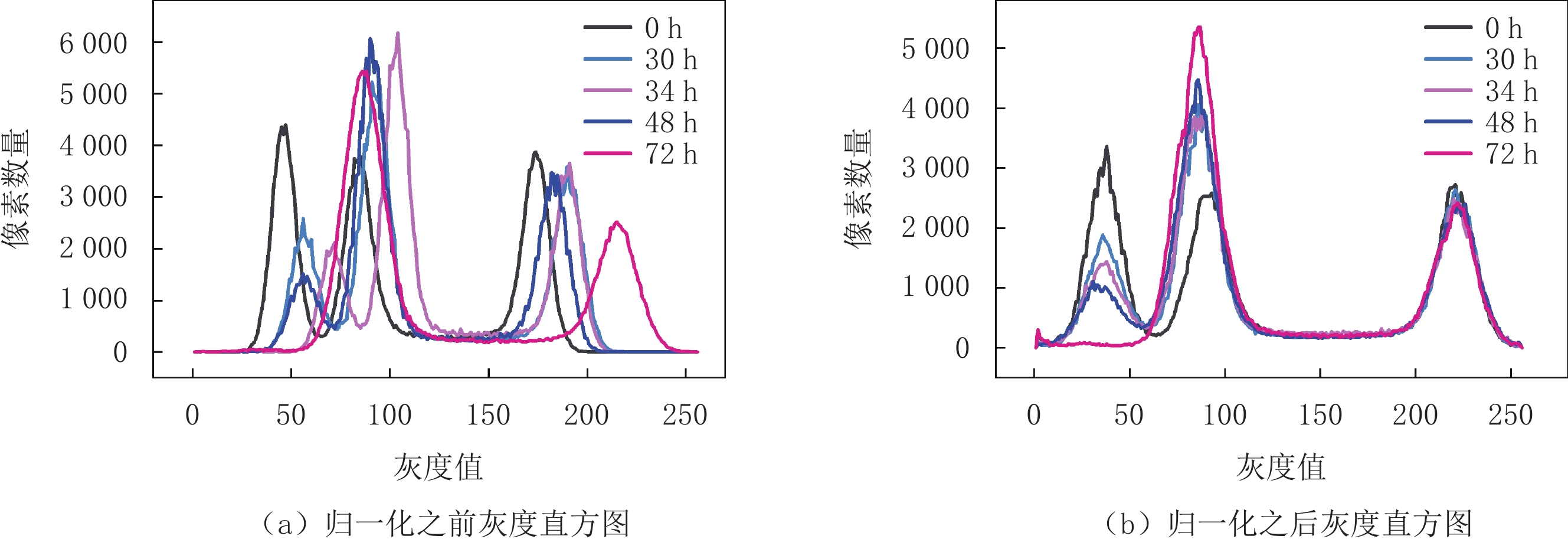
摘要: 微米X射线计算机断层扫描作为一种数字岩心探测手段,被广泛应用于研究含天然气水合物沉积物赋存形态,但由于水合物与水对X射线的衰减系数相近,二者在CT图像中灰度区间存在交集,导致在从CT图像上对水合物与水进行分割时存在强非唯一性。为提高对CT图像中水合物与水阈值分割的准确性,本文通过分析天然气水合物生长过程中不同时刻CT图像直方图特征,提出归一化CT图像及其直方图的方法。首先选定甲烷气与石英砂的峰值灰度基准;然后用高斯函数分别对当前CT图像直方图中的甲烷气与石英砂曲线进行拟合,得到当前CT图像直方图中的甲烷气与石英砂峰值灰度;再将当前CT图像直方图中的甲烷气峰值灰度与石英砂峰值灰度归一化到选定的峰值灰度基准;进而用归一化的直方图对CT图像进行归一化;最后根据归一化灰度直方图的变化趋势,定量统计得到CT图像中水合物增加和气-水减少的灰度区间,完成图像中不同组分的阈值划分。实验结果表明,提出的阈值分割方法能够为天然气水合物CT图像中水合物与水边界的确定和水合物饱和度计算提供定量的依据,具有实际的工程应用价值。Abstract: Micro-scale X-ray computed tomography (CT) has been widely used to study the occurrence forms of gas hydrate-bearing sediments. However, the similarity between the X-ray attenuation coefficient of hydrate and that of water leads to a strong non-uniqueness in their phase differentiation in CT images. To improve threshold segmentation accuracy between hydrate and water in CT images, this study proposes a CT image and histogram normalized method by analyzing the histogram characteristics of CT images at different times during the growth process of natural gas hydrate. First, the peak gray value baseline of methane gas and quartz sand was selected. Then, a Gaussian function was used to fit the curves corresponding to methane gas and quartz sand in the current CT image histogram to obtain the peak gray values. In addition, the peak gray values of methane gas and quartz sand in the current CT image histogram were normalized to the chosen peak gray baseline. Subsequently, the normalized histogram was used to normalize the corresponding CT images. Finally, according to the changing trend of normalized gray histogram curves, the increasing gray ranges of hydrate and decreasing gray ranges of gas-water in CT images were obtained quantitatively, which guided threshold segmentation of CT images. Experimental results show that the proposed threshold segmentation method can provide a basis for phase differentiation between hydrate and water in CT images, improving the threshold segmentation accuracy.
-
Keywords:
- CT image /
- gas hydrate /
- threshold segmentation /
- normalization
-
-
![]()
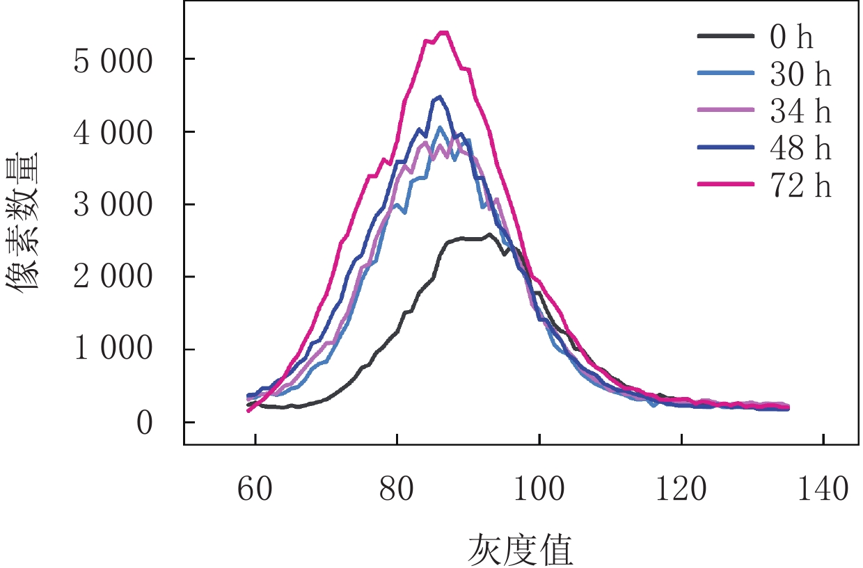
图 2 灰度直方图归一化对比(切片编号150)
Figure 2. Comparison of histogram curves before and after normalization(slice 150)
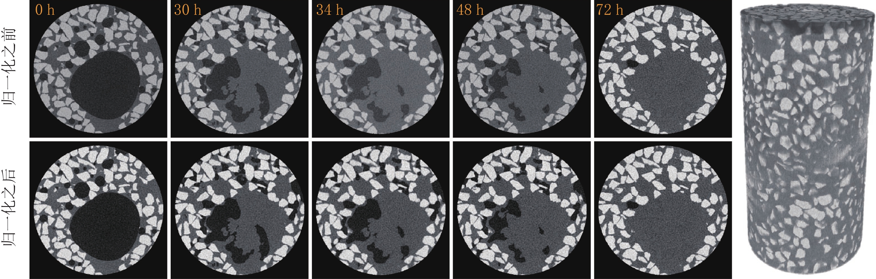
![]()
图 4 图像归一化对比(切片编号150)及三维数字岩心图像
Figure 4. Image normalization contrast (slice 150) and 3D digital core
![]()
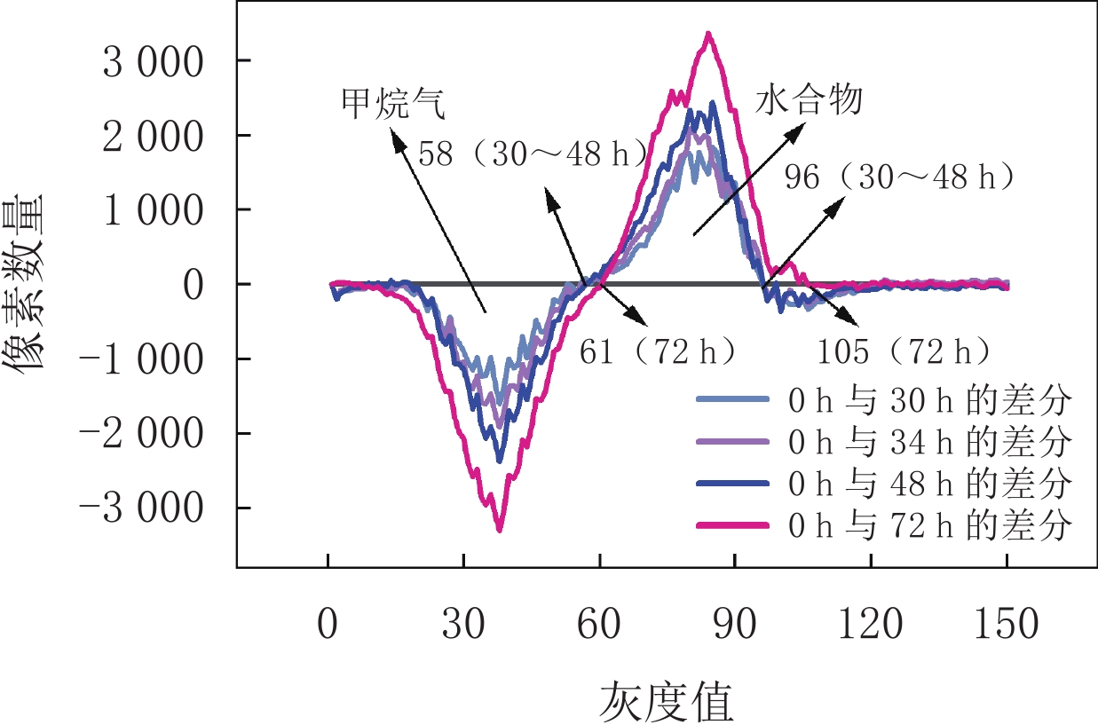
图 5 不同时刻CT图像直方图曲线差值(切片编号150)
Figure 5. Histogram curves of CT images subtracted at different times(slice 150)
![]()
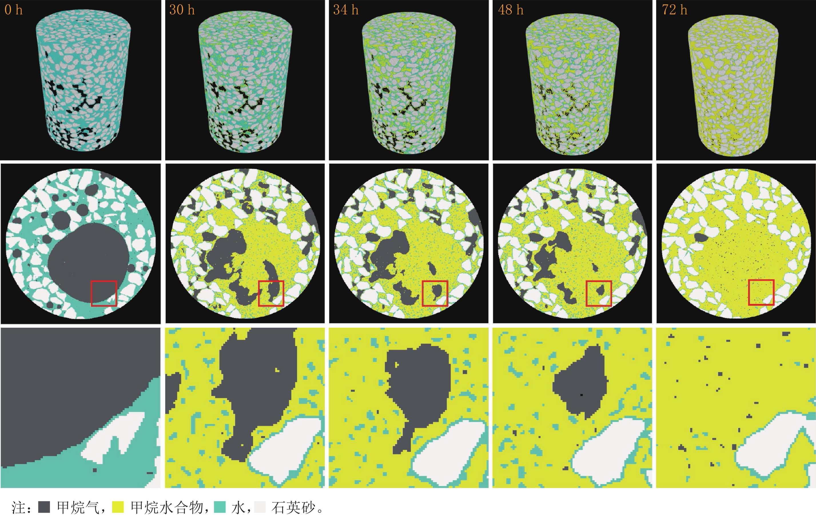
图 6 基于本文阈值分割方法得到的伪彩色三维数字岩心及图像的孔隙局部放大图像
Figure 6. Pseudo-color digital core and local pore magnification image obtained based on threshold segmentation method used in this study
表 1 CT图像中各种组分的灰度区间
Table 1 Gray intervals of various components of CT images
反应时间/h 灰度区间(0~255) 甲烷气 甲烷水合物 水 砂 0 0~58 - 59~156 157~255 30 0~58 59~96 97~156 157~255 34 0~58 58~96 97~156 157~255 48 0~58 59~96 97~156 157~255 72 0~61 62~105 106~156 157~255  下载: 导出CSV
-
[1] PRIEST J A, BEST A I, CLAYTON C R I, et al. A laboratory investigation into the seismic velocities of methane gas hydrate-bearing sand[J]. Journal of Geophysical Research: Solid Earth, 2005, 110: B04102.
[2] REN A R, LIU Y J, LIU Y X, et al. Acoustic velocity and electrical resistance of hydrate bearing sediments[J]. Journal of Petroleum Science and Engineering, 2010, 70: 52−56. doi: 10.1016/j.petrol.2009.09.001
[3] ZHANG L, GE K, WANG J, et al. Pore-scale investigation of permeability evolution during 364 hydrate formation using a pore network model based on X-ray CT[J]. Marine and Petroleum Geology, 2020, 113: 104157. doi: 10.1016/j.marpetgeo.2019.104157
[4] 李淑霞, 郭尚平, 陈月明, 等. 天然气水合物开发多物理场特征及耦合渗流研究进展与建议[J]. 力学学报, 2020,52(3): 828−842. doi: 10.6052/0459-1879-20-050 LI S X, GUO S P, CHEN M Y, et al. Research progress and suggestions on characteristics of multi-physical fields and coupled seepage in gas hydrate development[J]. Chinese Journal of Theoretical and Applied Mechanics, 2020, 52(3): 828−842. (in Chinese). doi: 10.6052/0459-1879-20-050
[5] LIU C L, MENG Q G, HU G W, et al. Characterization of hydrate-bearing sediments recovered from the Shenhu area of the South China Sea[J]. Interpretation, 2017, 5(3): 1−39.
[6] 叶建良, 秦绪文, 谢文卫, 等. 中国南海天然气水合物第二次试采主要进展[J]. 中国地质, 2020,47(3): 557−568. doi: 10.12029/gc20200301 YE J L, QIN X W, XIE W W, et al. Major process of second trial exploitation on gas hydrate in the South China Sea[J]. Geology in China, 2020, 47(3): 557−568. (in Chinese). doi: 10.12029/gc20200301
[7] SELL K, SAENGER E H, FALENTY A, et al. On the path to the digital rock physics of gas hydrate-bearing sediments-processing of in situ synchrotron-tomography data[J]. Solid Earth, 2016, 7(4): 1243−1258. doi: 10.5194/se-7-1243-2016
[8] LEI L, SEOL Y, JARVIS K. Pore-scale visualization of methane hydrate-bearing sediments with micro-CT[J]. Geophysical Research Letters, 2018, 45(11): 5417−5426. doi: 10.1029/2018GL078507
[9] LEE J Y, JUNG J W, LEE M H, et al. Pressure core based study of gas hydrates in the ulleung basin and implication for geomechanical controls on gas hydrate occurrence[J]. Marine & Petroleum Geology, 2013, 47: 85−98.
[10] KNEAFSEY T J, MORIDIS G J. X-ray computed tomography examination and comparison of gas hydrate dissociation in NGHP-01 expedition (India) and Mount Elbert (Alaska) sediment cores: Experimental observations and numerical modeling[J]. Marine and Petroleum Geology, 2014, 58: 526−539. doi: 10.1016/j.marpetgeo.2014.06.016
[11] JIN S, NAGAO J, TAKEYA S, et al. Structural investigation of methane hydrate sediments by microfocus X-ray computed tomography technique under high-pressure conditions[J]. Japanese Journal of Applied Physics, 2006, 45(24-28): L714−L716.
[12] 刘冬梅. 结合Retinex校正和显著性的主动轮廓图像分割[J]. 光学精密工程, 2019,27(7): 1593−1600. doi: 10.3788/OPE.20192707.1593 LIU D M. Active contour model for image segmentation based on Retinex correction and saliency[J]. Optics and Precision Engineering, 2019, 27(7): 1593−1600. (in Chinese). doi: 10.3788/OPE.20192707.1593
[13] 袁小翠, 吴禄慎, 陈华伟. 基于Otsu方法的钢轨图像分割[J]. 光学精密工程, 2016,24(7): 1772−1781. doi: 10.3788/OPE.20162407.1772 YUAN X C, WU L S, CHEN H W. Rail image segmentation based on Otsu threshold method[J]. Optics and Precision Engineering, 2016, 24(7): 1772−1781. (in Chinese). doi: 10.3788/OPE.20162407.1772
[14] KERKAR P B, KRISTINE H, KEITH W J, et al. Imaging methane hydrates growth dynamics in porous media using synchrotron X-ray computed microtomography[J]. Geochemistry, Geophysics, Geosystems, 2014, 15(12): 4759−4768. doi: 10.1002/2014GC005373
[15] TA X H, TAE S Y, BALASINGAM M, et al. Observations of pore-scale growth patterns of carbon dioxide hydrate using X-ray computed microtomography[J]. Geochemistry, Geophysics, Geosystems, 2015, 16(3): 912−924. doi: 10.1002/2014GC005675
[16] CHAOUACHI M, ANDRZEJ F, KATHLEEN S, et al. Microstructural evolution of gas hydrates in sedimentary matrices observed with synchrotron X-ray computed tomographic microscopy[J]. Geochemistry, Geophysics, Geosystems, 2015, 16(6): 1711−1722. doi: 10.1002/2015GC005811
[17] CHEN X Y, ESPINOZA D N. Ostwald ripening changes the pore habit and spatial variability of clathrate hydrate[J]. Fuel, 2018, 214: 614−622. doi: 10.1016/j.fuel.2017.11.065
[18] 李承峰, 胡高伟, 业渝光, 等. X射线计算机断层扫描测定沉积物中水合物微观分布[J]. 光电子•激光, 2013,24(3): 551−557. LI C F, HU G W, YE Y G, et al. The microscopic distribution observation of hydrate in sediments by X-ray computed tomography[J]. Journal of Optoelectronics Laser, 2013, 24(3): 551−557. (in Chinese).
[19] 胡高伟, 李承峰, 业渝光, 等. 沉积物孔隙空间天然气水合物微观分布观测[J]. 地球物理学报, 2014,57(5): 1675−1682. doi: 10.6038/cjg20140530 HU G W, LI C F, YE Y G, et al. Microscopic distribution of gas hydrate in pore sediments[J]. Chinese Journal of Geophysics, 2014, 57(5): 1675−1682. (in Chinese). doi: 10.6038/cjg20140530
[20] LI C F, LIU C L, HU G W, et al. Investigation on the multiparameter of hydrate-bearing sands using nano-focus X-ray computed tomography[J]. Journal of Geophysical Research:Solid Earth, 2019, 124(3): 2286−2296. doi: 10.1029/2018JB015849
[21] YANG L, ZHAO J, LIU W, et al. Microstructure observations of natural gas hydrate occurrence in porous media using micro-focus X-ray computed tomography[J]. Energy & Fuels, 2015, 29(8): 4835−4841.
[22] 李晨安, 李承峰, 刘昌岭, 等. CT图像法观测不同粒径多孔介质中水合物分布[J]. 核电子学与探测技术, 2018,(4): 545−551. doi: 10.3969/j.issn.0258-0934.2018.04.020 LI C A, LI C F, LIU C L, et al. Hydrate distribution observation in porous media with different particle sizes in CT image[J]. Nuclear Electronics & Detection Technology, 2018, (4): 545−551. (in Chinese). doi: 10.3969/j.issn.0258-0934.2018.04.020
[23] LIU Z C, KIM J C, LEI L, et al. 2019. Tetrahydrofuran hydrate in clayey sediments: Laboratory formation, morphology, and wave characterization[J]. Journal of Geophysical Research: Solid Earth, 124(4): 3307-3319.
[24] 李志军, 张佳璐, 解恺, 等. 含粉砂四氢呋喃水合物微观结构试验[J]. 油气储运, 2021,41(1): 84−90. LI Z J, ZHANG J L, XIE K, et al. Experimental study on microstructure of silty sand tetrahydrofuran hydrate[J]. Oil & Gas Storage and Transportation, 2021, 41(1): 84−90. (in Chinese).
-
期刊类型引用(1)
1. 唐淑慧,蔡晓娟. 探讨腹部外周性原始神经外胚层肿瘤的临床及CT、MR诊断. 现代医用影像学. 2022(07): 1254-1257 .  百度学术
百度学术
其他类型引用(0)
计量
- 文章访问数: 478
- HTML全文浏览量: 151
- PDF下载量: 54
- 被引次数: 1


