
Neck-chest-abdomen-pelvis Combined Enhanced CT in Breast Cancer Patients: Comparison between Dual-energy and Conventional Scanning Mode
-
摘要: 目的:通过与常规扫描方案比较,探讨能谱扫描方案在乳腺癌患者颈胸腹盆联合增强CT中的应用价值。方法:回顾性分析我院因乳腺癌行颈胸腹盆联合增强CT的女性患者40例,其中行常规颈胸腹盆增强CT患者20例,能谱颈胸腹盆增强CT患者20例。比较两种扫描方案动脉期和延迟期辐射剂量;通过测量颈胸部淋巴结的信号噪声比(SNR)及对比噪声比(CNR)以及主观评分,评价两种扫描方案的图像质量。结果:常规和能谱颈胸腹盆联合增强CT扫描方案的动脉期和延迟时期的辐射剂量具有统计学差异,能谱的辐射剂量更低;颈部动脉期淋巴结的SNR值、CNR值在常规和能谱扫描之间不存在统计学差异;胸部动脉期腋窝淋巴结的SNR值、CNR值在常规和能谱扫描之间存在统计学差异;颈部淋巴结常规和能谱扫描的主观评分不存在统计学差异,胸部腋窝淋巴结常规和能谱扫描的主观评分存在统计学差异,能谱主观评分更高,主观评分一致性较好,Kappa值为0.916。结论:能谱颈胸腹盆联合增强CT扫描方案的辐射剂量更低,扫描流程更简便,患者配合度更高,对于胸部腋窝淋巴结的显示更清晰,加之能谱扫描可以提供多参数的图像数据,因此具有重要的应用推广价值。Abstract: Objective: To investigate the application value of dual-energy scanning scheme in neck-chest-abdomen-pelvis combined enhanced CT in patients with breast cancer patients by comparing with the conventional scanning scheme. Methods: A retrospective analysis was performed on 40 female patients with breast cancer who underwent neck-chest-abdomen-pelvis combined enhanced CT in Beijing hospital, including 20 patients scanned by conventional protocol and 20 patients scanned by dual-energy protocol. The radiation doses in arterial phase and delayed phase were compared between the two modes. The image qualities were evaluated by measuring signal-to-noise ratio (SNR), contrast noise ratio (CNR) and subjective scoring. Results: The radiation doses in arterial phase and delayed phase of conventional and dual-energy protocol were statistically different, and the radiation dose of dual-energy protocol was lower. There was no significant difference between the two modes in SNR and CNR of cervical arterial lymph nodes. There was significant difference between conventional and dual-energy mode in SNR and CNR value of axillary lymph nodes in thoracic artery phase. There was no significant difference in the subjective scores of conventional and dual-energy protocol of cervical lymph nodes. However, there was significant difference in the subjective scores of conventional and dual-energy protocol of axillary lymph nodes. The subjective score of dual-energy protocol was higher and the consistency of subjective score was better with Kappa value of 0.916. Conclusion: Compared with conventional scanning scheme, dual-energy scanning scheme for neck-chest-abdomen-pelvis combined enhanced CT shows lower radiation dose, simpler scanning process, higher patient cooperation degree, clearer display of axillary lymph nodes, which has important clinical application and promotion value.
-
-
![]()
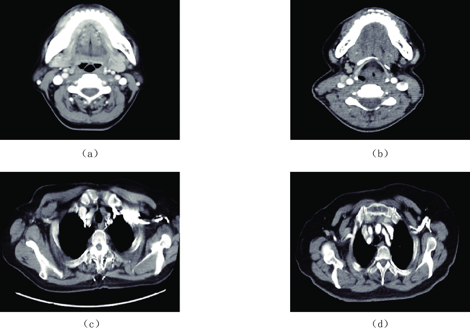
图 1 常规和能谱颈胸腹盆联合增强CT颈部和胸部淋巴结图像
(a)常规颈胸腹盆联合增强CT颈部淋巴结图像;(b)能谱颈胸腹盆联合增强CT颈部淋巴结图像;(c)常规颈胸腹盆联合增强CT胸部淋巴结图像,静脉中对比剂对淋巴结的显示产生影响;(d)能谱颈胸腹盆联合增强CT胸部淋巴结图像,基本去除静脉中对比剂对周围软组织的影响,腋窝淋巴结可以清晰显示。
Figure 1. Cervical and axillary lymph nodes images of conventional and dual-energy scanning process of neck-chest-abdomen-pelvis combined enhanced CT
表 1 常规颈胸腹盆增强CT扫描流程
Table 1 Conventional scanning process of neck-chest-abdomen-pelvis combined enhanced CT
项目 扫描启时间/s 患者姿势 扫描范围 胸腹盆动脉期 25~30 上举双手在头颅两侧 肺尖至耻骨联合 颈部动脉期 40 放手在身体两侧 胸锁关节至乳突 胸腹盆静脉期 60 上举双手在头颅两侧 肺尖至耻骨联合 腹盆延时期 180 上举双手在头颅两侧 肝顶至耻骨联合  下载: 导出CSV
表 2 能谱颈胸腹盆增强CT扫描流程
Table 2 Dual-energy scanning process of neck-chest-abdomen-pelvis combined enhanced CT
项目 扫描启动时间/s 患者姿势 扫描范围 颈胸腹盆动脉期 25~30 上举双手在头颅两侧 乳突至耻骨联合 颈胸腹盆静脉期 60 上举双手在头颅两侧 乳突至耻骨联合 腹盆延时期 180 上举双手在头颅两侧 肝顶至耻骨联合
下载: 导出CSV
表 3 常规和能谱颈胸腹盆联合增强CT扫描方案动脉期和延时期辐射剂量统计表
Table 3 Statistical table of radiation dose in arterial phase and extended phase of conventional and dual-energy scanning process of neck-chest-abdomen-pelvis combined enhanced CT
项目 指标 辐射剂量 统计检验 常规 能谱 统计值 P 动脉期 DLP 804.84±67.12 676.62±87.02 -5.218 <0.001 CTDvol 9.40±0.60 8.33±0.79 -4.851 <0.001 ED 12.07±1.01 10.15±1.31 -5.218 <0.001 延迟期 DLP 378.48±80.64 307.80±16.44 -3.841 0.001 CTDvol 8.14±1.37 6.25±0.26 -6.038 <0.001 ED 5.68±1.21 4.62±0.25 -3.841 0.001
下载: 导出CSV
表 4 常规和能谱颈胸腹盆联合增强CT扫描方案动脉期颈部和胸部淋巴结客观图像质量评价表
Table 4 Evaluation table of objective image quality of cervical and thoracic lymph nodes in arterial phase in conventional and dual-energy scanning process of neck-chest-abdomen-pelvis combined enhanced CT
指标 项目 客观图像质量 统计检验 常规 能谱 统计值 P CT值 颈部淋巴结 52.47±13.03 45.29±9.58 -1.985 0.055 颈部同层面脂肪 -101.58±4.96 -100.27±5.26 -0.813 0.421 胸部淋巴结 36.92±16.09 39.35±15.94 0.480 0.634 胸部同层面脂肪 -106.72±8.46 -103.84±5.36 1.288 0.206 SD 颈部淋巴结 10.58±2.46 9.65±2.40 -1.206 0.235 颈部同层面脂肪 6.83±2.22 6.55±1.51 -0.463 0.647 胸部淋巴结 36.92±16.09 39.35±15.94 -4.871 0.001 胸部同层面脂肪 9.15±1.79 6.96±1.06 -4.722 <0.001 SNR 颈部淋巴结 5.26±1.99 5.12±2.12 -0.217 0.830 胸部淋巴结 2.88±1.48 4.32±1.84 2.726 0.010 CNR 颈部淋巴结 25.02±8.57 23.20±4.96 -0.825 0.416 胸部淋巴结 16.27±3.84 20.99±3.63 3.996 <0.001
下载: 导出CSV
表 5 常规和能谱颈胸腹盆联合增强CT扫描方案主观图像质量评分表
Table 5 Subjective image quality evaluation table of conventional and dual-energy scanning process of neck-chest-abdomen-pelvis combined enhanced CT
项目 主观评分者 主观图像质量 统计检验 3分 4分 5分 Z P 颈部 观察者1 6 13 21 -0.763 0.495 观察者2 7 12 21 -0.596 0.602 胸部 观察者1 9 22 9 -3.724 0.001 观察者2 8 22 10 -3.756 <0.001
下载: 导出CSV
-
[1] 耿雪. 全视野器官剂量调制技术在女性化疗患者颈胸腹盆CT增强中的应用研究[D]. 保定: 河北大学, 2021. GENG X. Application of full-field organ dose modulation technique in CT enhancement of neck-chest-abdomen-pelvis in female chemotherapy patients[D]. Baoding: Hebei University, 2021. (in Chinese).
[2] 王小英. 多层螺旋CT双期增强扫描诊断乳腺癌淋巴结转移的价值分析[J]. 基层医学论坛, 2020,24(19): 2766−2767. DOI: 10.19435/j.1672-1721.2020.19.059. WANG X Y. Value analysis of dual-phase enhanced multi-slice spiral CT in diagnosis of lymph node metastasis of breast cancer[J]. Forum of Primary Medicine, 2020, 24(19): 2766−2767. DOI: 10.19435/j.1672-1721.2020.19.059. (in Chinese).
[3] 徐秋贞, 邵海磊, 吕燕, 等. 胸部能谱成像模式与常规CT扫描的辐射剂量及图像质量的仿真体模研究[J]. 中华放射医学与防护杂志, 2017,5(12): 957−961. DOI: 10.3760/cma.j.issn.0254-5098.2017.12.015. XU Q Z, SHAO H L, LV Y, et al. A study on radiation dose and image quality of chest spectral imaging and conventional CT in anthropomorphic phantom[J]. Chinese Journal of Radiological Medicine and Protection, 2017, 5(12): 957−961. DOI: 10.3760/cma.j.issn.0254-5098.2017.12.015. (in Chinese).
[4] 林晓珠, 沈云, 陈克敏. CT能谱成像的基本原理与临床应用研究进展[J]. 中华放射学杂志, 2011,45(8): 798−800. DOI: 10.3760/cma.j.issn.1005-1201.2011.08.028. LIN X Z, SHEN Y, CHEN K M. Spectral CT imaging: Principle, clinical application and research[J]. Chinese Journal of Radiology, 2011, 45(8): 798−800. DOI: 10.3760/cma.j.issn.1005-1201.2011.08.028. (in Chinese).
[5] 张文军, 田明华, 张洪胜, 等. 能谱CT在乳腺癌诊断中的初步应用研究[J]. 中国医师进修杂志, 2015,38(4): 262−266. DOI: 10.3760/cma.j.issn.1673-4904.2015.04.008. ZHANG W J, TIAN M H, ZHANG H S, et al. A preliminary study on the application of energy spectrum CT in the diagnosis of breast cancer[J]. Chinese Journal of Postgraduates of Medicine, 2015, 38(4): 262−266. DOI: 10.3760/cma.j.issn.1673-4904.2015.04.008. (in Chinese).
[6] 赵慧萍, 吕培杰, 张丽英, 等. 基于能谱CT智能匹配技术的半剂量能谱CT联合自适应统计迭代重建技术在肥胖患者上腹部扫描中的应用价值[J]. 中华医学杂志, 2017,97(47): 3681−3686. DOI: 10.3760/cma.j.issn.0376-2491.2017.47.001. ZHAO H P, LV P J, ZHANG L Y, et al. Application of half-dose spectral CT based on the automatic spectral imaging mode selection and adaptive statistical iterative reconstruction in the CT examination of upper abdomen in obese patients[J]. National Medical Journal of China, 2017, 97(47): 3681−3686. DOI: 10.3760/cma.j.issn.0376-2491.2017.47.001. (in Chinese).
[7] 郭锬, 周诚, 陈涓, 等. 颈部能谱CT与常规CT扫描辐射剂量与图像质量的对比研究[J]. 中华放射学杂志, 2015,49(4): 279−282. DOI: 10.3760/cma.j.issn.1005-1201.2015.04.010. GUO T, ZHOU C, CHEN J, et al. Comparative study of radiation dose and image quality between spectral CT scanning and conventional scanning on neck[J]. Chinese Journal of Radiology, 2015, 49(4): 279−282. DOI: 10.3760/cma.j.issn.1005-1201.2015.04.010. (in Chinese).
[8] KAZUHIRO, MATSUMOTO, MASAHIRO, et al. Virtual monochromatic spectral imaging with fast kilovoltage switching: Improved image quality as compared with that obtained with conventional 120-kVp CT[J]. Radiology, 2011, 259(1): 257−262. DOI: 10.1148/radiol.11100978.
[9] 中华医学会放射学分会质量管理与安全管理学组. CT辐射剂量诊断参考水平专家共识[J]. 中华放射学杂志, 2017,51(11): 817−822. DOI: 10.3760/cma.j.issn.1005-1201.2017.11.001. Quality Management and Safety Management Group, Radiology Society, Chinese Medical Association. CT radiation dose in the diagnosis of reference level expert consensus[J]. Chinese Journal of Radiology, 2017, 51(11): 817−822. DOI: 10.3760/cma.j.issn.1005-1201.2017.11.001. (in Chinese).
[10] VALENTIN J. Managing patient dose in multi-detector computed tomography (MDCT). ICRP Publication 102[J]. Annals of the ICRP, 2007, 37(1): 1−79. DOI: 10.1088/0952-4746/28/3/b01.
[11] 刘丹丹, 牛延涛. 相同CT剂量指数时管电压对表浅辐射敏感器官剂量的影响[J]. 中华放射学杂志, 2018,52(12): 957−961. DOI: 10.3760/cma.j.issn.1005-1201.2018.12.015. LIU D D, NIU Y T. The study on the effect of tube voltage on the dose of superficial radiation sensitive organ in the same CT dose index[J]. Chinese Journal of Radiology, 2018, 52(12): 957−961. DOI: 10.3760/cma.j.issn.1005-1201.2018.12.015. (in Chinese).
[12] 王贵生, 高建华, 赵帅, 等. 肝脏增强扫描门静脉期能谱CT与传统多层螺旋CT辐射剂量和图像质量的比较[J]. 中华放射学杂志, 2013,47(4): 340−343. DOI: 10.3760/cma.j.issn.1005-1201.201304.011. WANG G S, GAO J H, ZHAO S, et al. Comparing radiation dose and image quality between spectral CT and conventional multi-slice CT in imaging liver[J]. Chinese Journal of Radiology, 2013, 47(4): 340−343. DOI: 10.3760/cma.j.issn.1005-1201.201304.011. (in Chinese).
[13] 赵永霞, 左紫薇, 索红娜, 等. 肾动脉CT血管成像中采用常规扫描和能谱成像扫描方案对图像质量和辐射剂量的影响[J]. 中华放射学杂志, 2017,51(4): 304−307. DOI: 10.3760/cma.j.issn.1005-1201.2017.04.014. ZHAO Y X, ZUO Z W, SUO H N, et al. Comparison of spectral imaging and conventional CT in CT angiography of the kidney: Image quality and radiation dose[J]. Chinese Journal of Radiology, 2017, 51(4): 304−307. DOI: 10.3760/cma.j.issn.1005-1201.2017.04.014. (in Chinese).
[14] 李原, 张剑, 马俊丽, 等. 能谱CT迭代重建算法在腹部增强扫描中降低辐射剂量与改善图像质量的可行性研究[J]. 宁夏医学杂志, 2022,44(4): 321−324. DOI: 10.13621/j.1001-5949.2022.04.0321. LI Y, ZHANG J, MA J L, et al. Feasibility study of the spectral CT multi-model iterative reconstruction algorithm in reducing the radiation dose and improving the image quality in abdominal enhanced CT scan[J]. Ningxia Medical Journal, 2022, 44(4): 321−324. DOI: 10.13621/j.1001-5949.2022.04.0321. (in Chinese).
[15] 付珺, 胡俊岭. 颈部能谱CT与常规CT扫描辐射剂量与图像质量的差异分析[J]. 临床医学研究与实践, 2016,1(2): 48. doi: 10.19347/j.cnki.2096-1413.2016.02.034 FU J, HU J L. Analysis of radiation dose and image quality difference between neck dual-energy CT and conventional CT scan[J]. Clinical Research and Practice, 2016, 1(2): 48. (in Chinese). doi: 10.19347/j.cnki.2096-1413.2016.02.034
[16] 熊祖坤, 付晓伟, 吕律, 等. 能谱CT技术去除胸部增强CT扫描患者腋静脉和锁骨下静脉对比剂伪影[J]. 中华放射学杂志, 2016,50(11): 825−828. DOI: 10.3760/cma.j.issn.1005-1201.2016.11.004. XIONG Z K, FU X W, LV L, et al. The application research of spectral CT for reducing beam-hardening artifacts around axillary vein and subclavian vein due to the utility of contrast agent in the chest enhanced CT scan[J]. Chinese Journal of Radiology, 2016, 50(11): 825−828. DOI: 10.3760/cma.j.issn.1005-1201.2016.11.004. (in Chinese).
[17] 赵永霞, 左紫薇. 能谱CT在消除硬化伪影中的应用[J]. 医学研究与教育, 2013,(6): 26−29. DOI: 10.3969/j.issn.1674-490X.2013.06.006. ZHAO Y X, ZUO Z W. Application of spectral CT imaging in reducing beam-hardening artifacts[J]. Medical Research and Education, 2013, (6): 26−29. DOI: 10.3969/j.issn.1674-490X.2013.06.006. (in Chinese).
[18] HE T, QIAN X, ZHAI R, et al. Computed tomography number measurement consistency under different beam hardening conditions: Comparison between dual-energy spectral computed tomography and conventional computed tomography imaging in phantom experiment[J]. Journal of Computer Assisted Tomography, 2015, 39(6): 981−985. DOI: 10.1097/RCT.0000000000000287.
[19] 沈云. 宝石CT能谱成像原理及其扫描射线剂量[J]. 中国医疗设备, 2012,27(9): 13−16. DOI: 10.3969/j.issn.1674-1633.2012.09.003. SHEN Y. Principle and radiation dose of energy imaging for discovery CT[J]. China Medical Devices, 2012, 27(9): 13−16. DOI: 10.3969/j.issn.1674-1633.2012.09.003. (in Chinese).
[20] 林桐. CT能谱成像对乳腺癌腋窝淋巴结转移的诊断效能评价[J]. 现代医用影像学, 2020,29(4): 704−706. doi: 10.3969/j.issn.1006-7035.2020.04.034 LIN T. Evaluation of CT dual-energy imaging in the diagnosis of axillary lymph node metastasis of breast cancer[J]. Modern Medical Imagelogy, 2020, 29(4): 704−706. (in Chinese). doi: 10.3969/j.issn.1006-7035.2020.04.034
-
期刊类型引用(2)
1. 李洋森,王伟,李炳颖,毛云新,刘晓晖. 分频AVO技术在西湖凹陷中深层薄储层评价中的应用. CT理论与应用研究(中英文). 2025(03): 409-418 .  百度学术
百度学术
2. 朱焱辉. 基于压缩感知的地震频带拓宽方法——以珠江口盆地东部惠州地区为例. 中外能源. 2023(06): 44-52 . 百度学术
其他类型引用(0)
计量
- 文章访问数: 398
- HTML全文浏览量: 195
- PDF下载量: 17
- 被引次数: 2


