
In Vivo Study of the Influence of CT Acquisition and Reconstruction Parameters on Chest CT Number
-
摘要: 目的:探讨不同扫描参数及重建算法对在体胸部各组织CT值的影响。方法:在不同CT扫描条件下测量人体胸部的气管、血管、肺、椎体与肌肉的CT值。分别设定6组不同扫描参数及重建算法:S1层厚5 mm、50%多模型自适应迭代重建技术(ASIR-V)、低剂量;S2层厚5 mm、滤波反投影(FBP)、常规剂量;S3层厚1.25 mm、50%ASIR-V、低剂量;S4层厚1.25 mm、50% ASIR-V、常规剂量;S5层厚1.25 mm、FBP、低剂量;S6层厚1.25 mm、FBP、常规剂量。扫描的辐射剂量采用两种噪声指数(NI)来进行控制,包括低剂量(NI=40)和常规剂量(NI=10)。采用t检验或秩和检验分析比较不同的两个组之间 CT值的差异。结果:扫描剂量仅对气管CT值的影响具有统计学意义,对其他组织CT值无影响;扫描层厚与重建算法对胸部各个组织CT值的影响均未见统计学差异。结论:人体胸部组织CT值受CT层厚、重建算法和CT剂量的影响小,具有良好的稳定性。Abstract: Objective: To explore the influence of different CT acquisition and reconstruction parameters on the CT number of the chest in vivo. Methods: The CT number of the trachea, blood vessels, lungs, vertebral bodies, and muscles of the human chest were measured under different CT scanning parameters. Six groups of different scanning parameters and reconstruction algorithms were set respectively: slice thickness 5 mm, 50% multi-model adaptive statistical iterative reconstruction Veo (ASIR-V) and low-dose for S1; slice thickness 5 mm, filtered back projection (FBP) and standard-dose for S2; slice thickness 1.25 mm, 50% ASIR-V and low-dose for S3; slice thickness 1.25 mm, 50% ASIR-V and standard-dose for S4; slice thickness 1.25 mm, FBP, low-dose for S5; slice thickness 1.25 mm, FBP, standard-dose for S6. The radiation dose of the scan was controlled using two noise indexes (NI), including low-dose (NI=40) and standard-dose (NI=10). Differences in CT number between two groups were compared using t-test or rank-sum test. Results: Significant differences of CT number of the trachea were detected between low-dose and standard-dose, but no significant differences of CT number of other tissues were detected between low-dose and standard-dose. No significant differences of CT number of chest tissues were detected between either 5 mm thickness and 1.25 mm thickness or 50% ASIR-V and FBP. Conclusion: The CT number of human chest tissues showed well stability which was scarcely influenced by slices thickness, reconstruction algorithm and scan dose.
-
Keywords:
- CT scan parameters /
- reconstruction algorithms /
- CT number /
- scan dose
-
坏死性筋膜炎(necrotizing fasciitis,NF)是危及生命的侵袭性软组织感染,其坏死区域主要累及筋膜及肌肉。这是一种罕见的疾病,常好发于躯干、会阴及四肢,而对于颈部坏死性筋膜炎(cervical necrotizing fasciitis,CNF)来说,由于发病部位的血供丰富,因此其发病率更低,仅占NF的1%~10%[1]。牙源性以及扁桃体感染是常见的发病原因,颌下以及咽旁间隙是最好发的感染部位,发生于鼻咽部较为罕见。
CNF如果不及时诊断以及治疗,病变区域有可能沿着颈深区域向纵隔蔓延,从而导致严重的后果。不受控制的高血糖会导致患者的免疫系统随着年龄的增长越来越虚弱,这往往是该疾病的主要诱发因素[2]。由于CNF在发病初期并无特异性的体征以及实验室指标的异常,因此影像学检查,尤其是CT检查在该病的精准诊断中显得尤为重要。
1. 病史资料
1.1 临床资料
患者男性,58岁,1年余前无明显诱因出现左侧头痛,伴有左耳流脓,无鼻塞、流涕、涕中带血,无耳痛、耳内流血等不适。于当地医院诊断为“慢性化脓性中耳炎”,予以抗炎治疗后头痛稍有减轻。此后患者上述症状持续存在,均予以抗炎治疗,但头痛并未缓解。
10月前患者自觉上述症状加重,并出现左侧面瘫及吞咽困难,之后对症治疗后均未见好转。既往双侧慢性化脓性中耳炎50余年;糖尿病史5年,合并糖尿病性视网膜病史,平时胰岛素控制;高血压病史4年。
1.2 实验室检查
血常规:红细胞3.70×10¹²/L(下降),血红蛋白108 g/L(上升),红细胞压积32.6%(下降),淋巴细胞15.9%(下降),其余正常。血生化:谷丙转氨酶6.3 U/L(下降),谷草转氨酶10.6 U/L(下降),总胆汁酸6.8 umol/L(上升),总蛋白61.3 g/L(下降),白蛋白33.6 g/L(下降),肌酐101 umol/L(上升),葡萄糖4.39 mmol/L,糖化血红蛋白21.9%(上升),其余正常。肿瘤指标:AFP、CEA、CA199、CA125、CA724、PSA、fPSA、fPSA/PSA、CTFRA21-1、SCC、CA50均阴性。
1.3 影像学表现
CT图像显示鼻咽腔基本对称,双侧咽隐窝略变浅(图1);两侧咽旁间隙及咽后间隙软组织肿胀,颅底斜坡骨质破坏,蓝色箭头处可见无强化的筋膜征象(图1(d))。
MRI图像示鼻咽腔基本对称,双侧咽隐窝略变浅(图2);两侧咽旁间隙及咽后间隙软组织肿胀,肌间脂肪消失,增强后咽旁间隙可见散乱不均匀的异常强化,病变沿肌间隙走行,鼻咽粘膜未见异常增厚。蓝色箭头见颅骨斜坡右侧及左下可见稍长T1稍短T2信号影,增强后强化不均匀,周围见强化的软组织信号影(图2(e)和图2(f)),两侧乳突气房可见液性信号影。
PET/CT图像显示FDG异常代谢区域位于鼻咽深部软组织伴邻近颅底斜坡骨质破坏(图3),鼻咽粘膜未见异常代谢(图3(a)~图3(f));头颈部PET图像显示颈部未见肿大淋巴结影(图3(g))。CT、MRI以及PET/CT检查,均误诊为鼻咽癌。
1.4 诊疗过程
患者行鼻内镜下鼻咽部肿物活检术,于鼻咽部可见包裹性脓腔及坏死组织,取部分病理组织送病理及脓性分泌物送细菌培养,广泛彻底清除病灶至正常黏膜。
病理诊断:局部见炎性渗出,局部纤维结缔组织增生及粘液变性,胶原变性。
1.5 随访
患者出院1月后门诊复查,疼痛及吞咽困难的症状缓解,面瘫有所好转。半年后电话跟踪随访,患者疼痛及吞咽困难的症状消失,面瘫基本缓解,仅左眼睑闭合不佳。
2. 讨论
2.1 病因及发病机制
NF是一种组织坏死并且进展迅速的疾病,病原体入侵软组织并且引起血管血栓,最终导致脂肪组织、筋膜及皮肤坏死[3]。CNF更是一种罕见的累及颈部筋膜的微生物感染,它的易感因素包括糖尿病、不良的口腔卫生、酗酒、肿瘤以及静脉吸毒[4]。
CNF最常见原因是牙源性感染(27.5%),其次是扁桃体疾病(22.5%)、皮肤感染(8.75%)和腮腺感染(6.25%)[5]。最常见的并发症是气道阻塞以及下行性坏死性纵隔炎[6]。纵隔炎的预后非常差,因此它与感染性休克一样都是CNF最为严重的并发症[7-8]。
2.2 临床特征
由于CNF预后不良,及时诊断并且早期干预就显得尤为重要。压痛、发热和皮肤红斑是早期NF的常见体征[9]。回顾本病例,可能是由于发病位置的特殊(鼻咽部),患者并无上诉症状,而是表现为左侧头痛伴左耳流脓。
Wong等[10]为了对包括CNF在内的NF进行早期诊断,提出了坏死性感染实验室风险指标(LRINEC)评分。LRINEC评分是以6项实验室指标的异常进行评分,其中包括血清C反应蛋白(>150 mg/L)、白细胞(WBC)计数(>15000/μL)、血红蛋白(<13.5 g/dL)、血清钠水平(<135 mmol/L)、血清肌酐水平(142 mmol/L)和血清葡萄糖水平(10 mmol/L)。LRINEC评分大于等于8分,发生NSTI的风险为75%。虽然之后一些研究对LRINEC评分进行评估,证实了该评分在NF感染初期诊断中的有效性,但是近期的一些研究又发现该指标的敏感性较差,并不能作为排除NF的有效手段[11,12-13]。通过回顾本例患者的实验室指标也验证了这一结论,患者的LRINEC评分仅为2分,远没有达到诊断NF的水平。
2.3 影像学表现
对于临床体征及实验室指标均不明确的早期NF患者,影像学检查可以发挥重要作用。如怀疑为NF,CT扫描是一个有价值的影像学工具。一项关于坏死性筋膜炎的CT研究发现,CT的敏感性达到了100%,特异性为98%,因此CT阴性结果可以有效的排除坏死性筋膜炎,CT阳性结果对诊断坏死性筋膜炎具有很高的价值[14]。当CT图像中出现脂肪受累、沿着筋膜平面走行的液体以及气体聚集,尤其是增强图像出现无强化的筋膜增厚等征象,需要考虑NF[14]。
而MR则被认为是诊断NF最佳的影像学检查,当T2加权像上出现深筋膜增厚>3 mm并伴有多个肌筋膜室受累,此为诊断NF的重要征象[15]。虽然MRI的表现优于CT,但是MRI在某些紧急情况下难以进行,因此不建议将其作为首选的影像学检查技术[11]。我们回顾该患者CT及MRI图像,图像中虽然出现两侧咽旁间隙及咽后间隙肿胀,伴双侧欠对称,咽隐窝变浅,合并颅底骨破坏等表现,这些都是与鼻咽癌相同的征象,但是图像中另外可见典型的深筋膜增厚的表现,尤其是MR上可见散乱不均匀的异常强化,病变沿肌间隙走行,这些均提示需要与坏死性筋膜炎进行鉴别。
在该患者的PET/CT图像上,虽然鼻咽部肿胀伴有较大范围的FDG代谢增高,同时合并颅底骨的破坏,但是FDG异常代谢的区域主要局限在鼻咽深部区域,粘膜并未见FDG异常代谢,这点从MR中信号正常的鼻咽粘膜中得到了印证(图2(c)和图2(d))。
2.4 治疗及转归
本例患者通过鼻咽肿物活检术发现鼻咽部包裹性浓腔及坏死组织,并且病灶进行广泛彻底地清除,完成对CNF的诊断以及治疗。术后患者症状明显缓解,出院后继续接受头孢曲松抗感染治疗。
2.5 诊断与鉴别诊断
目前NF的诊断主要依赖症状学、实验室指标、影像学以及侵入性诊断。然而早期的NF体征与症状几乎没有特异性,通常难以明确诊断。因此当出现肿胀、发热以及与症状不成比例的剧烈疼痛时,需要高度怀疑NF。既往LRINEC评分曾经作为NF诊断的重要依据,但是该评分诊断的敏感性较低,并不能作为排除NF的有效手段。当NF诊断不明确时,影像学检查可以提供相对有价值的信息。
一项包含23项研究总计纳入5982名患者的META分析评估了体格检查、影像学检查以及LRINEC评分在NF诊断中的准确性[13]。该研究发现影像学检查具有敏感性及特异性,尤其是CT的敏感性为88.5%,特异性为93.3%;而体格检查以及LRINEC评分敏感性较差,均不能应用于排除NF。侵入性诊断——手术探查是诊断NF的金标准,当手指可轻易分离筋膜(手指实验阳性)、组织缺血坏死以及恶臭分泌物,均提示NF的诊断[16]。由于该例患者CNF发生于鼻咽部,需要与以下疾病进行鉴别。
2.5.1 鼻咽癌
鼻咽癌病理类型目前以未分化癌及鳞状细胞癌为主,EB病毒感染与鼻咽癌发病率密切相关。早期鼻咽癌基本无症状,也可能因为咽鼓管阻塞引起一系列症状,其中包括鼻塞、鼻出血、中耳炎、听力下降等。影像学表现:CT及MR表现基本相似,表现为鼻咽部两侧欠对称,局部见软组织肿块突入鼻腔内,咽隐窝变浅或消失;当咽旁间隙受累时,其脂肪间隙消失,再向外可累及翼腭窝及颞下窝,向后可累及颅底;增强扫描多表现为不均匀强化。PET/CT表现为FDG高代谢。
2.5.2 鼻咽淋巴瘤
鼻咽淋巴瘤是仅次于鼻咽癌第2常见的鼻咽部恶性肿瘤,它的发病率与EB病毒感染也密切相关。影像学表现,它好发于鼻咽顶壁咽扁桃体和咽鼓管扁桃体附近粘膜内聚集的淋巴小结,往往表现为一种弥漫对称分布的肿块[17]。同时它通常沿着粘膜或脂肪间隙扩散至口咽部及下咽扁桃体,极少累及深层结构,因此鼻咽淋巴瘤很少累及颅底。增强扫描多为均匀性的强化表现,PET/CT也表现为FDG高代谢。
3. 结论
鼻咽部坏死性筋膜炎缺乏早期诊断的特征性表现,但是影像学检查,尤其是CT对该病的早期诊断具有重要价值。
-
![]()
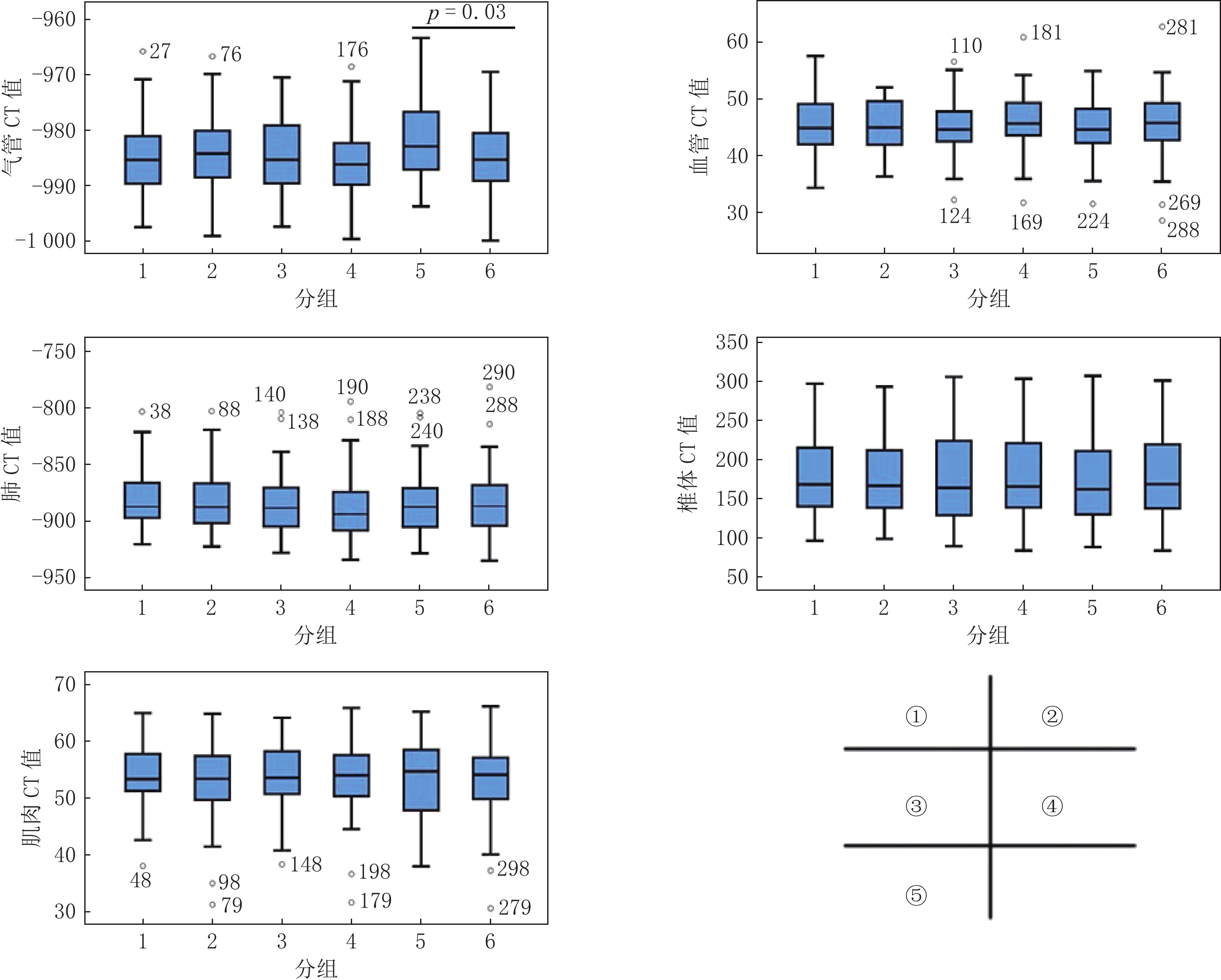
图 1 不同胸部组织在不同序列CT值分布
①气管 CT值在1-6序列中的分布,②血管 CT值在1-6序列中的分布,③肺 CT值在1-6序列中的分布,④椎体 CT值在1-6序列中的分布,⑤肌肉 CT值在1-6序列中的分布;通过对CT值两两比较,发现气管CT值在S5与S6两组的差异具有统计学意义,余各组织各序列相互比较均未见显著统计学差异。
Figure 1. Distribution of CT number measured in different sequences of each tissue
表 1 胸部CT扫描参数及重建方法
Table 1 Chest CT Scanning and reconstruction parameters
序列号 层厚/mm 重建算法 辐射剂量 S1 5.00 50% ASIR-V 低剂量 S2 5.00 FBP 常规剂量 S3 1.25 50% ASIR-V 低剂量 S4 1.25 50% ASIR-V 常规剂量 S5 1.25 FBP 低剂量 S6 1.25 FBP 常规剂量  下载: 导出CSV
表 2 两名研究者测量CT值一致性检验
Table 2 Interobserver variability test of CT number measured by two doctors
项目 ICC 95% CI F P S1-气管CT值 0.90 0.83~0.94 19.05 <0.001 S2-气管CT值 0.75 0.59~0.85 6.85 <0.001 S3-气管CT值 0.95 0.91~0.97 39.26 <0.001 S4-气管CT值 0.93 0.87~0.96 25.72 <0.001 S5-气管CT值 0.68 0.49~0.80 5.19 <0.001 S6-气管CT值 0.64 0.44~0.78 4.58 <0.001 S1-血管CT值 0.96 0.93~0.97 23.42 <0.001 S2-血管CT值 0.85 0.74~0.92 6.78 <0.001 S3-血管CT值 0.83 0.71~0.91 6.03 <0.001 S4-血管CT值 0.89 0.80~0.94 8.95 <0.001 S5-血管CT值 0.85 0.73~0.91 6.46 <0.001 S6-血管CT值 0.84 0.71~0.91 6.14 <0.001 S1-肺CT值 0.73 0.52~0.84 3.61 <0.001 S2-肺CT值 0.91 0.84~0.95 11.13 <0.001 S3-肺CT值 0.92 0.86~0.96 12.65 <0.001 S4-肺CT值 0.81 0.66~0.89 5.16 <0.001 S5-肺CT值 0.91 0.84~0.95 10.75 <0.001 S6-肺CT值 0.93 0.88~0.96 118.26 <0.001 S1-椎体CT值 0.99 0.96~0.99 118.26 <0.001 S2-椎体CT值 0.98 0.97~0.99 55.34 <0.001 S3-椎体CT值 0.90 0.83~0.95 10.75 <0.001 S4-椎体CT值 0.90 0.81~0.94 9.26 <0.001 S5-椎体CT值 0.93 0.88~0.96 15.17 <0.001 S6-椎体CT值 0.88 0.78~0.93 8.03 <0.001 S1-肌肉CT值 0.84 0.72~0.91 6.23 <0.001 S2-肌肉CT值 0.87 0.77~0.93 7.77 <0.001 S3-肌肉CT值 0.75 0.56~0.86 3.99 <0.001 S4-肌肉CT值 0.88 0.79~0.93 8.47 <0.001 S5-肌肉CT值 0.80 0.64~0.88 4.90 <0.001 S6-肌肉CT值 0.88 0.79~0.93 8.37 <0.001
下载: 导出CSV
表 3 各部位不同序列测量CT值的分布情况
Table 3 Distribution of CT number measured in different sequences of each tissue
序列 气管 血管 肺 椎体 肌肉 S1 -985.15±7.11 45.58±5.00 -887.22(-897.64~-865.48) 168.44(140.10~215.58) 53.57±5.85 S2 -983.95±7.11 41.94(44.98~49.66) -887.48(-902.30~-866.45) 166.67(138.18~214.14) 53.40(49.62~57.46) S3 -984.57±7.03 44.84±4.80 -884.84±27.11 177.88±50.97 53.83±6.09 S4 -985.93±6.49 45.58±5.41 -893.63(-908.19~-872.49) 178.76±51.34 138.60(165.74~221.41) S5 -981.90±6.81 45.11±4.78 -884.07±28.33 162.20(129.89~213.69) 53.58±6.57 S6 -984.89±6.54 45.52±6.01 -884.09±30.24 179.34±52.86 54.14(49.78~57.23)
下载: 导出CSV
表 4 胸部各组织不同序列CT值比较
Table 4 Comparison of different sequence CT values of chest tissues
组织 S1 vs S3 S2 vs S6 S3 vs S4 S5 vs S6 S3 vs S5 S4 vs S6 z/t P z/t P z/t P z/t P z/t P z/t P 气管 -0.41 0.68 0.69 0.49 1.00 0.32 2.23 0.03 -1.93 0.06 -0.80 0.43 血管 0.75 0.46 -0.72 0.47 -0.98 0.33 -0.38 0.70 -0.28 0.78 0.29 0.78 肺 -0.64 0.52 -0.43 0.66 -0.61 0.54 0.00 >0.99 -0.14 0.89 -0.44 0.66 椎体 0.05 0.96 -0.06 0.95 -0.09 0.93 -0.38 0.70 -0.26 0.79 -0.06 0.96 肌肉 -0.21 0.83 -0.29 0.77 -0.18 0.86 -0.38 0.71 0.19 0.85 -0.11 0.91
下载: 导出CSV
-
[1] National Lung Screening Trial Research Team, ABERLE D R, ADAMS A M, et al. Reduced lung-cancer mortality with low-dose computed tomographic screening[J]. The New England Journal of Medicine, 2011, 365(5): 395−409. doi: 10.1056/NEJMoa1102873
[2] HANSELL D M, BANKIER A A, MacMAHON H, et al. Fleischner society: Glossary of terms for thoracic imaging[J]. Radiology, 2008, 246(3): 697−722. doi: 10.1148/radiol.2462070712
[3] GODOY M C, NAIDICH D P. Subsolid pulmonary nodules and the spectrum of peripheral adenocarcinomas of the lung: Recommended interim guidelines for assessment and management[J]. Radiology, 2009, 253(3): 606−622. doi: 10.1148/radiol.2533090179
[4] 李琼, 于红, 张丽, 等. 迭代重建技术对低管电压胸部CT增强扫描图像质量的影响[J]. 实用放射学杂志, 2012,28(10): 1615−1618. doi: 10.3969/j.issn.1002-1671.2012.10.033 LI Q, YU H, ZHANG L, et al. The effect of iterative reconstruction on image quality of contrast-enhanced chest CT with low tube voltage settings[J]. Journal of Practical, 2012, 28(10): 1615−1618. (in Chinese). doi: 10.3969/j.issn.1002-1671.2012.10.033
[5] 彭文献, 彭天舟, 夏顺仁, 等. X线管电压对生物组织CT值影响的实验研究[J]. 放射学实践, 2013,28(11): 1102−1104. PENG W X, PENG T Z, XIA S R, et a1. The effects of X-ray tube voltage on tissue CT value[J]. Radiologic Practice, 2013, 28(11): 1102−1104. (in Chinese).
[6] 彭文献, 彭天舟, 叶小琴, 等. CT扫描参数对人体组织CT值影响的研究[J]. 中华放射医学与防护杂志, 2010,30(1): 79−81. doi: 10.3760/cma.j.issn.0254-5098.2010.01.026 PENG W X, PENG T Z, YE X Q, et al. Effect of CT scanning parameters on CT number[J]. Chinese Journal of Radiological Medicine and Protection, 2010, 30(1): 79−81. (in Chinese). doi: 10.3760/cma.j.issn.0254-5098.2010.01.026
[7] 赵雷, 刘波. 重建算法及射束硬化伪影对CT值测量的影响[J]. 影像研究与医学应用, 2019,3(14): 88−90. [8] 韩萍, 于春水, 余永强, 等. 医学影像诊断学[M]. 4版. 北京: 人民卫生出版社, 2017: 4-5. [9] 朱明, 刘贵霞, 李敬玉. CT扫描参数对CT值影响因素的实验研究[J]. 中国中西医结合影像学杂志, 2017,15(4): 91−93. doi: 10.3969/j.issn.1672-0512.2017.04.029 [10] AOKAGE K, MIYOSHI T, ISHII G, et al. Clinical and pathological staging validation in the eighth edition of the TNM classification for lung cancer: Correlation between solid size on thin-section computed tomography and invasive size in pathological findings in the new T classification[J]. Journal of Thoracic Oncology, 2017, 12(9): 1403−1412. doi: 10.1016/j.jtho.2017.06.003
[11] HENSCHKE C I, YIP R, SMITH J P, et al. CT screening for lung cancer: Part-solid nodules in baseline and annual repeat rounds[J]. American Journal of Roentgenology, 2016, 207(6): 1176−1184. doi: 10.2214/AJR.16.16043
[12] YANKELEVITZ D F, YIP R, SMITH J P, et al. CT screening for lung cancer: Nonsolid nodules in baseline and annual repeat rounds[J]. Radiology, 2015, 277(2): 555−64. doi: 10.1148/radiol.2015142554
[13] 张丽, 吴宁, 李蒙, 等. Ⅰ期浸润性肺腺癌磨玻璃成分定量分析与附壁样生长的相关性研究[J]. 中华肿瘤杂志, 2017,39(4): 269−273. doi: 10.3760/cma.j.issn.0253-3766.2017.04.006 ZHANG L, WU N, LI M, et al. The correlation study of ground glass opacity and lepidic growth pattern component in stage I lung invasive adenocarcinoma[J]. Chinese Journal of Oncology, 2017, 39(4): 269−273. (in Chinese). doi: 10.3760/cma.j.issn.0253-3766.2017.04.006
[14] GAO C, LI J, WU L, et al. The natural growth of subsolid nodules predicted by quantitative initial CT features: A systematic review[J]. Frontiers in Oncology, 2020: 10318.
[15] 蒋宇, 贾晓民, 赵杰. 肺亚实性结节CT定量分析对肺腺癌病理侵袭性诊断价值[J]. 放射学实践, 2021,36(10): 1232−1237. doi: 10.13609/j.cnki.1000-0313.2021.10.007 JIANG Y, JIA X M, ZHAO J. Diagnostic value of quantitative CT analysis of pulmonary subsolid nodules to lung adenocarcinoma[J]. Radiologic Practice, 2021, 36(10): 1232−1237. (in Chinese). doi: 10.13609/j.cnki.1000-0313.2021.10.007
-
期刊类型引用(1)
1. 司武杰,何韬,陈萍,张家奎. 光谱CT不同单能量图像在头部CT检查患者的应用及对比. 中国实用神经疾病杂志. 2025(01): 73-77 .  百度学术
百度学术
其他类型引用(0)


计量
- 文章访问数: 696
- HTML全文浏览量: 426
- PDF下载量: 371
- 被引次数: 1


