
CT Lymphangiography (CTL) in Different Type of Primary Intestinal Lymphangiectasia (PIL): A Comparative Study
-
摘要: 目的:探讨CT淋巴管成像(CTL)在不同分型的原发性小肠淋巴管扩张症(PIL)患者的临床与影像特征比较和应用价值。方法:收集2016年1月至2019年12月确诊为PIL的所有患者,回顾分析其CTL图像,由两名放射科医师盲法评价,评价指标包括:性别、首发年龄、临床症状和体征、血清白蛋白、肠壁是否增厚、肠系膜密度增高、浆膜腔是否积液、异常淋巴管分布的部位及范围、淋巴液是否返流、淋巴瘘的有无,腹腔内是否有淋巴结和颈部是否有异常扩张的淋巴管。基于对比剂是否分布于小肠壁和肠系膜这一特异影像学征象,首次提出将PIL分型为Ⅰ型(阳性组)与Ⅱ型(阴性组),比较两组患者的临床和影像学特征。结果:34例PIL患者中,Ⅰ型15例,Ⅱ型19例。Ⅰ型首次发病年龄较大(15.3±9.4 vs.8.3±3.8),腹泻发生率较高,而肢体和/或颜面部肿胀的比例低于Ⅱ型,两组性别比和白蛋白水平差异无统计学意义;影像征象方面,Ⅰ型腹膜后异常扩张淋巴管和淋巴瘘的比例高于Ⅱ型,肠系膜密度增高和腹腔淋巴结出现的比例低于Ⅱ型。两组间肠壁增厚、浆膜腔积液、返流样表现和颈部异常扩张淋巴管的差异无统计学意义。结论:CTL可以评价PIL患者异常淋巴管分布的部位、范围和程度,基于CTL提出了PIL的影像分型,不同类型的PIL具有不同的临床和影像特征。
-
关键词:
- CT /
- 淋巴管成像 /
- 原发性小肠淋巴管扩张症 /
- 分型
Abstract: Objective: To explore the clinical and imaging characteristics and application value CT lymphangiography (CTL) in patients with different types of primary intestinal lymphangiectasia (PIL). Methods: Patients diagnosed as PIL in our center were recruited in this retrospective study from January 2016 to December 2019, All CTL data were blindly reviewed by two radiologists separately, and the evaluation indicators included: sex, onset age, symptoms and signs, serum albumin, wall thickening, serous cavity effusion, abnormal distribution of lymphatics, lymph reflux, lymph nodes, fistula and abnormal lymphatics around neck area. Based on the abnormal lymphatics in intestinal wall and/or mesentery, PIL was classified into type Ⅰ (positive type) and type Ⅱ (negative type). The clinical and imaging features were compared between the two groups. Results: 34 PIL patients were recruited in this study, including 15 cases of Ⅰ and 19 cases of Ⅱ. Type I showed older age of first onset (15.3±9.4 vs. 8.3±3.8), higher rate of diarrhea, and lower rate of limb and/or facial edema than type Ⅱ, with no statistical difference in sex and serum albumin. For imaging features comparisons, type Ⅰ demonstrated higher rate of abnormal dilated lymphatics and fistula, lower rate of increased attenuation of mesentery and lymph nodes, while no statistical difference was found between wall thickening, serous cavity effusion, lymph reflux and abnormal dilated lymphatics around neck area. Conclusion: CTL demonstrated capability of evaluation in detection of location, distribution and range of abnormal lymphatics in PIL. Based on CTL, the imaging classification of PIL was proposed. Different types of PIL showed different clinical and imaging features, which was useful for therapeutic adoptions.-
Keywords:
- CT /
- lymphangiography /
- primary intestinal lymphangiectasia /
- type
-
-
![]()
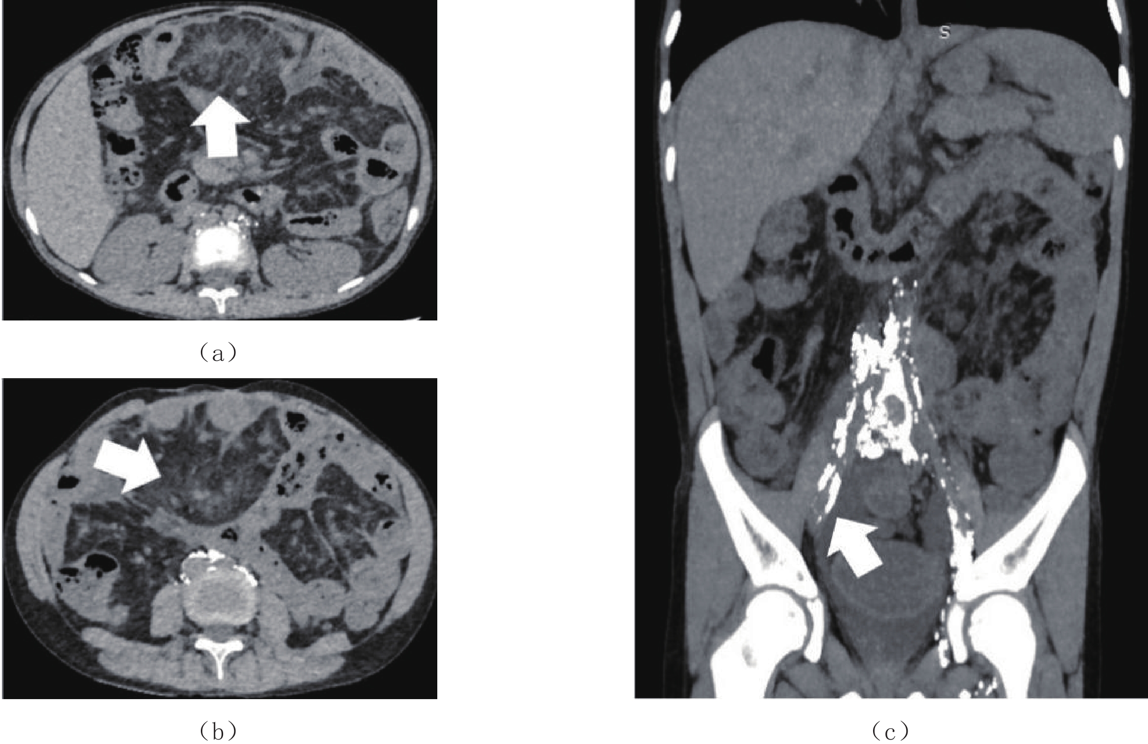
图 1 Ⅰ型PIL,多发对比剂异常分布
Figure 1. Type Ⅰ PIL with diffusive abnormal contrast media in abdomen
表 1 不同类型PIL的临床特征比较分析
Table 1 Comparison of clinical characteristics in different types of PIL
临床特征 类型 统计检验 Ⅰ型(n=15) Ⅱ型(n=19) t/χ2 P 首次发病年龄 15.3±9.4 8.3±3.8 2.995 <0.010 性别比(女/男) 7/8 10/9 0.119 0.730 腹泻 11(73.3%) 6(31.6%) 5.846 0.016 颜面和/或肢体肿胀 8(53.3%) 19(100%) 11.165 <0.010 血清白蛋白/(g/L) 17.1±4.4 18.6±6.3 -0.935 0.357  下载: 导出CSV
表 2 不同类型PIL的影像特征比较分析
Table 2 Comparison of CTL imaging features in different types of PIL
影像特征 类型 统计检验 Ⅰ型(n=15) Ⅱ型(n=19) χ2 P 小肠改变 肠壁环形增厚 2(13.3%) 1(5.3%) 0.679 0.410 肠系膜密度增高 7(46.7%) 18(94.7%) 9.952 0.002 浆膜腔积液 仅腹腔积液 1(6.7%) 3(15.8%) 0.672 0.412 腹腔+胸腔积液 1(6.7%) 4(21.1%) 1.383 0.240 返流样表现 8(53.3%) 7(36.8%) 0.925 0.336 淋巴瘘 5(33.3%) 0(0.0%) 7.425 0.006 腹腔内淋巴结 2(13.3%) 12(63.2%) 8.591 0.003 腹膜后淋巴管扩张 13(86.7%) 0 26.660 0.000 颈部扩张淋巴管 5(33.3%) 12(63.2%) 2.982 0.084
下载: 导出CSV
-
[1] GONZALEZ N R, DOMINGUEZ C A, ALCOBA V L, et al. Primary intestinal lymphangiectasia: A rare disease as a cause of protein-losing enteropathy[J]. Revista Espanola de Enfermedades Digestivas, 2022. DOI:10.17235/reed.2022.8673/2022. (Online ahead of print).
[2] KHAYAT A A. Primary intestinal lymphangiectasia presenting as limb hemihyperplasia: A case report and literature review[J]. BMC Gastroenterol, 2021, 21(1): 225. doi: 10.1186/s12876-021-01813-6
[3] BORZUTZKY A, ESPINO A, ALBERTI G, et al. Primary intestinal lymphangiectasia (Waldmann's Disease)[J]. American Journal of Gastroenterology, 2019, 114(2): 197. doi: 10.1038/s41395-018-0413-0
[4] YANG C, DEHNER L P. Protein-losing enteropathy with intestinal lymphangiectasia in skeletal dysplasia with Lys650 Met mutation[J]. American Journal of Medical Genetics Part A, 2016, 170(11): 2993−2997. doi: 10.1002/ajmg.a.37756
[5] CRUTZEN B, PONCELET P A. Protein-losing enteropathy in primary lymphangiectasia[J]. Journal of the Belgian Society of Radiology, 2020, 104(1): 34. doi: 10.5334/jbsr.2136
[6] LOPEZ R N, DAY A S. Primary intestinal lymphangiectasia in children: A review[J]. Journal of Paediatrics and Child Health, 2020, 56(11): 1719−1723. doi: 10.1111/jpc.14837
[7] MANORIA P, GULWANI H V. Primary intestinal lymphangiectasia in a middle-aged female[J]. Saudi Jorunal Medicine & Medical Sciences, 2021, 9(3): 280−281.
[8] HUBER R, SEMMLER G, MAYR A, et al. Primary intestinal lymphangiectasia in an adult patient: A case report and review of literature[J]. World Journal of Gastroenterology, 2020, 26(48): 7707−7718. doi: 10.3748/wjg.v26.i48.7707
[9] PRASAD D, SRIVASTAVA A, TAMBE A, et al. Clinical profile, response to therapy, and outcome of children with primary intestinal lymphangiectasia[J]. Digestive Diseases, 2019, 37(6): 458−466. doi: 10.1159/000499450
[10] CRAVEN M D, WASHABAU R J. Comparative pathophysiology and management of protein-losing enteropathy[J]. Journal of Veterinary Internal Medicine, 2019, 33(2): 383−402. doi: 10.1111/jvim.15406
[11] KAKIUCHI T, MIZUOCHI T, KOJI A, et al. Long-term endoscopic findings in pediatric primary intestinal lymphangiectasia[J]. Clinical Case Reports, 2021, 9(2): 1029−1030. doi: 10.1002/ccr3.3619
[12] CUNNINGHAM J M, NEPAL S, TRUESDALE A E. Primary intestinal lymphangiectasia diagnosed by video capsule endoscopy in a patient with immunodeficiency presenting with morganella morganii bacteraemia[J]. BMJ Case Reports, 2020, 13(9): e235898. doi: 10.1136/bcr-2020-235898
[13] TOMINAGA K, TSUCHIYA A, KAWATA Y, et al. Novel magnified single-balloon enteroscopy enables observation of jejunal white spots associated with lymphangiectasia[J]. Digestive Diseases, 2019, 37(2): 170−174. doi: 10.1159/000493578
[14] Van der REIJDEN S M, van WIJK M P, JACOBS M, et al. Video capsule endoscopy to diagnose primary intestinal lymphangiectasia in a 14-month-old child[J]. Journal of Pediatric Gastroenterology and Nutrition, 2017, 64(6): e161. doi: 10.1097/MPG.0000000000001586
[15] BROWNELL J N, BIKO D M, MAMULA P, et al. Dynamic contrast magnetic resonance lymphangiography localizes lymphatic leak to the duodenum in protein-losing enteropathy[J]. Journal of Pediatric Gastroenterology and Nutrition, 2022, 74(1): 38−45. doi: 10.1097/MPG.0000000000003287
[16] DONG J, XIN J, SHEN W, et al. Unipedal diagnostic lymphangiography followed by sequential ct examinations in patients with idiopathic chyluria: A retrospective study[J]. American Journal of Roentgenology, 2018: 210(4): 792-798.
[17] 董健, 信建峰, 霍萌, 等. 直接淋巴管造影和CT淋巴管成像在乳糜尿中的诊断价值[J]. 临床放射学杂志, 2018,37(5): 798−802. doi: 10.13437/j.cnki.jcr.2018.05.020 DONG J, XIN J F, HUO M, et al. Direct lymphangiography with sequential CT lymphangiography in chyluria: A retrospective study[J]. Clinical Radiology, 2018, 37(5): 798−802. (in Chinese). doi: 10.13437/j.cnki.jcr.2018.05.020
[18] 董健, 沈文彬, 信建峰, 等. 联合应用CT淋巴管成像与直接淋巴管造影诊断原发性小肠淋巴管扩张症的价值[J]. 中华放射学杂志, 2017,51(5): 362−365. doi: 10.3760/cma.j.issn.1005-1201.2017.05.008 DONG J, SHEN W B, XIN J F, et al. Value of CT lymphangiography combined with direct lymphangiography in diagnosing primary intestinal lymphangiectasia[J]. Journal of Chinese Radiology, 2017, 51(5): 362−365. (in Chinese). doi: 10.3760/cma.j.issn.1005-1201.2017.05.008
[19] DONG J, XIN J, SHEN W, et al. CT lymphangiography (CTL) in primary intestinal lymphangiectasia (PIL): A comparative study with intraoperative enteroscopy (IOE)[J]. Academic Radiology, 2019, 26(2): 275−281. doi: 10.1016/j.acra.2018.04.023
[20] 董健, 信建峰, 张春燕, 等. 直接淋巴管造影术后CT在原发性小肠淋巴管扩张症中的回顾性研究[J]. 临床放射学杂志, 2018,37(9): 1510−1514. DONG J, XIN J, ZHANG C Y, et al. Post-lymphangiographic CT (PLCT) in primary intestinal lymphangiectasia: A retrospective study[J]. Clinical Radiology, 2018, 37(9): 1510−1514. (in Chinese).
[21] 董健, 信建峰, 霍萌, 等. 直接淋巴管造影与CT淋巴管成像在继发性小肠淋巴管扩张症的回顾性研究[J]. 临床放射学杂志, 2018,37(3): 419−423. doi: 10.13437/j.cnki.jcr.2018.03.014 DONG J, XIN J, HUO M, et al. Direct lymphangiography with sequential CT lymphangiography in secondary intestinal lymphangiectasia: A retrospective study[J]. Clinical Radiology, 2018, 37(3): 419−423. (in Chinese). doi: 10.13437/j.cnki.jcr.2018.03.014
计量
- 文章访问数: 383
- HTML全文浏览量: 193
- PDF下载量: 20


