
Application of Real Size Film Printing Technology in Costal Cartilage CT Image
-
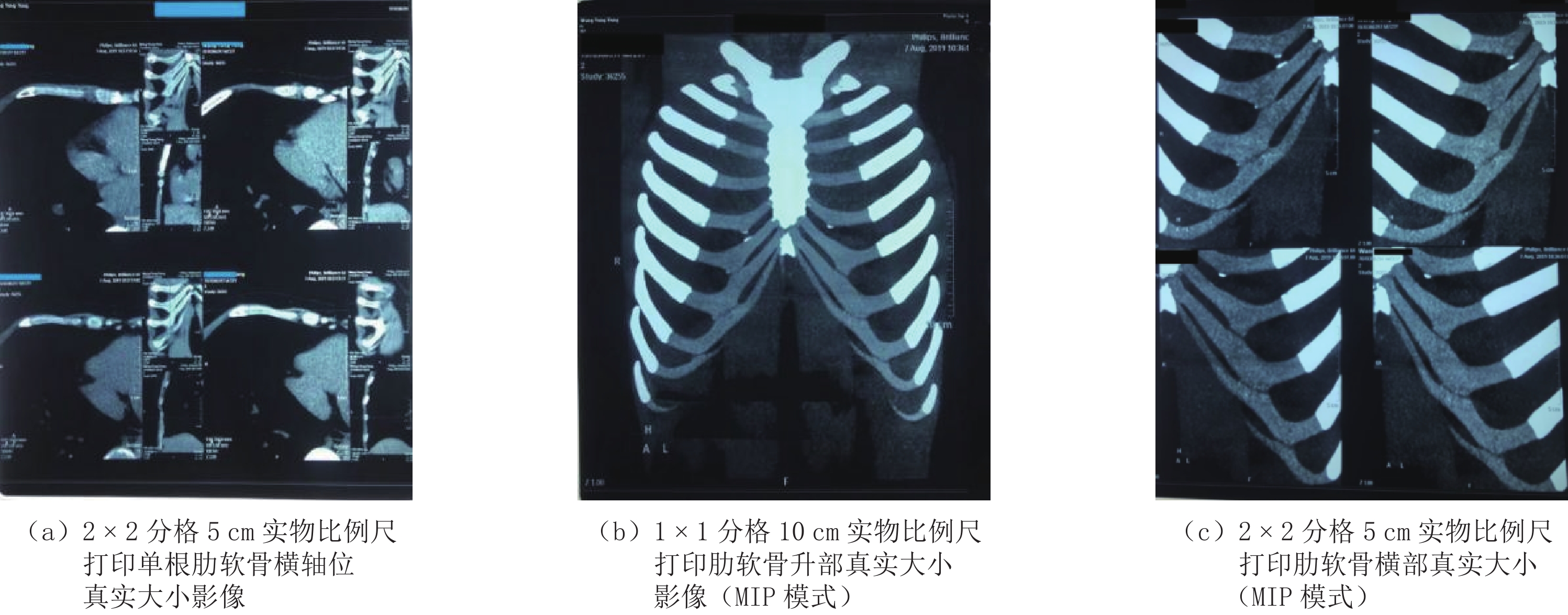
摘要: 目的:探索在医用胶片上对肋软骨CT影像显示其真实大小的胶片打印技术。方法:选取31例患者肋软骨CT影像资料,经影像后处理,胶片采用14英寸×17英寸(35 cm×43 cm)规格,计算EBW工作站显示屏主影像显示区和14×17胶片上实际影像显示区之间的比例关系,制成10 cm和5 cm两个长度规格的实物比例尺。打印时将胶片以2×2分格呈4幅图像布局,选取5 cm实物比例尺打印单根肋软骨横轴面或矢状面影像;以1×1分格呈单幅图像布局,选取10 cm实物比例尺打印全部前肋弓(含肋软骨)的三维影像。选取右侧第6肋软骨升部、横部的长度,升部横部连接处宽度、厚度,肋软骨胸骨端、肋软骨肋骨端的厚度等6个指标的胶片测量值和手术中实体测量值进行比较,并做统计学分析。结果:①对已打印的胶片进行测量,胶片上所示10 cm和5 cm比例尺与直尺的实际尺寸相等;②胶片上肋软骨影像的 6组测量值和手术中的肋软骨实体测值对比,测量值差异无统计学意义。结论:基于DICOM协议下的打印技术可以实现肋软骨CT影像在胶片上的真实大小打印,术者从胶片中所获取的目标组织的形态数据可靠。Abstract: Objective: To explore the film printing technology for displaying the true size of costal cartilage on medical film. Methods: The CT images of costal cartilage of 31 patients were selected and processed 14×17 inches (35×43 cm), calculate the main image display area and 14×17 the scale between the actual image display areas on the film shall be made into physical scales of 10 cm and 5 cm in length. Print the film in 2×2. Four images are arranged in the grid, and the transverse or sagittal images of single rib cartilage are printed on a 5 cm physical scale; with 1×1 The grid is a single image layout, and the 3D images of all the front rib arches (including costal cartilage) are printed on a 10 cm physical scale. The film measurement values of six indicators, including the length of the ascending and transverse parts of the right sixth costal cartilage, the width and thickness of the transverse junction of the ascending part, the thickness of the sternal end of the costal cartilage and the rib end of the costal cartilage, were compared with the solid measurement values during the operation, and statistical analysis was made. Results: (1) The printed film was measured, and the 10 cm and 5 cm scales shown on the film were equal to the actual size of the ruler. (2) There was no significant difference between the measured values of six groups of costal cartilage images on film and the measured values of costal cartilage entities during surgery. Conclusion: The printing technology based on DICOM protocol can realize the real size printing of costal cartilage CT image on film. The morphological data of the target tissue obtained by the operator from the film are reliable.
-
Keywords:
- CT Image /
- costal cartilage /
- true size /
- film printing
-
近年来,岩性复合型油气藏勘探已逐渐成为东海油气扩储增产的重要来源,但储集体普遍呈现低孔渗特征,流体引起的弹性差异微弱,空间非均质性展布规律显著。N构造位于西湖凹陷中央洼陷反转构造带中南部,紧邻区域生烃主洼,在轴向物源和西侧物源双源汇聚的背景下,受局部地貌和区域断裂条件影响,发育多种类型砂岩岩性体,周边已钻井揭示纵向发育多层系多套油气层。源-断-圈-储高效匹配具备有利油气成藏条件,开展致密砂岩储层流体描述,对于打开该构造背景上的岩性复合型油气藏勘探的突破口具有重要意义。
利用地震资料直接判断岩层含油气情况是国内外地球物理学家亟需攻克的热点课题[1]。相比于岩石固体骨架,孔隙流体所激发的地震信号微弱,通常借助地震波的运动学、动力学、形态学、弹性以及黏滞性等特征合理放大异常响应差异[2]。由此而产生了“亮点”技术、频率域吸收衰减属性以及流体因子等方法,其中以后者最为聚焦[3]。特别是随着振幅随偏移距的变化(amplitude variation with offset,AVO)技术的诞生和发展,基于叠前地震信息的流体描述技术得到了广泛关注和深入研究。
Ostrander[4]对岩心进行孔隙流体替换后发现叠前AVO响应特征存在明显差异;Chiburis[5]通过消除深浅层反射振幅能量差异,突出流体所引起的AVO响应,成功实现了叠前地震流体检测。Smith等[6]研究发现纵横波反射率的加权叠加能够有效区分流体,由此开启了利用“流体因子”进行油气检测的先河;Gidlow等[7]、Fatti等[8]和Wallace等[9]研究认为如何减弱背景因素对地震反射特征的影响是此类流体因子构建的关键,并给出了纵横波阻抗反射率的优化方案;Smith等[10]提出了“流体因子角”思想,与在阻抗域进行坐标旋转的泊松阻抗和扩展弹性阻抗理念相符;Goodway等[11-12]分析认为拉梅参数相比于岩石速度更具本质优势,实现了利用弹性模量开展烃类检测的跨越。此后,Hedlin[13]从岩石弹性模量中解耦出孔隙流体空间模量用以直接表征流体的弹性变化;Batzle[14]通过实验室测量发现利用体积模量与剪切模量的差值可进一步压制背景因素的影响提高流体识别能力;在双相多孔弹性介质理论的框架下,Russell等[15-16]对岩石流体项和固体项分别实现了数学形态解耦,发现流体项在时空域具有较高的流体描述精度。然而,研究对象的差异化导致每种方法均存在一定的局限性和适用性,必须结合实际工区优选高灵敏的烃检因子[17]。
叠前地震反演是从地震数据中提取岩石弹性信息的有效手段。苏世龙等[18]基于叠前纵横波阻抗同步反演开展储层含油气性预测;王保丽等[19]探讨了拉梅参数弹性阻抗方程的标准化形式,取得了较好的流体识别效果;宗兆云等[20]给出了流体项的纵横波模量表达形式,并利用弹性阻抗反演实现了流体敏感信息的直接提取;印兴耀等[21]深入解析了如何利用两项弹性阻抗反演提升深层 Russell流体因子可靠性的策略;杨培杰等[22]在贝叶斯理论框架下求解基于 Russell近似的AVO模型参数化方程实现了流体项的直接反演;邓炜等[23]以多孔弹性介质岩石物理模型为驱动,利用弹性阻抗反演实现了等效流体体积模量的直接反演,该方法对地震数据要求高,在致密储层存在求解不稳定问题;贾凌云等[24]结合广义弹性阻抗方程,利用新构建的敏感流体因子,对致密砂岩储层流体识别进行了尝试;周家雄等[25]将约束最小二乘与信赖域算法融合,通过对储层物性参数(粘土含量和孔隙度)的直接岩石物理反演进行“甜点”储层评价。但含水饱和度预测的收敛精度仍受制约;周林等[26]构建了精确佐普利兹方程的流体因子和泊松比的直接反演方法,提升了烃类检测精度和可靠性;Ma等[27]、Pan等[28]以岩石物理模型为驱动,探索了各向异性参数的直接反演方法。但预测结果对低频模型依赖度高,横向边界刻画不清晰。此外,传统叠前反演方法的初始模型导致地震振幅横向相对变化特征易被“模糊化”,并且地震子波对反演结果的影响程度较大[29]。
针对研究区致密砂岩储层地震流体识别所面临的岩石物理特征叠置严重、储层非均质性变化剧烈以及深层地震数据缺乏大角度信息等问题,本文结合岩石物理定性和定量判析技术,优选高灵敏烃检因子。以两项AVO模型参数化方程为基础,利用有色反演技术实现拉梅参数的直接提取,最终得到高灵敏烃检因子的地震流体敏感弹性信息。此成果应用于研究区对构造-岩性复合型气藏致密砂岩储层流体描述具有较高精度和适用性。
1. 方法原理
1.1 基于拉梅参数的两项AVO模型参数化方程
地震波反射系数的佐普利兹方程精确解及其近似式是对全弹性波方程的简化,为从叠前AVO信息中直接提取能够反映地层特性的弹性参数搭建了重要桥梁[30]。针对深层缺乏大角度信息的问题,刘晓晶等[31]推导了基于Russell近似的包含流体项f 和剪切模量μ的两项AVO近似式。当干岩石纵横波速度比的平方取为2.0时,就退化为包含拉梅模量λ和剪切模量μ的两项AVO近似式。可表示如下:
$$ \begin{aligned} {R_{{\rm{pp}}}}\left( \theta \right) = \,\Biggr( {\left( {\frac{1}{2} - \frac{1}{{\gamma _{{\rm{sat}}}^2}}} \right){{\sec }^2}\theta \frac{{\gamma _{{\rm{sat}}}^2{l_1} - 2{l_2}}}{{\left( {2{l_1} + 1} \right)\gamma _{{\rm{sat}}}^2 - 2\left( {2{l_2} + 1} \right)}}} \Biggr)\frac{{\Delta \lambda }}{\lambda } + \\ \Biggr( {\frac{1}{2} - \frac{2}{{\gamma _{{\rm{sat}}}^2}}{{\sin }^2}\theta - \left( {1 - \frac{1}{{\gamma _{{\rm{sat}}}^2}}{{\sec }^2}\theta } \right)\frac{{{l_2}}}{{1 + 2{l_2}}}} \Biggr)\frac{{\Delta \mu }}{\mu } \text{,}\; \end{aligned}$$ (1) 式中
$\gamma_{{\rm{sat}}} $ 表示饱和岩石纵横波速度比;l1和l2分别表示实际工区的密度与纵波速度、横波速度反射系数之间的拟合系数,θ表示阻抗差界面入射角度和透射角度之和的一半,λ和μ分别表示反射界面两侧拉梅参数模量和剪切模量的均值,Δλ和Δμ分别表示阻抗差界面相应拉梅参数模量和剪切模量的差值。表1为文献[26]中建立的双层砂岩模型弹性参数,其中关键参数
$\gamma ^2_{{\rm{dry}}} $ 和$\gamma ^2_{{\rm{sat}}} $ 的平均值分别取2.31和4.0。结合此模型开展佐普利兹方程精确解、Russell近似式和λ-μ近似式的反射系数精度对比分析。表 1 含气砂岩和含水砂岩模型弹性参数Table 1. Parameters of the sandstone model地层 Kdry/GPa μ/GPa $\gamma ^2_{{\rm{dry}}}$ $\gamma ^2_{{\rm{sat}}} $ $\rho $/(g/c3) VP/(m/s) VS/(m/s) 含水砂(上层) 3 3 2.32 4.48 1.9 2680 1265 含气砂(下层) 3 3 2.30 3.51 1.7 2520 1345 从图1中可以看到,λ-μ近似式与 Russell近似式的整体匹配度较高。特别地,在入射角小于20° 时两者与佐普利兹方程精确解的误差均较小;当入射角大于20° 时,相对误差均逐渐增大。因此,利用λ-μ近似式能够满足小角度入射条件下地震反演的精度需求。
![]() 图 1 含水砂岩底界面反射系数对比Figure 1. Comparison of reflection coefficients of the bottom interface of water-bearing sandstone
图 1 含水砂岩底界面反射系数对比Figure 1. Comparison of reflection coefficients of the bottom interface of water-bearing sandstone1.2 叠前有色反演技术
有色反演技术是传统递归反演的改进算法,其本质思想是在频率域建立井间弹性参数与地震信息的映射关系,从而实现地震反射率体到弹性参数体的直接转换[32]。
地震记录利用褶积模型可数学表征为:
$$ s(t) = r(t) * w(t) \text{,} $$ (2) 式中:
$ s(t) $ 表示地震记录,$ r(t) $ 表示反射系数,$ w(t) $ 表示地震子波。将(2)式由时间域变换到频率域,即:
$$ S(\omega)=R(\omega) \cdot W(\omega) \text{,} $$ (3) 式中:
$ S(\omega ) $ 表示地震振幅谱,$ R(\omega ) $ 表示反射系数振幅谱,$ W(\omega ) $ 表示子波振幅谱。将(3)式两边取对数,得:
$$ \ln S(\omega)=\ln R(\omega)+\ln W(\omega) 。 $$ (4) 将界面反射系数变换到频率域,即:
$$ R(\omega)=\Big(Z_{2}(\omega)-Z_{1}(\omega)\Big) \Big/\Big(Z_{2}(\omega)+Z_{1}(\omega)\Big) 。 $$ (5) 将式(5)带入式(4)中,求得频率域匹配算子
$ W(\omega ) $ ,进一步通过傅里叶反变换即可获得时间域有色反演匹配算子$ w(t) $ 。该反演方法可有效减少稀井区井插值低频模型约束所造成的模型化问题,不仅能够保持反演结果在测井数据的量纲范围内波动,而且更好地反映了地震数据的细节特征。实现关键环节为[29]:①抽取靶区井旁地震道测井数据与地震数据分别进行振幅谱统计分析;②利用最小二乘拟合算法构建频率域匹配算子,进而转换得到时间域相位旋转 -90° 的有色算子;③结合上述算子将弹性反射率体直接转换为相对弹性数据体,开展无井反演;④通过频率域融合等算法,即可进一步实现层间绝对弹性数据体的有井反演。
1.3 拉梅参数弹性信息直接提取
叠前弹性参数直接反演技术是开展地震储层流体描述的重要手段,有效规避了间接计算的误差积累,具有更高的参数提取精度。利用(1)式建立叠前地震反射系数与拉梅模量λ和剪切模量μ在不同入射角情况下的映射关系:
$$ \left( {\begin{array}{*{20}{c}} \\ {{R_{PP}}({\theta _1},t)} \\ {{R_{PP}}({\theta _2},t)} \\ \vdots \\ {{R_{PP}}({\theta _n},t)} \\ \end{array}} \right) = \left( {\begin{array}{*{20}{c}} \\ {\begin{array}{*{20}{c}} {A({\theta _1},t)} \\ {A({\theta _2},t)} \\ \vdots \\ {A({\theta _n},t)} \\ \end{array}}&{\begin{array}{*{20}{c}} \\ {B({\theta _1},t)} \\ {B({\theta _2},t)} \\ \vdots \\ {B({\theta _n},t)} \end{array}}\\ \end{array}} \right)\left( {\begin{array}{*{20}{c}} \\ {\Delta \lambda /\lambda } \\ {\Delta \mu /\mu } \\ \end{array}} \right) \text{,} $$ (6) 式中
$ A\left( {{\theta _i},t} \right) $ 和$ B\left( {{\theta _i},t} \right) $ 分别表示方程系数,i表示地震数据不同采样点。通过反演算法求解上述矩阵方程组,即可从叠前道集中分别获得拉梅模量反射率
$ {{\Delta \lambda } \mathord{\left/ {\vphantom {{\Delta \lambda } \lambda }} \right. } \lambda } $ 和剪切模量反射率$ {{\Delta \mu } \mathord{\left/ {\vphantom {{\Delta \mu } \mu }} \right. } \mu } $ 的AVO属性体。进一步结合叠前有色反演直接获得相应的层间弹性信息,通过简单数学运算即可提取敏感流体因子弹性数据体[33]。关键实现操作流程,如图2所示。![]() 图 2 利用拉梅参数直接反演方法进行烃类检测流程图Figure 2. The flow chart of hydrocarbon detection using lame parameters direct inversion
图 2 利用拉梅参数直接反演方法进行烃类检测流程图Figure 2. The flow chart of hydrocarbon detection using lame parameters direct inversion2. 实例应用
N构造位于西湖凹陷中央洼陷-反转构造带,是中新世晚期东西向挤压应力作用下形成的宽缓低幅背斜构造,属于典型的“洼中隆”。研究区发育缓坡背景下的河流-三角洲沉积体系,整体以三角洲平原分流河道砂体为主,主要目的层M组(M1、M2和M3)分布于3800~4100 m。受埋深压实作用、成岩作用和地质沉积环境影响,岩层岩石物理特征叠置严重,横向非均质性强,孔渗关系复杂。研究发现工区深层实际地震有效角度最大为25°;岩芯和壁芯资料表明储层孔隙度主要分布在0.8%~14.2%,平均值10.9%;渗透率主要分布在0.01~11.6 mD,平均值3.1 mD;总体为低孔低渗,局部呈特低孔特低渗特征。
实践证明该区存在低孔低渗背景下“一块多藏,一砂一藏”的构造-岩性复合型气藏模式,落实致密砂岩储层流体分布范围是打开该区新领域勘探突破的重要保障。本文利用拉梅参数直接反演技术实现了烃检因子地震流体弹性信息的直接提取,为致密砂岩储层含气性检测提供指导。
高质量的地震资料是开展深层致密储层烃类检测的有力保障。采用多步保真噪音压制技术有效压制涌浪噪音、线性及绕射噪音。针对浅水海域鬼波时空变剧烈问题,利用自适应参数优化方法实现震源及电缆鬼波的综合压制。通过τ-p域自相关扫描优化海底深度模型,实现水体速度校正和浅水多次波压制。数据规则化后,利用高精度 Radon变换压制残余多次波突出有效信号。基于波动方程建立包含振幅和相位的吸收补偿递推关系,实现叠前时间偏移道集的反Q补偿,提升地震资料分辨率。为后续地震流体识别奠定了坚实基础。
2.1 岩石物理敏感度判析
目前判别不同流体因子的识别能力主要采用岩石物理交会定性分析和数值化指标定量分析两种技术[34]。结合研究区已钻探井A井、B井和C井主力层的实测井数据以及解释成果,进行了地震岩石物理交会。从图3中可以看出,纵波阻抗ZP、横波阻抗ZS、体积密度ρ、剪切模量μ以及密度乘以剪切模量ρμ 对于含烃层和非烃层存在严重的叠置;泊松比σ、密度乘以拉梅模量ρλ、拉梅模量λ和扩展弹性阻抗 EEI 30° 对于含烃层和非烃层的区分度提升,但仍存在一定的叠置区间。而拉梅参数比值λ/μ的叠置区间最小,区分度最优。
流体敏感指示系数[35]通常作为衡量某种弹性参数对不同流体识别敏感度的定量判别标准,其数值越大对应识别灵敏度越高,如(7)式所示:
$$ H = \frac{{\left| {{E_{\rm{w}}} - {E_{\rm{g}}}} \right|}}{{{D_{\rm{g}}}}} \text{,} $$ (7) 其中,H表示流体敏感指示系数,其数值越大表示弹性参数对不同流体的区分能力越强;Dg表示储层含气时弹性参数的标准方差值;Ew表示储层含水时弹性参数的期望值;Eg表示储层含气时弹性参数的期望值。
这里对研究区12种弹性参数敏感度进行了统计计算。图4显示常规参数纵、横波阻抗和体积密度的敏感指示系数最低;泊松比、弹性模量及其密度组合和扩展弹性阻抗等整体敏感指示系数相对较高,其中剥离岩石骨架影响的拉梅参数比值灵敏度最高。因此,通过井间岩石物理判析表明拉梅参数比值
$ {\lambda \mathord{\left/ {\vphantom {\lambda \mu }} \right. } \mu } $ 能够有效满足对致密砂岩储层烃类检测的精度要求。![]() 图 4 多种弹性参数的流体敏感指示系数1-AI;2-SI;3-σ;4-ρ;5-K;6-M;7-λ;8-μ;9-λ/μ;10-EEI30°;11-ρλ;12-ρμ。Figure 4. Fluid sensitivity indicator coefficient for various elastic parameters
图 4 多种弹性参数的流体敏感指示系数1-AI;2-SI;3-σ;4-ρ;5-K;6-M;7-λ;8-μ;9-λ/μ;10-EEI30°;11-ρλ;12-ρμ。Figure 4. Fluid sensitivity indicator coefficient for various elastic parameters2.2 弹性模量岩石物理量板分析
以实测数据为基础,结合Xu-White模型建立了拉梅参数的岩石物理量板。黏土含量变化范围为0%~50%,间隔25%;含水饱和度变化范围为0%~100%,间隔10%;孔隙度变化范围为0%~35%,间隔5%;其中流体相态混合模式采用更贴合实际的Brie经验公式。从图5中可以看到,剪切模量μ与孔隙度相关性较高,对流体的变化不敏感。在岩石高孔隙度时,拉梅模量λ能够有效识别流体;但随着孔隙度的减小,低孔气砂和高孔水砂弹性特征叠置严重,流体敏感性降低(图5(a))。而拉梅模量比值
$ {\lambda \mathord{\left/ {\vphantom {\lambda \mu }} \right. } \mu } $ 整体对于流体识别能力更强,叠置区间更小(图5(b))。因此,从岩石物理正演角度验证了该参数对致密储层流体识别的有效性。2.3 致密砂岩储层烃类检测
本研究区共有3口探井A井、B井和C井。主要目的层M组中M1层和M2层为低孔渗构造-岩性复合型气藏,M3层气饱含量低为含气水层。岩层阻抗差异小,叠后地震数据不能有效指示含烃层(图6(a))。本文分别开展常规扩展弹性阻抗反演(EEI 30°)和拉梅参数直接反演的烃类检测,并对比分析利用叠前同步反演间接估算拉梅参数比值
$ {\lambda \mathord{\left/ {\vphantom {\lambda \mu }} \right. } \mu } $ 的属性差异。图6(b)和图6(c)中红黄色代表低值含气性,蓝色代表高值含水性。在图中B井黑色虚方框处,M2层钻遇气层,M3层由两套相邻的含气水层砂体组成。在图中C井黑色虚椭圆框处,M1层揭示薄水层。对比可知,常规扩展弹性阻抗反演(EEI 30°)流体识别能力有限,目标区预测结果存在较强多解性。基于叠前同步反演间接计算的拉梅参数比值λ/μ 属性优于前者,但在黑色虚框处同样无法有效预测流体性质。主要是由于在缺乏大角度信息情况下利用三项式不能稳定提取弹性参数,并且间接计算会产生累计误差降低精度。而拉梅参数比值 λ/μ 直接反演结果清晰刻画了储集体内部流体的变化特征,整体与测井解释成果匹配度更高。主力层 M1层在该构造南部发育一条晚期南北向分流河道,切割北东向早期分流河道(图7)。地质油藏综合评价认为早期分流河道西南部发育构造-岩性复合型气藏,并且烃检存在强异常显示,设计部署了一口定向井。此外,工区南部为区域另一条河道主砂体发育部位,烃检异常明显(图7黑色实方框处),可作为后续该区滚动勘探开发的潜力目标区。
![]() 图 7 主力层M1沿层拉梅参数比值 λ/μ 烃类检测平面属性Figure 7. The plane attribute of λ/μ hydro- carbon detection of main layer M1
图 7 主力层M1沿层拉梅参数比值 λ/μ 烃类检测平面属性Figure 7. The plane attribute of λ/μ hydro- carbon detection of main layer M13. 总结与认识
为了落实研究区致密砂岩储层流体展布规律,本文引入一种基于拉梅参数直接反演的烃类检测方法。通过岩石物理定性和定量判析,确定拉梅参数比值对于研究区致密砂岩储层流体性质具有较高的识别能力。针对深层地震数据缺乏大角度信息的问题,采用两项AVO近似方程能够满足深层小角度入射的精度需求。充分利用叠前道集信息直接反演拉梅参数地震反射率数据体,并结合有色反演技术实现拉梅参数的直接估算,减小了间接估算的累计误差。有色反演技术有效保持了地震振幅的横向相对变化特征,使弹性参数地震提取更加客观。
该方法能够对研究区致密砂岩储层流体分布范围进行有效刻画,为后续勘探开发设计部署提供重要参考,于致密储层地震流体描述具有一定的应用价值和推广意义。
-
![]()
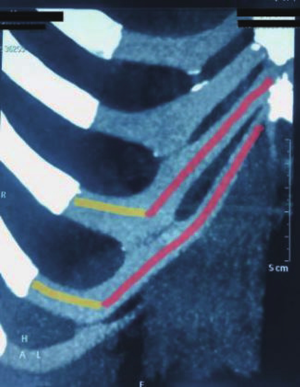
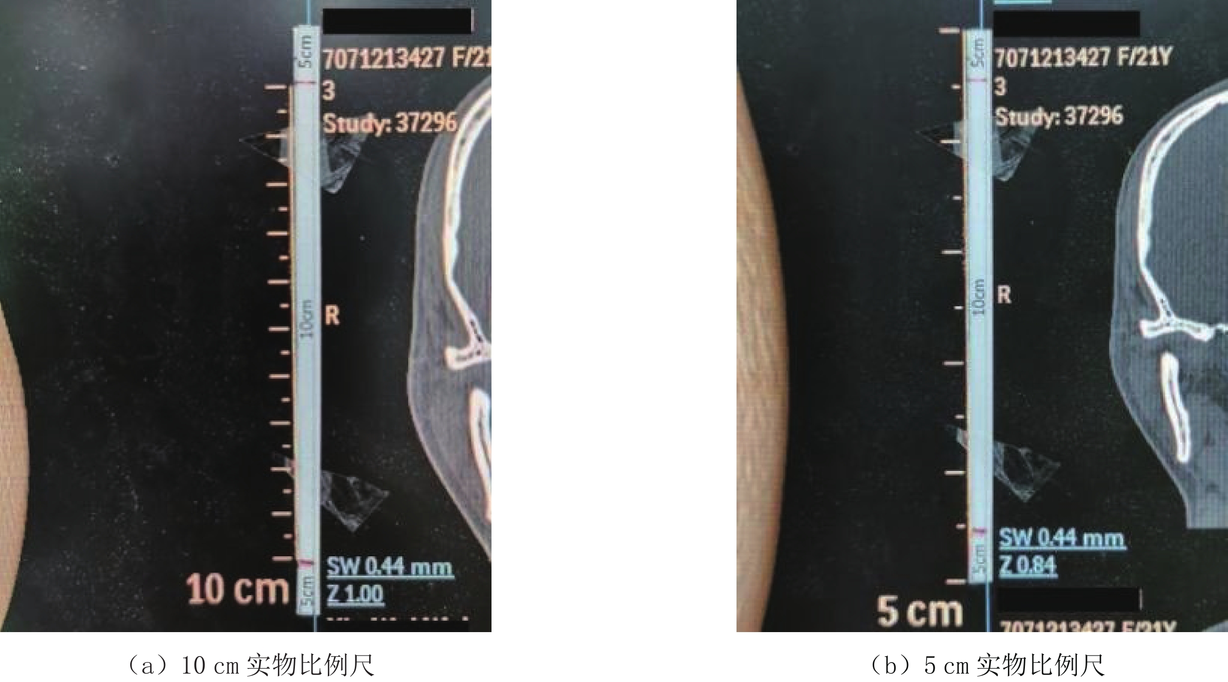
图 3 利用5 cm和10 cm实物比例尺在胶片上打印肋软骨真实大小影像
Figure 3. Print the true size image of costal cartilage on filsm with 5 cm and 10 cm physical scales
![]()
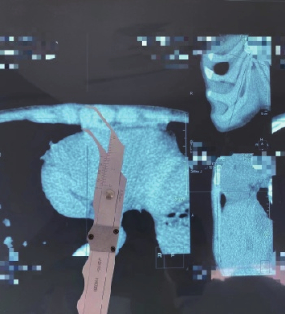
图 5 在胶片上测量右侧第6肋软骨胸骨端厚度(矢冠轴3图以十字线同步定位)
Figure 5. The measure the thickness of the sternal end of the sixth costal cartilage on the film
![]()
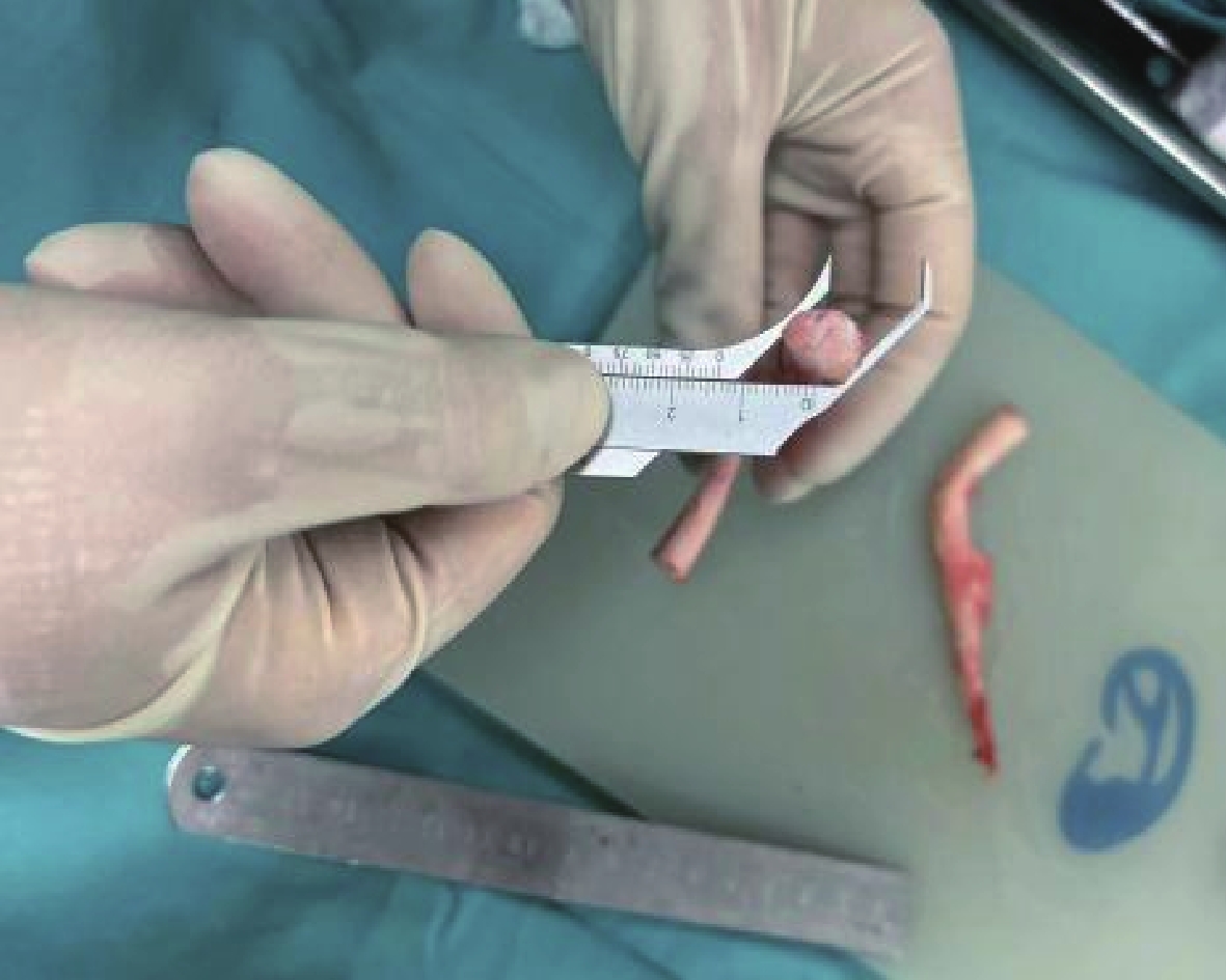
图 6 右侧第6肋软骨胸骨端术中实体测量厚度
Figure 6. The measured thickness of the sternal end of the sixth costal cartilage on the right during operation
表 1 肋软骨6个相同位置胶片测量值和人体测量值差异统计
Table 1 Difference statistics of film measurement value and human measurement value at five same positions of costal cartilage
分类 胶片测量值(均数±标准差) 统计检验 实体 胶片 t P 肋软骨升部长度/cm 77.46±13.46 77.84±13.45 -0.07 0.94 肋软骨横部长度/cm 33.44±5.49 33.24±5.85 0.09 0.93 升部横部连接处宽度/cm 15.83±1.94 15.80±2.24 0.03 0.98 升部横部连接处厚度/cm 5.95±1.77 5.96±1.77 -0.01 0.99 肋软骨胸骨端厚度/cm 12.05±2.50 12.02±2.49 0.03 0.98 肋软骨肋骨端厚度/cm 8.48±1.11 8.49±1.02 -0.04 0.97  下载: 导出CSV
-
[1] VON G H, FISCHER H, EPPSSTEIN R, et al. Framework fabrication with rib eartilage in partial and total nasal reconstruction[J]. Facial Plastic Surgery, 2014, 30(3): 306−317. DOI: 10.1055/s-0034-1376876.
[2] 周佳宇, 林琳, 蒋海越, 等. 个性化肋软骨采集及其耳支架雕刻[J]. 中华耳科学杂志, 2013,11(4): 502−505. DOI: 10.3969/j.issn.1672-2922.2013.04.008. ZHOU J Y, LIN L, JIANG H Y, et al. Individualized fabrication and application of autogenous cartilage framework in auricular reconstruction[J]. Chinese Journal of Otology, 2013, 11(4): 502−505. DOI: 10.3969/j.issn.1672-2922.2013.04.008. (in Chinese).
[3] 王少健, 陈娇, 丁忠祥, 等. 螺旋CT三维后处理技术在肋软骨骨折诊断中的价值[J]. 中华创伤杂志2020, 36(1): 78-81. DOI: 10.3760/cma.j.issn.1001-8-50.2020.01.016. WANG S J, CHEN J, DING Z X, et al. The value of spiral CT three-dimensional post-processing technique in the diagnosis of costal cartilage fracture[J]. Chinese Journal of Trauma, 2020, 36(1): 78-81. DOI:10.3760/cma.j.issn.1001-8-50.2020.01.016. (in Chinese).
[4] 何乐人, 杨庆华, 蒋海越, 等. 小耳畸形八大处法耳廓再造术−团队10年经验[J]. 中华整形外科杂志, 2017,33(8): 30−35. DOI: 10.3760/cma.j.issn.1009-4598.2017.s1.007. HE L R, YANG Q H, JIANG H Y, et al. Ear reconstruction with Ba-Da-Chu method: Ten-year experiences of our team[J]. Chinese Journal of Plastic Surgery, 2017, 33(8): 30−35. DOI: 10.3760/cma.j.issn.1009-4598.2017.s1.007. (in Chinese).
[5] 毛小明, 蒋廷宠. 低场MR对肋软骨损伤的检查价值[J]. 实用放射学杂志, 2011,27(4): 644−645. DOI: 10.3969/j.issn.1002-1671.2011.04.050. MAO X M, JIANG T C. Costal cartilage injury: Evaluation by low-field magnetic resonance imaging[J]. Journal of Practical Radiology, 2011, 27(4): 644−645. DOI: 10.3969/j.issn.1002-1671.2011.04.050. (in Chinese).
[6] 王永振, 何乐人, 刘雳, 等. 多层螺旋CT扫描及三维重建技术在肋软骨组织量评估中的应用研究[J]. 中国修复重建外科杂志, 2014,28(10): 1266−1269. DOI: 10.7507/1002-1892.20140274. WANG Y Z, HE L R, LIU L, et al. Evaluationof multi-slice spiral CT scanand image reconstruction technology ineetimating costal cartilage volume[J]. Chinese Journal of Reparative and Reconstructive Surgery, 2014, 28(10): 1266−1269. DOI: 10.7507/1002-1892.20140274. (in Chinese).
[7] 刘雳, 李博, 曹捷, 等. 儿童肋软骨MSCT扫描后三种三维成像技术的比较[J]. 中华整形外科杂志, 2017,33(5): 363−366. DOI: 10.3760/cma.j.issn.1009-4598.2017.05.009. LIU L, LI B, CAO J, et al. Comparison of three 3D imaging techniques in children with costal cartilage MSCT scan[J]. Chinese Journal of Plastic Surgery, 2017, 33(5): 363−366. DOI: 10.3760/cma.j.issn.1009-4598.2017.05.009. (in Chinese).
[8] 魏忠荣, 陈涛, 戴维思, 等. 多层螺旋CT及Cardiac 1预设重建模式对肋软骨骨折的诊断价值[J]. 实用放射学杂志, 2020,36(3): 472−474, 490. DOI: 10.3969/j.issn.1002-1671.2020.03.033. WEI Z R, CHEN T, DAI W S, et al. The diagnostic value of MSCT and the preset reconstruction model of cardiac 1 in costal cartilage fractures[J]. Journal of Practical Radiology, 2020, 36(3): 472−474, 490. DOI: 10.3969/j.issn.1002-1671.2020.03.033. (in Chinese).
[9] 路涛, 蒲红, 杨诚, 等. 多层螺旋CT容积重建技术在儿童鸡胸中的应用价值[J]. 实用放射学杂志, 2016,32(7): 1088−1091. DOI: 10.3969/j.issn.1002-1671.2016.07.025. LU T, PU H, YANG C, et al. Value of volume rendering technique of MSCT in the diagnosis of pediatric pectus carinatum[J]. Journal of Practical Radiology, 2016, 32(7): 1088−1091. DOI: 10.3969/j.issn.1002-1671.2016.07.025. (in Chinese).
[10] 王立振, 李秀涛, 吕涵青. 多层螺旋CT三维重建在肋骨及肋软骨的应用体会[J]. 中国CT和MRI杂志, 2018,16(2): 124−126. DOI: 10.3696/j.issn.1672-5131.2018.03.039. WANG L Z, LI X T, LV H Q. Multislice spiral CT three-dimensional reconstruction of bone in rib and costal cartilage experience[J]. Chinese Journal of CT and MRI, 2018, 16(2): 124−126. DOI: 10.3696/j.issn.1672-5131.2018.03.039. (in Chinese).
[11] 周晨钟. CR系统在四肢床旁片应用价值的研究[J]. 临床和实验医学杂志, 2012,11(1): 63−64. DOI: 10.3969/j.issn.1671-4695.2012.01.032. ZHOU C Z. Study on the application value of CR system in bedside films of limbs[J]. Journal of Clinical and Experimental Medicine, 2012, 11(1): 63−64. DOI: 10.3969/j.issn.1671-4695.2012.01.032. (in Chinese).
[12] 宋玉全, 何志诚, 伍筱梅, 等. 斜射线摄影对直接数字化X线摄影系统影像质量的影响评价[J]. 中华放射学杂志, 2005,39(10): 1084−1087. DOI: 10.3760/j.issn:1005-1201.2005.10.017. SONG Y Q, HE Z C, WU X M, et al. Effect of oblique ray on image quality of directdigitized radiography system[J]. Chinese Journal of Radiology, 2005, 39(10): 1084−1087. DOI: 10.3760/j.issn:1005-1201.2005.10.017. (in Chinese).
[13] 汤婷, 张颖佳, 王继华, 等. 多层螺旋CT容积再现技术在肋软骨切取术中的应用[J]. 中华整形外科杂志, 2017,33(1): 57−60. DOI: 10.3760/cma.j.issn.1009-4598.2017.01.014. TANG T, ZHANG Y J, WANG J H, et al. Application of multi-slice spiral CT volume reconstruction technique in costal cartilage resection[J]. Chinese Journal of Plastic Surgery, 2017, 33(1): 57−60. DOI: 10.3760/cma.j.issn.1009-4598.2017.01.014. (in Chinese).
[14] 徐遄, 吴勇, 贾克斌, 等. 数字医学影像与通信的重要标准−DICOM标准[J]. 中国医学影像技术, 2002,18(9): 952−954. doi: 10.13929/j.1003-3289.2002.09.058 [15] 邢文珊, 钱瑾, 胡金天, 等. 肋软骨等比例打印在耳廓支架构建中的应用[J]. 中华整形外科杂志, 2018,(3): 206−209. DOI: 10.3760/cam.j.issn.1009-4598.2018.03.010. XING W S, QIAN J, HU J T, et al. The application of a 2D printing of rib cartilage in personalized ear framework fabrication[J]. Chinese Journal of Plastic Surgery, 2018, (3): 206−209. DOI: 10.3760/cam.j.issn.1009-4598.2018.03.010. (in Chinese).
[16] 苏扬, 刘静, 王江玥. 多层螺旋CT骨三维重建在肋骨及肋软骨骨折的诊断价值[J]. 中国CT和MRI杂志, 2016,14(7): 124−126. DOI: 10.3696/j.issn.1672-5131.2016.07.041. SU Y, LIU J, WANG J Y. The diagnostic value of multi-slice spiral CT 3D bone reconstruction on rib and rib cartilage fractures[J]. Chinese Journal of CT and MRI, 2016, 14(7): 124−126. DOI: 10.3696/j.issn.1672-5131.2016.07.041. (in Chinese).
[17] SUNWOO W, CHOL H, KIM D, et al. Characteristics of rib cartilage al calcification in Asian[J]. JAMA Facial Plastic Surgery, 2014, 16(2): 102−106. DOI: 10.1001/jamafacial.2013.2031.


计量
- 文章访问数: 394
- HTML全文浏览量: 195
- PDF下载量: 302


