
Current Situation and Future of Medical Imaging in the Diagnosis of Lymphatic Diseases
-
-
淋巴系统(lymphatic system)又称淋巴循环系统,是一个完整的独立于人体血液循环系统之外的具有输送淋巴液、产生淋巴细胞和主导免疫防御功能的重要体液循环系统,组织解剖学上由淋巴管(lymphatic channels)、淋巴液(lymphatic fluid)、淋巴细胞(lymphocytes)、淋巴组织(lymphoid tissue)和淋巴器官(lymphatic organs)等构成。淋巴系统相关的病理改变和疾病谱包括两大类:淋巴增生性疾病(lymphoproliferative disorders,LPDs)和淋巴回流障碍性疾病(lymphatic reflux disease,LRDs)。
多模态影像学检查作为一组广泛普及、无创伤性、高分辨率和精准评判形态及功能的成像方法,在淋巴系统疾病的诊断和治疗中发挥着重要的价值[1-3]。本文结合文献,就淋巴系统组织解剖和病理改变的特殊性,对不同类型影像学检查的现状和未来发展进行回顾性总结分析,旨在提高临床、影像和病理医师对此系统疾病的认知能力。
1. 淋巴系统的组织解剖和病理改变的特殊性
淋巴系统的5个构成部分依据解剖和生理功能分为两大类,即由大量淋巴细胞组成的淋巴组织或器官和类似毛细血管及静脉回流的内含淋巴液的淋巴管道系统。
1.1 淋巴细胞和淋巴组织
淋巴细胞是构成淋巴组织和淋巴器官的主要成分,依据来源和功能分为4种亚型:即胸腺依赖淋巴细胞(T细胞)、骨髓依赖淋巴细胞(B细胞)、杀伤性淋巴细胞(K细胞)和自然杀伤性淋巴细胞(NK细胞)。
(1)T细胞系一些淋巴干细胞迁移到胸腺内增殖分化而成,属小型淋巴细胞且数量最多,占外周血液淋巴细胞总数的75%~80%。T细胞不产生抗体而直接作用于抗原,故称作细胞免疫。
(2)B细胞由骨髓中的淋巴干细胞分化而成,体积略大但数量较少,占外周血淋巴细胞总数的10%~15%,经外周血迁出进入脾脏和淋巴结。B细胞受抗原刺激后增殖分化为大量浆细胞,浆细胞可合成和分泌抗体并在血液或体液中循环,故称为体液免疫。
(3)K细胞直接从骨髓中的淋巴干细胞分化发育而成,中型大小,占血液中淋巴细胞总数的5%~10%。K细胞多分布于腹腔渗出液和脾脏中,需在抗体协助下才具有免疫杀伤作用,故又称抗体依赖性细胞毒细胞,主要攻击比微生物大的靶细胞。
(4)NK细胞由骨髓中淋巴细胞分化而来,体积较大,外周血中占淋巴细胞总数的15%,广泛分布于外周血、脾脏、淋巴结、中空器官的管壁固有层和一些实质性器官的间质中。NK细胞可直接杀伤被病毒感染的细胞、肿瘤细胞、较大病原体和同种异体移植的器官等靶细胞,正常情况下,这些淋巴细胞在淋巴组织之间和血液中反复循环和交换,与外周淋巴组织内抗原递呈细胞或靶细胞等发生免疫反应,最后经输出淋巴管和胸导管再入血循环,又称淋巴细胞再循环。
淋巴组织又称免疫组织(immune tissue),为含有大量淋巴细胞和少量巨噬细胞、网状细胞、浆细胞和成纤维母细胞的网状结缔组织,细胞间微细的网状纤维支架由胶原纤维和弹性纤维构成。淋巴组织主要以弥散淋巴组织和淋巴小结等两种形态广泛分布于空腔脏器的黏膜和淋巴器官内,前者多为黏膜相关淋巴组织(mucosa-associated lymphoid tissue,MALT),组织学特征为网状细胞和网状纤维形成的支架网孔中分布有大量松散的淋巴细胞、少量浆细胞和巨噬细胞及肥大细胞等,与周围结缔组织分界不清,其内可见毛细血管后微静脉,是淋巴细胞由血液进入淋巴组织的重要通道。当弥散淋巴组织受抗原或炎症刺激时,可出现淋巴小结(lymphoid nodule)。
淋巴小结又称淋巴滤泡(lymphoid follicle),是反映体液免疫应答的重要形态学标志,呈圆形或椭圆形、直径为0.2~1.0 mm、境界清晰的结节状密集淋巴组织,内有大量B细胞和少量T细胞及巨噬细胞。淋巴小结根据形态结构又分为初级和次级淋巴小结等两种类型,可单独孤立存在或由10~40个左右成群存在的集合淋巴小结。
淋巴器官(lymphoid organ)系以淋巴组织为主的具有免疫功能的实性器官,根据发生和功能的不同分为中枢淋巴器官和周围淋巴器官等两大类。前者又称初级淋巴器官,系淋巴细胞产生和发育成熟的场所,包括胸腺、腔上囊类器官(卵黄囊)、肝脏和骨髓等;后者又称次级淋巴器官,是成熟的免疫活性细胞定居、增殖以及与外来抗原免疫应答的重要场所,包括淋巴结、脾脏、扁桃体和黏膜内淋巴组织。
1.2 淋巴管道系统
淋巴管道系统,系源于静脉系统引流淋巴液的脉管网络,是构成淋巴循环系统的重要组成部分,对淋巴的生成和转运、信息和物质及能量的传递以及机体微环境的稳定具有重要意义。淋巴管道呈密集支脉状广泛分布于人体组织和脏器内,根据起源、走向和管径大小,分为淋巴管前通路(毛细淋巴管前结构)、毛细淋巴管(初始淋巴管)、淋巴管丛(初始后淋巴管和微收集淋巴管)、集合淋巴管、淋巴干和淋巴导管等6级,其中前3级构成淋巴微循环,具有运送淋巴和吸收液体、蛋白、脂类、大分子物质及细胞等双重功能;后3级构成淋巴大循环,以淋巴回流入静脉为主。
在组织结构和生理功能上,淋巴管道与毛细血管和静脉有诸多类似之处,但又有很大差异和特殊性。两者的相同之处为毛细管均由单层内皮细胞构成,且较大的管道均有内膜、中膜、外膜3层结构以及突向腔内的瓣膜等。
淋巴管道的特殊性表现包括:①淋巴管较血管腔大、壁薄、瓣膜数量多;②毛细淋巴管壁外无周细胞(平滑肌细胞)和基底膜;③淋巴管的内皮细胞在电镜下呈叠瓦状排列,重叠的边缘可以自由向内浮动,细胞间隙可达0.5 μm以上,内皮细胞外还有锚状纤维附着并借组织间隙中的透明质酸凝胶与周围组织细胞相连;④淋巴内皮细胞的胞质外缘呈指状突,胞质内有多达500个以上的囊泡或吞饮小泡,有助于大分子物质的吸收;⑤毛细淋巴管基于以上结构特征呈现更大的通透性等。集合淋巴管以上的大淋巴管呈现较多的瓣膜、完整的平滑肌和具有节律性收缩等功能,有助于淋巴液的向心性回流。80%~85% 的集合淋巴管经淋巴结过滤淋巴液后形成输出淋巴管,但15%~20% 的集合淋巴管直接流入静脉,所以淋巴结增生性病变时多不伴有淋巴回流障碍。
淋巴干(lymphatic trunk)为集合淋巴管交织汇合成的9条较粗大的淋巴管,即左右成对的颈淋巴干、锁骨下淋巴干、支气管纵膈淋巴干和腰淋巴干以及单一的肠淋巴干,管径约1~2 mm,其管壁结构有无间隙的内皮细胞、富有弹力组织的平滑肌细胞和完整连续的结缔组织外膜等3层结构,腔内有多个瓣膜和肌肉组织。
淋巴导管(lymphatic duct)由全身9条淋巴干汇合成两条粗大的淋巴导管,即右淋巴导管和胸导管。其中右淋巴导管由右颈干、右锁骨下干和右支气管纵膈干汇合而成,位于右颈根部的锁骨下区域,多为2~3支的多干型,约20% 呈典型的单干型,其收纳人体1/4区域的淋巴,包括右侧头颈部、右上肢、右侧半胸壁、右肺和心脏右半部等。胸导管(thoracic duct)是人体内最粗大的淋巴管道,下端起始于第1腰椎前方的乳糜池,上行经主动脉裂孔入胸腔,沿脊柱前面上行至第4~5胸椎处转向左上方,于食管左侧上行经胸廓上口达颈部并呈弓形向下外弯曲注入左静脉角。长度成人约27~41 cm、儿童9.5~21 cm,内径3~5 mm、管壁厚度0.1~0.3 mm。常见变异类型有正常型(84.7%)、双干型(10.7%)、分叉型、右位型和左位型等5种。胸导管末端入口处有局部膨大的胸导管壶腹(thoracic duct ampulla)和腔内有一对游离缘朝向静脉的瓣膜结构。胸导管收纳人体3/4区域的淋巴,包括左侧头颈部、左上肢、左半胸部、腹盆部和双下肢等。
淋巴液源于血浆由毛细血管壁近端滤过而形成的组织间液,大部分的组织间液(90%)被毛细血管远端重吸收,小部分(约10%)的大分子蛋白质和大颗粒状物质或细胞等经毛细淋巴管前通道流入淋巴管而形成淋巴液,来自于饮食后小肠黏膜的淋巴液因富含脂肪或甘油三脂又称为乳糜(chyle或chylus)。淋巴液化学成分包括体液和细胞等两部分,即蛋白质、各种凝血因子、电解质、脂质或脂蛋白、糖类或氨基酸及肌酐等大颗粒物质和不同类型细胞成分等。
正常成年人在安静状态下机体每日产生的淋巴液总量为2000~3000 mL,相当于全身的血浆总量,每小时的淋巴回流量约为120 mL,其中经胸导管的回流量约100 mL,每天由淋巴液带回到血液的蛋白质多达75~200 g,所以淋巴液的吸入和回流对于稳定机体组织微环境的液体平衡和免疫常态具有重要意义。
淋巴液的回流机制与静脉内血液存在较大差异,静脉回流主要取决于外周静脉与中心静脉之间的压力差、静脉对血流的阻力以及体位、骨骼肌挤压和呼吸运动等因素;而淋巴液的回流主要依靠:①淋巴不断生成的推动力;②淋巴管本身的自主节律性收缩和淋巴管泵(lymphatic pump);③淋巴管外的压力改变;④单向活瓣样结构-瓣膜的作用等。
2. 淋巴系统相关的病理改变和疾病谱
淋巴系统的病理改变与其组织起源和构成密切相关,主要分为淋巴增生性疾病和淋巴回流障碍性疾病等两大类,前者为淋巴细胞或组织异常增生引起的良性或恶性的增殖性或浸润性疾病,常见疾病谱有淋巴瘤、结节病、淋巴细胞性间质性肺炎(LIP)、Castleman’s病、移植后或胶原病相关的淋巴组织增生、IgG4相关性疾病和癌性淋巴管炎等10余种原发或继发性病变;后者是先天发育或继发损伤引起的淋巴管道异常(如梗阻、狭窄或扩张、瓣膜失能和管壁增生等)和淋巴液倒流、瘀滞、堆积或漏出等病理改变,常见疾病谱包括淋巴管畸形、淋巴管扩张症、淋巴管肌瘤病、淋巴水肿和乳糜漏等。
3. 淋巴系统相关的多模态影像学
目前临床使用的淋巴系统相关的多模态影像学方法包括常规成像和淋巴管特殊成像,其中常规成像方式为常规X线检查、常规CT平扫和增强及能谱成像、常规MR平扫和增强及DWI成像、常规超声检查、常规SPECT和PETCT等,其扫描方式和成像参数在临床工作和文献报道中极为经典,此文不做赘述。淋巴管特殊成像主要指淋巴管造影、CT淋巴管造影成像、MR淋巴管成像、超声淋巴管成像、核素淋巴显像和淋巴管近红外荧光显像等。
3.1 淋巴管造影
淋巴管造影(lymphangiography),由Kinmonth等于1952年首先提出并应用于临床。根据阳性碘对比剂(iodinated contrast agent,ICA)的注射路径分为3种方式[3-5]:①直接淋巴管造影术(direct lymphangiography,DLG),即通过高压注射器将ICA直接注入足背淋巴管或腹股沟淋巴结内,在X线透视下实时动态观察全身显影淋巴管的形态学和对比剂的流动,尤其适合于评价集合淋巴管、淋巴干和淋巴导管等淋巴大循环;②间接淋巴管造影术(indirect lymphangiography,ILG),将ICA注入皮肤的真皮间质内,经组织间液回流入毛细淋巴管和集合淋巴管,此方法主要用于显示病变局部的细小初级淋巴管,尤其适合寻找可吻合的小淋巴管和评价原发性淋巴水肿治疗前后淋巴微循环的改善情况;③逆行淋巴管造影术(retrograde lymphangiography,RLA),经胸导管末端出口将导丝和导管逆行置入胸导管内或乳糜池水平,再注入ICA以显示整个胸导管及其分支,主要用于判断胸部淋巴管和胸导管及其分支的异常。
淋巴造影是唯一能动态观察淋巴管道内部结构和淋巴液回流的影像方法,一直被临床认为是诊断淋巴管道系统疾病的金标准。
3.2 CT淋巴管造影成像
CT淋巴管造影成像(CT lymphangiography,CTL),是指DLG后30~60 min或延迟(48~72 h)行全身多排螺旋CT扫描(MSCT),扫描范围上缘自锁骨上5 cm处至腹股沟下方5 cm处,扫描后将原始图像传至随机工作站,行MPR、CPR、MIP、SSD及VR等三维重建后处理[6-8]。
CTL是LG后某一个时间段的静止图像,可清晰显示淋巴管内的对比剂回流情况、有无返流或漏出以及病变的部位和程度,同时可显示扫描范围内所有脏器有无相关器质性病变,对于淋巴回流障碍性疾病的定位、定量和定性诊断以及临床分期或分级、疗效评价和风险因素预警等可提供重要的影像学依据。
3.3 MR淋巴管成像
MR淋巴管成像(MR lymphography,MRL),包括不注射对比剂的MRL和增强的MRL等两种成像方式。前者采用T2加权脂肪抑制序列,即磁共振淋巴管水成像,利用扩张迂曲的淋巴管内淋巴液的流速缓慢或静止的特性,在T2加权像上淋巴管表现为明显的高信号;在抑制脂肪组织的序列上,淋巴管显示更加清楚并形成“自身造影”的作用。
增强的MRL通过注入磁共振对比剂以显示淋巴管和淋巴结的方法,根据对比剂注射方式分为皮下、静脉和皮内注射等3种类型[9-10],其中,①皮下注入法系在病变周围或者肢体远端皮下注入造影剂,注入后每间隔5~10 min分别扫描淋巴引流区域,可清楚显示淋巴管和引流淋巴结。②静脉法注入造影剂后扫描延时较长,正常淋巴管主干表现为光滑均匀线样强化,内部造影剂通过顺利,无“皮肤返流”现象,而淋巴结均匀强化。如出现淋巴管阻塞,则淋巴管内造影剂稀疏;肿瘤淋巴结转移者,在淋巴结内可见环形充盈缺损区,此方法主要用于肿瘤前哨淋巴结的检出而淋巴管较难显示。③皮内注射法又叫 MR间质淋巴造影,常规沿l、2、3、4指蹼皮肤进行皮内穿刺,注射时间控制在30 s内并均匀按摩注射部位约30 s,然后行淋巴管水成像和淋巴管造影动态增强成像,扫描结束后对原始图像进行三维最大密度投影(MIP)重建。
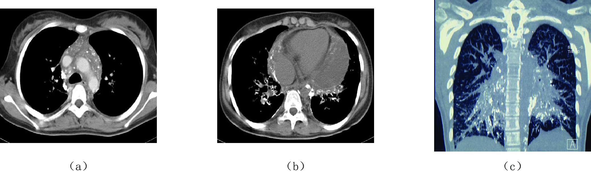
![]() 图 3 MR淋巴管成像(a)显示为胸导管影和胸导管末端分支畸形,右淋巴管导管分支畸形和纵隔淋巴管扩张;(b)和(c)显示纵隔、肺门和支气管血管束弥漫性淋巴管扩张、呈T2高信号影,中轴间质显著增厚。
图 3 MR淋巴管成像(a)显示为胸导管影和胸导管末端分支畸形,右淋巴管导管分支畸形和纵隔淋巴管扩张;(b)和(c)显示纵隔、肺门和支气管血管束弥漫性淋巴管扩张、呈T2高信号影,中轴间质显著增厚。图像分析包括平扫成像、动态增强成像和MIP图像,根据增强脉管的形态和大小以及信号强度区分淋巴管和静脉,淋巴管表现为信号强度高、显影清晰、脉管增强后呈串珠状改变;而静脉表现为管壁光滑和显影时间早于淋巴管。MRL对于淋巴管的形态和结构、有无梗阻扩张或发育畸形以及淋巴水肿的定位、定量和定性诊断等具有重要意义。
3.4 超声淋巴管成像
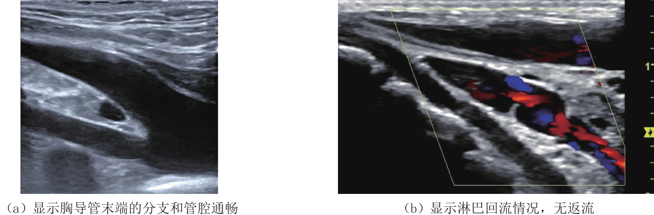
超声淋巴管成像(ultrasonic lymphography,ULG),特指采用高频超声探头获取颈部胸导管、胸导管末段及出口显像的影像学方法[11],由于胸导管的结构细长、管壁菲薄、管径狭小且位置隐蔽,常规低频超声的波长较长和图像分辨率较低而显示不佳,而高频超声的短波长明显提高了图像分辨力,可较清晰的显示颈段胸导管的管腔结构、胸导管末端的解剖形态学类型、有无狭窄和淋巴回流以及流入静脉的情况等。
根据高频超声显像特点,胸导管末端可分为5种类型:Ⅰ型,正常型;Ⅱ型,弥漫缩窄性型;Ⅲ 型,末端梗阻型;Ⅳ 型,血栓型;Ⅴ型,反流型。
3.5 核素淋巴成像
核素淋巴成像,又称同位素淋巴闪烁成像(lymphoscintigram,LSG),是临床最常用的间接淋巴成像技术[12-13],常用方法包括99 Tcm-右旋糖酐淋巴显像(99 Tcm-Dextran lymphoscintigraphy,简称99 Tcm-DXL)、99 Tcm-人血蛋白淋巴显像(99 Tcm-HAS lymphoscintigraphy,简称99 Tcm-HASL)和99 Tcm-67 Ga胶体等,其原理是于双足趾蹼皮下组织注入放射性示踪剂,用外部辐射探测器分布于注射后10 min、1 h、3 h和6 h行从足到头的全身显像,从而获得放射性示踪剂分布的二维投影图像,观察淋巴管和淋巴结的结构和功能。LSG具有方法简便、图像清晰、灵敏度和特异性高等优点。
主要观察内容包括:①肠道是否显影及最初显影的时间、位置、范围和放射性随时间发生变化的情况;②腹腔区域是否有放射性异常分布;③有无其他伴随放射性异常分布。
3.6 淋巴管近红外荧光显像
淋巴管近红外荧光显像(near infrared fluorescence lymphangiography,NIFL),又称吲哚菁绿红外显像(infrared imaging with indocyanine green),是荧光探针在特定波长红光激发下发出的波长为700~900 nm的NIF。
NIFL最先由日本学者Unno等于2007年首先提出,采用吲哚青绿(indocyanine green,ICG)作为NIFL的显像剂,在淋巴水肿区域的远端皮内和/或皮下注射ICG后15 min,采用红外探头显像系统在体外照射激发皮下的吲哚菁绿显像,进行双侧肢体的动态采集,再现出组织内部荧光分布的显像技术[14-15]。
NIFL是近年来的分子影像研究热点之一,尤其在前哨淋巴结活检、淋巴水肿临床分级诊断、术前评价淋巴管的通畅度、位置标记以及准确选择手术切口等呈现显著的可行性和优势。
-
![]()
图 3 MR淋巴管成像
(a)显示为胸导管影和胸导管末端分支畸形,右淋巴管导管分支畸形和纵隔淋巴管扩张;(b)和(c)显示纵隔、肺门和支气管血管束弥漫性淋巴管扩张、呈T2高信号影,中轴间质显著增厚。
-
[1] DONG J, XIN J, SHEN W, et al. Unipedal diagnostic lymphangiography followed by sequential CT examinations in patients with idiopathic chyluria: A retrospective study[J]. American Journal of Roentgenology, 2018, 210(4): 792−798. DOI: 10.2214/AJR.17.18936.
[2] JI Y, CHEN S, PENG S, et al. Kaposiform lymphangiomatosis and kaposiform hemangioendothelioma: Similarities and differences[J]. Orphanet Journal of Rare Diseases, 2019, 14(1): 165. DOI: 10.1186/s13023-019-1147-9.
[3] JIN D, SUN X, SHEN W, et al. Diagnosis of lymphangiomatosis: A study based on CT lymphangiography[J]. Academic Radiology, 2020, 27(2): 219−226. DOI: 10.1016/j.acra.2019.03.024.
[4] TAGHINIA A H, UPTON J, TRENOR C C, et al. Lymphaticovenous bypass of the thoracic duct for the treatment of chylous leak in central conducting lymphatic anomalies[J]. Journal of Pediatric Surgery, 2019, 54(3): 562−568. DOI: 10.1016/j.jpedsurg.2018.08.056.
[5] 王仁贵, 陈孝柏, 段永利, 等. MSCT 直接淋巴管造影在弥漫性肺淋巴管瘤病中的诊断价值[J]. 中国医学影像技术, 2012,28(2): 185−189. DOI: 10.13929/j.1003-3289.2012.02.046. WANG R G, CHEN X B, DUAN Y L, et al. Diagnostic value of MSCT direct lymphangiography in diffuse pulmonary lymphangiomatosis[J]. Chinese Journal of Medical Imaging Technology, 2012, 28(2): 185−189. DOI: 10.13929/j.1003-3289.2012.02.046. (in Chinese).
[6] O'LEARY C, ITKIN M, ROSHKOVAN L, et al. CT Features of lymphatic plastic bronchitis in adults: Correlation with multimodality lymphatic imaging[J]. Radiology Cardiothorac Imaging, 2022, 4(2): e210048. DOI: 10.1148/ryct.210048.
[7] PATEL S, HUR S, KHADDASH T, et al. Intranodal CT lymphangiography with water-soluble iodinated contrast medium for imaging of the central lymphatic system[J]. Radiology, 2022, 302(1): 228−233. DOI: 10.1148/radiol.2021210294.
[8] PIEPER C C, FEISST A, SCHILD H H. Contrast-enhanced interstitial transpedal MR lymphangiography for thoracic chylous effusions[J]. Radiology, 2020, 295(2): 458−466. DOI: 10.1148/radiol.2020191593.
[9] NAKAMURA F, KATO H, OZEKI M, et al. CT and MRI findings of focal splenic lesions and ascites in generalized lymphatic anomaly, kaposiform lymphangiomatosis, and Gorham-Stout disease[J]. Journal of Clinical Imaging Science, 2021, 11: 44. DOI: 10.25259/JCIS_101_2021.
[10] ZHENG Q, ITKIN M, FAN Y. Quantification of thoracic lymphatic flow patterns using dynamic contrast-enhanced MR lymphangiography[J]. Radiology, 2020, 296(1): 202−207. DOI: 10.1148/radiol.2020192337.
[11] RICCI K W, IACOBAS I. How we approach the diagnosis and management of complex lymphatic anomalies[J]. Pediatric Blood Cancer, 2021: e28985. DOI: 10.1002/pbc.28985.
[12] MUNN L L, PADERA T P. Imaging the lymphatic system[J]. Microvascular Research, 2014, 96: 55−63. DOI: 10.1016/j.mvr.2014.06.006.
[13] HOU G, JIANG Y, JING H, et al. Usefulness of 99mTc-ASC lymphoscintigraphy and SPECT/CT in the evaluation of rare lymphatic disorders Gorham–Stout disease, lymphangioma, and lymphangioleiomyomatosis[J]. Medicine, 2020, 99(39): 7−7. DOI: 10.1097/MD.0000000000022414.
[14] YAMAMOTO T, YAMAMOTO N, DOI K, et al. Indocyanine green-enhanced lymphography for upper extremity lymphedema: A novel severity staging system using dermal backflow patterns[J]. Plastic and Reconstructive Surgery, 2011, 128(4): 941−947. DOI: 10.1097/PRS.0b013e3182268cd9.
[15] NARUSHIMA M, YAMAMOTO T, OGATA F, et al. Indocyanine green lymphography findings in limb lymphedema[J]. Journal of Reconstructive Microsurgery, 2016, 32: 79. DOI: 10.1055/s-0035-1564608.
-
期刊类型引用(2)
1. 张怡梦,孙小丽,王仁贵. 中央淋巴系统的解剖变异及多模态影像研究进展. 临床放射学杂志. 2024(10): 1822-1826 .  百度学术
百度学术
2. 陈传智,龚秀茹,都业弘,王轶彬,路青. 核磁共振淋巴造影在胸腹腔乳糜漏中的诊断价值研究. 上海医学. 2024(11): 692-700 . 百度学术
其他类型引用(0)
 下载:
下载:


计量
- 文章访问数: 428
- HTML全文浏览量: 122
- PDF下载量: 42
- 被引次数: 2


