
The Value of Spectral CT in Differential Diagnosis and Radiotherapy Localiation of Central Lung Cancer with Obstructive Atelectasis
-
摘要: 目的:探讨能谱CT扫描在诊断中央型肺癌合并阻塞性肺不张及放疗前定位鉴别肿瘤边界、精准勾画靶区中的应用价值。方法:回顾性分析我院65例经病理证实的中央型肺癌伴阻塞性肺不张患者的放疗前定位 Ⅲ期能谱CT扫描的影像资料,比较分析Ⅲ期增强CT扫描不同参考指标(BMI、BMI-ICM及Eff-Z、PI)显示瘤-肺交界面评分值差异,比较肿瘤与不张肺组织IC值及CT值差异。结果:Ⅲ期增强CT扫描瘤-肺边界评分4项指标(BMI、BMI-ICM及Eff-Z、PI)间存在统计学差异,BMI-ICM最高,PI最低;动脉期、静脉期及延迟期 Ⅲ期扫描中,显示瘤-肺边界的主观评分有差异,动脉期最高;肿瘤与不张肺组织IC值及CT值差异具有统计学意义,IC值比CT值差别更大。结论:能谱CT扫描的不同指标能帮助鉴别中央型肺癌与不张肺组织的边界,利用最佳指标实现放疗前精准勾画放疗靶区,实现放疗计划最优化。Abstract: Objective: To investigate the value of spectral CT in differential central lung cancer from obstructive atelectasis for achieving the accurate delineation and radiotherapy localiation. Methods: The CT radiotherapy localiation images of three phase contrast enchancement by spectral CT of 65 patients confirmed by pathology with central lung cancer and obstructive atelectasis with were collected and analyzed. The scores and detection rates of tumor-lung interface between the polychromatic image (PI), the best monochromatic image (BMI), the effective atomic number (Eff-Z) and the best monochromatic image combined with iodine concentration map (BMI-ICM) were compared. Results: Statistical differences in the scores of tumor-lung interface were observed between PI, BMI, Eff-Z and BMI-ICM; the pairwise comparison showed that the subjective score of BMI-ICM was the highest, and PI was the lowest. The pulmonaty artery phase detection rates of tumor-lung interface of three phase contrast enchancement were the highest. There was statistical difference of iodine concentration (IC) and CT score between lung cancer and obstructive atelectasis. The difference of IC was higher than the CT score. Conclusion: Spectral CT is helpful to distinguish central lung cancer delineation from atelectasis. It provides a new method to delineate the radiotherapy localiation for radiotherapy plan of optimization.
-
Keywords:
- spectral CT /
- lung tumor /
- pulmonary atelectasis /
- radiotherapy
-
弯刀综合征(scimitar syndrome,SS)是一种罕见的先天性心肺异常,发病率约为1~3/100000[1],其特征是部分或全部一侧肺静脉异常引流至下腔静脉、肝静脉、门静脉或右心房较低部位,此异常引流静脉解剖形状似“弯刀”状,因此也被称为弯刀静脉(scimitar vein,SV),左侧异常较右侧罕见[2]。弯刀综合征分为婴儿型和儿童型/成人型。婴儿型患者合并的畸形较多,预后差,迫切需要多学科联合早期精准诊疗。儿童型/成人型大部分患儿无症状或症状轻微,即使不进行医学干预也可正常生活[2]。
鉴于此,本文旨在分析弯刀综合征的CT影像学表现,比较二型的影像学差异及其临床意义,为临床及时精准诊治提供影像学依据。
1. 材料与方法
1.1 临床资料
搜集2019年1月至2024年1月经我院确诊的弯刀综合征的患儿共28例,其中男10例,女18例。婴儿型弯刀综合征15例,男5例,女10例,平均发病年龄约(6.78±3.50)月,其中3例因重症肺炎就诊,1例因先天性回肠闭锁,1例因出生后无肛就诊,1例因纵隔占位就诊,1例因哭闹后口周青紫明显就诊,8例因心脏病变就诊。
成人型弯刀综合征13例,男5例,女8例,平均年龄约(5.53±2.72)岁。其中5例因肺炎就诊,3例为发现心脏杂音来诊,2例为已经明确诊断2年余来院手术,1例因胸廓畸形来诊,1例因肺隔离症就诊,1例因一侧肺间质病变伴轻度肺动脉高压就诊(表1)。
表 1 婴儿型与成人型弯刀综合征患者的临床资料Table 1. Clinical characteristics of adult and Infantile types of patients with scimitar syndrome (SS)临床资料 组别 统计检验 婴儿型(n=15) 成人型(n=13) 统计值 P 男 5 5 — 0.554* 女 10 8 平均年龄/月 6.78±3.50 66.36±32.64 3.085 <0.001 就诊原因 重症肺炎 3例 肺炎 5例 先天性肠闭锁 1例 心脏杂音 3例 无肛 1例 胸廓畸形 1例 纵隔占位 1例 肺隔离症 2例 哭闹后口周青紫 1例 肺间质病变 1例 心脏病变 8例 明确诊断 2例 肺动脉高压/例 2 1 手术治疗/例 5 3 3.589 <0.001 介入治疗/例 0 2 死亡/例 0 0 <0.001 注:*为Fisher确切检验。 本文为回顾性研究,经医院伦理委员会批准进行。
1.2 检查方法
使用GE Revolution 256排螺旋CT扫描仪进行胸部、心脏检查。不能合作患儿,扫描前30分钟至1小时口服10%水合氯醛(剂量:0.5 mL/kg,最多不超过10 mL)镇静后进行。对比剂为欧乃派克(320 mg/mL),剂量1.6~2.0 mL/kg,流率0.9~1.8 mL/s。注射后再以1.0~2.0 mL/s的流率注入生理盐水(使用量为对比剂的1/2~2/3),用以减少对比剂的硬化伪影。
患儿仰卧于检查床上,双臂上举,身体位于检查床正中,头先进,定位像为前后正位像,肺尖为扫描基线,从胸廓入口至肺底进行螺旋扫描。扫描参数见表2。
表 2 不同年龄段低剂量CT扫描参数(胸部增强CT、心脏CT)Table 2. Low-dose CT parameters used for different age groups(contrast-enhanced chest CT, cardiac CT)年龄/岁 管电压/kV 噪声指数 管电流/mA 螺距 管球旋转时间/s 层厚/mm 后处理算法 ≤1 100 11 30~100 1.375 0.35 0.625 40% ASIR-V 1~7 100 13 60~120 1.375 0.35 0.625 40% ASIR-V ≥7 100 15 80~200 1.375 0.35 0.625 40% ASIR-V 注:ASIR-V为多模型的迭代重建算法(adaptive statistical iterative reconstructions-V)。 1.3 影像征象
对婴儿型及成人型弯刀综合征的患者的胸部增强CT扫描图像进行分析:①肺静脉异常引流的位置、有无狭窄梗阻;②体动脉分支供血的情况;③合并肺发育不良的情况;④是否伴发其他畸形包括心脏大血管畸形及其他系统畸形。
1.4 统计方法
应用SPSS 22.0统计软件进行分析。以(
$\bar x \pm s$ )表示服从正态分布的计量资料,采用t检验进行组间比较;分类资料用百分比描述,组间比较采用卡方检验。以P<0.05为差异有统计学意义。2. 结果
28例患儿中婴儿型弯刀综合征15例,男5例,女10例,平均发病年龄约(6.78±3.50)月;成人型弯刀综合征13例,男5例,女8例,平均年龄约(5.53±2.72)岁(表3)。
表 3 婴儿型与成人型弯刀综合征患者的影像学比较Table 3. Comparison of imaging features of patients with adult and infantile types of SS合并异常疾病 组别 统计检验 婴儿型(n=15)(%) 成人型(n=13)(%) $\chi^2/t $ P 右侧肺静脉异位引流 15(100) 13(100) — — 主支气管左侧镜像 15(100) 11(84.6) — — 肺发育不良 15(100) 13(100) — — 合并右位心 15(100) 13(100) — — 合并右肺动脉发育不良 15(100) 13(100) — — 体循环供应右肺 15(100) 13(100) — — 完全性右肺静脉异位引流 10(66.7) 7(53.8) 1.470 0.269 右下肺静脉异位引流 5(33.3) 6(46.1) 1.512 0.283 引流入下腔静脉 14(93.3) 13(100) 1.897 1.000* 引流入肝静脉 1(6.7) 0(0) 1.897 1.000* 合并心脏畸形 6(40.0) 0(0) 11.495 <0.001* 合并ASD 2(13.3) 0(0) 1.746 <0.001* 合并动脉导管未闭PDA 1(6.7) 0(0) 1.897 0.226* 异位引流的肺静脉狭窄(梗阻) 3(20.0) 0(0) — — 合并心外畸形 4(26.7) 1(7.7) 1.791 0.191 注:*为Fisher确切检验。 28例患儿右肺较左肺体积小,心影纵隔右移。其中26例患儿的支气管树为左侧镜像结构即左右主支气管均为两叶支气管结构,2例成人型患儿的支气管树为左二右三叶支气管的结构,但右上叶支气管较正常纤细,另外两例婴儿型患儿限局性气管狭窄;增强扫描本组28例患儿均为右侧肺静脉异位引流,右肺动脉均较左肺动脉细。28例患儿均可见弯刀静脉,26例降主动脉动脉分支供血右下肺,2例降主动脉动脉分支供血叶外型隔离肺。
婴儿型弯刀综合征15例患儿中10例为完全右肺静脉异位引流至下腔静脉,其中3例异位引流入下腔静脉处狭窄梗阻(图1),该3例患儿行部分肺静脉异位连接弯刀综合征矫治术,术后患儿随诊复查无不适,1例合并房间隔缺损及肺动脉高压,1例合并房间隔缺损、动脉导管未闭及肺动脉高压,该2例患儿均行房间隔修补术的同时行部分肺静脉异位连接弯刀综合征矫治术,合并动脉导管未闭的患儿还行动脉导管结扎术,其余5例患儿未行手术。
![]() 图 1 女孩3个月,气促、喘息10天注:(a)胸部CT增强冠状位重建;(b)CT平扫轴位;(c)最小密度投影;(d)容积重建,示右肺静脉异位引流,引流处狭窄(红箭所指处)。Figure 1. Female, 3 months old, admitted to hospital with shortness of breath and wheezing for 10 days
图 1 女孩3个月,气促、喘息10天注:(a)胸部CT增强冠状位重建;(b)CT平扫轴位;(c)最小密度投影;(d)容积重建,示右肺静脉异位引流,引流处狭窄(红箭所指处)。Figure 1. Female, 3 months old, admitted to hospital with shortness of breath and wheezing for 10 days4例为右下肺静脉异位引流至下腔静脉,均因合并其他畸形及肺部感染变行影像检查时发现,其中3例因回肠闭锁、无肛、纵隔占位行相关手术,该4例均未行异位引流肺静脉矫正术。
1例为右上肺静脉异位引流至奇静脉并右下肺静脉引流至右心房底部,利用MPR及VR重建观察到该患儿存在多枚椎体畸形,该例患儿并未行异位引流肺静脉矫正术。
成人型弯刀综合征13例中7例为完全右肺静脉异位引流至下腔静脉(图2),其中1例因胸廓畸形(鸡胸)行部分肺静脉异位连接弯刀综合征矫治术;6例为右下肺静脉异位引流至下腔静脉,其中2例明确诊断后,药物对症治疗期间反复肺炎行部分肺静脉异位连接弯刀综合征矫治术,2例合并叶外型隔离肺行介入栓堵治疗体动脉供血。
![]() 图 2 女孩,10岁,发现胸廓畸形、心脏位置异常5年余注:(a)CT增强冠状位最大密度投影;(b)CT增强轴位;(c)容积重建,弯刀静脉及异位引流的右肺静脉,引流至右心房较低部位与下腔静脉交界处(红箭所指处),并右下肺有异常主动脉分支供血及右肺动脉细小(白箭所指处)。Figure 2. Female, 10 years old, was found to have chest deformities and abnormal cardiac position for over 5 years
图 2 女孩,10岁,发现胸廓畸形、心脏位置异常5年余注:(a)CT增强冠状位最大密度投影;(b)CT增强轴位;(c)容积重建,弯刀静脉及异位引流的右肺静脉,引流至右心房较低部位与下腔静脉交界处(红箭所指处),并右下肺有异常主动脉分支供血及右肺动脉细小(白箭所指处)。Figure 2. Female, 10 years old, was found to have chest deformities and abnormal cardiac position for over 5 years3. 讨论
弯刀综合征又称肺发育不良综合征,其发病机制尚不清楚,可能与胚胎早期整个肺芽的发育障碍有关[3]。其特点为心脏右移右旋,小右肺、右肺动脉发育不良、部分性右肺静脉回流异常即弯刀静脉及异常体动脉的分支供血(通常是供应右下肺基底段或肺隔离症);同时存在肺叶、气管支气管分支异常,支气管树可呈左侧镜像(左-异构现象),部分患儿合并先天性心脏异常,以及心外畸形[4]。故弯刀综合征涉及多个系统及组织结构的畸形包括心脏畸形、胸腹腔大血管畸形、肺及气道畸形、部分病人还合并骨骼、消化道等其他的心外畸形。
另外,弯刀综合征患儿的血液动力学是心房水平的左向右分流,同时伴有异常体动脉的供血,导致右心容量负荷增加,其增加程度的不同、肺发育不良的程度、以及是否合并心内、心外畸形,是导致临床表现差异很大的原因,因此弯刀综合征可以在婴儿、儿童或成年人中被发现或发病。通常婴儿型患儿发病时间较早,是由于其心房水平的左向右分流量较大、和/或存在较粗的异常体动脉分支供血、和/或存在异位引流肺静脉有狭窄梗阻[5],或合并其他心内、心外畸形[6-7]。与此同时弯刀综合征的治疗手段亦不相同,包括药物治疗、介入治疗与外科治疗[6]。
CT扫描的最大优势是避免了影像的重叠,可以逐层观察图像,并且随着CT技术的发展,扫描速度明显加快,CT图像的空间、时间分辨率也明显提高,以及无间隔容积扫描、后处理重建技术的广泛应用,并结合造影剂的使用,利用平扫、增强及多种图像重建后处理方法可以精准、直观、多方位以及三维立体的显示肺、气道、血管、心脏及骨骼形态结构及病变,使之成为诊治弯刀综合征首选检查方法。
本组28例弯刀综合征的患儿均经胸部CT检查,并对图像进行重建后处理,确诊弯刀综合征。并为治疗方法的选择提供依据,其中18例患儿仅药物治疗即可保证患儿的病情稳定及正常的生长发育,8例行部分肺静脉异位连接弯刀综合征矫治术,2例行介入栓堵术,栓堵肺外型隔离肺的体循环供血动脉,解除体肺分流引起的症状。
CT图像的重建是基于前期的胸部CT平扫及增强的无间隔容积扫描的图像数据,图像重建分为二维图像重建及三维图像重建,针对弯刀综合征常用的图像重建后处理方法有多平面重建(multiplanar reformation,MPR)属于二维图像重建;以及三维图像重建包括最大密度投影(maximum intensity projection,MIP)、最小密度投影(minimum intensity projection,MIN)及容积成像(volume rendering,VR),在显示心脏、血管畸形方面几乎可以替代血管造影。
MPR是本病最基本的重建方法,可以任意平面、任意角度显示扫描范围内解剖结构的形态及走行,观察每支肺血管、弯刀静脉、气管支气管、肺叶形态及分割、异常体动脉分支的形态、粗细、走行,异常引流处的具体部位、是否狭窄以及是否合并的其他部位畸形,还可以重建出标准的矢、冠、轴位图像。MIP和MIN分别是指投影方向内最大密度和最小密度体素所参与的成像,均为部分重叠影像,MIP是基于CT增强扫描后的重建方法,用于显示血管及心脏。MIN主要基于CT平扫后的图像数据,用于显示气道或含气肺组织结构。VR为三维立体成像,为伪彩仿真立体图,按临床的要求提供心脏、大血管、气道及骨骼的整体三维影像,多方位、多角度地观察各解剖结构之间的关系,对于术前了解畸形部位特征和制定手术方案有指导作用。
本组28例患儿的胸部CT平扫的轴位图像显示这些患儿右肺体积小,右肺纹理粗细不等,且部分肺纹理走行紊乱,右肺透光度较左肺增高/减低,心影纵隔右移,部分患儿合并肺炎。之后行MPR及MIN重建发现患儿的气管支气管畸形及肺叶分割畸形,其中有1例利用MPR及VR重建观察到该患儿还存在多枚椎体畸形。
另外,由于弯刀综合征存在程度不等的心脏及血管畸形,对于分流量大于50%、异位引流肺静脉狭窄、肺动脉高压和隔离肺时需手术治疗[8]。有文献报道可根据CT增强后的MPR测量弯刀静脉汇入下腔静脉角度的大小采用不同的手术方法[9]。本组28例患儿均行胸部CT增强分期扫描,分别于动脉期及静脉期进行扫描,利用增强后的图像数据进行MPR及MIP重建对心脏、血管进行评估,其中临床症状较重的婴儿型患儿有3例在MPR及MIP图像上可见异位引流处狭窄梗阻(图1),2例可见合并了心内畸形—房间隔缺损及肺动脉主干明显增粗,其中1例还合并了较粗大的动脉导管未闭,并经手术证实,术后患儿情况良好。2例成人型患儿经MPR及MIP重建清晰显示了于膈肌水平主动脉分别发出1分支及2支血管供应肺外型隔离肺(图2),之后行介入手术,栓堵的该异常体动脉,术后患儿恢复良好。
婴儿型弯刀综合征患儿发病早的另一个主要原因是由于伴发其他致命畸形来医院就诊,而偶然被发现,本组有3例分别因无肛、回肠闭锁和纵隔占位(经病理证实为异位肝)来院准备做相应的手术,在术前CT平扫时怀疑合并有弯刀综合征,故此3例患儿经增强CT扫描和MPR、MIP、MIN及VR重建进一步证实均为右下肺静脉的单支肺静脉异位引流,分流量不大,肺动脉压未增高,由弯刀综合征所引发的症状并不严重,3例均未接受肺静脉异位连接弯刀综合征矫治术。
目前国际上报道了两例VACTERL联合征合并弯刀综合征[10],VACTERL联合征是一组非随机出现的先天性联合畸形,包括脊柱畸形(vertebral defects,V)、肛门闭锁(anal atresia,A)、气管食管瘘(trachea esophageal fistula,TE)、伴食管闭锁(esophageal atresia)、肾发育畸形(renal defects,R)、桡骨发育不良(radial dysplasia,R)、心脏发育缺陷(cardiac defects,C)和肢体畸形(1imb anomalies,L),存在3个或3个以上的先天发育异常称为VACTERL联合征[11]。本组弯刀综合征的患儿中,有6例合并上述相关畸形,其中2例合并消化道畸形为肛门闭锁伴阴道瘘和回肠闭锁、2例合并心脏畸形伴肺动脉高压、1例合并胸廓畸形(鸡胸)、1例合并椎体畸形,这几例患儿均因合并其他畸形就诊。因此在临床工作中,当弯刀综合征患儿出现上述畸形时需注意患儿是否存在VACTERL联合征。
综上,婴儿型和成人型弯刀综合征患儿的临床表现有很大差异,利用CT检查的多种成像模式,结合强大重建后处理功能对弯刀综合征患儿进行全面评估,可以为临床治疗方法的选择、手术方案的制定、改善预后及提高生活质量提供依据。
-
![]()
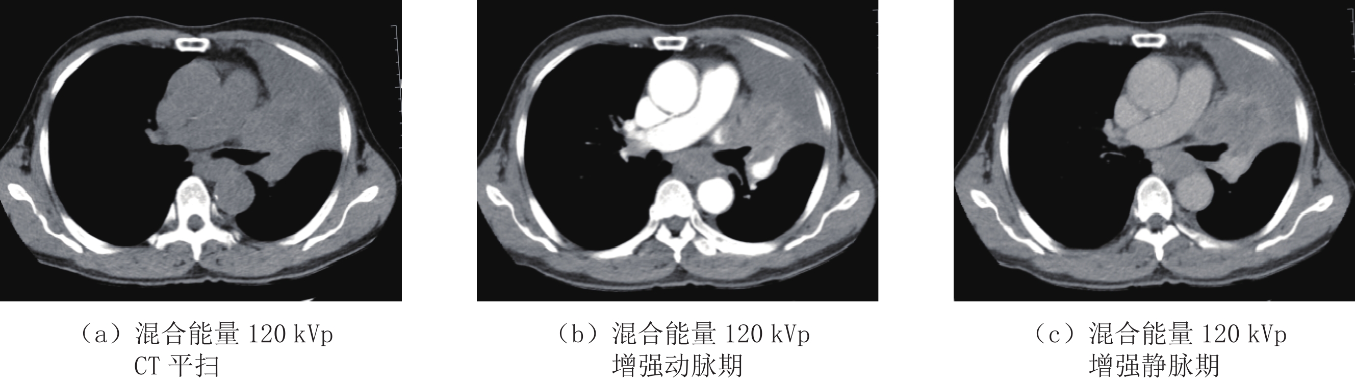
图 1 左肺上叶中央型肺癌并左肺上叶不张混合能量平扫+增强扫描
Figure 1. The left upper lobe central lung cancer with obsrtuctive atelectasis of polychromatic image flat scan and enhancement
![]()
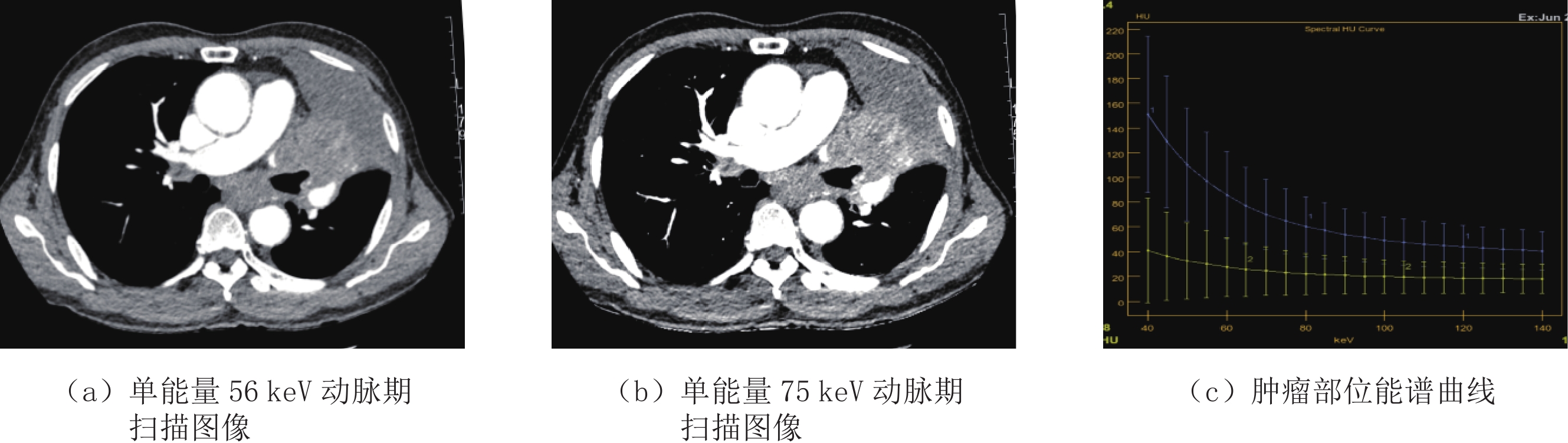
图 2 左肺上叶中央型肺癌并左肺上叶不张单能量增强扫描及能谱曲线
Figure 2. The Spectral HU curve of the same patient and monochromatic image of 56 keV and 75 keV
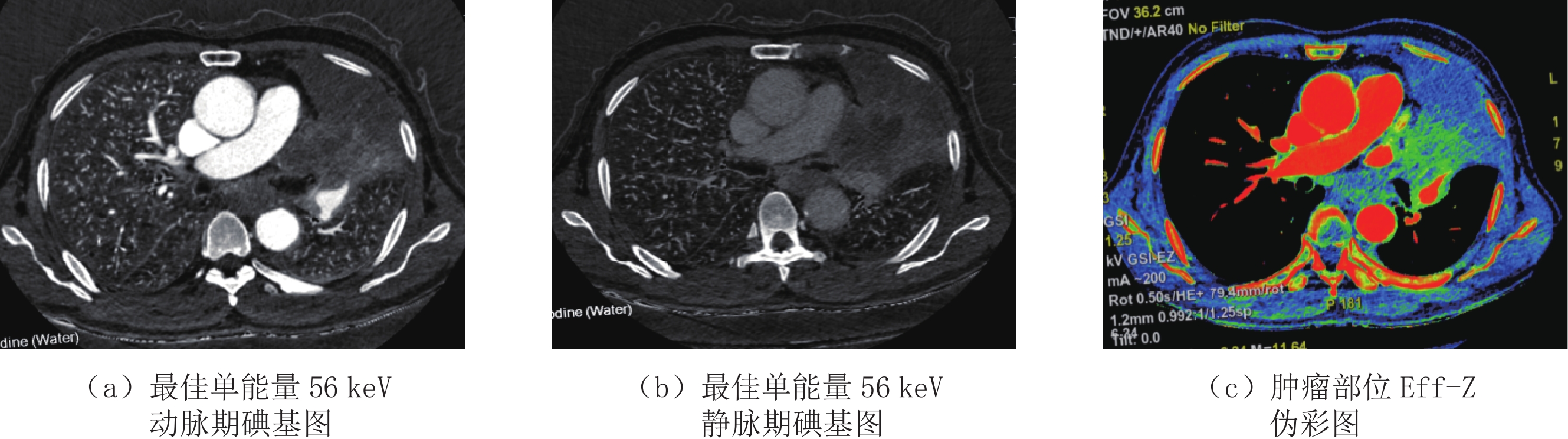
![]()
图 3 左肺上叶中央型肺癌并左肺上叶不张最佳单能量56 keV增强扫描碘基图及有效原子序数图
Figure 3. The best monochromatic enhancement image combined with iodine concentration map and effective atomic number image
表 1 增强CT扫描各期PI、BMI及BMI-ICM瘤-肺边界主观评分的比较
Table 1 The scores and detection rates of tumor-lung interface of PI, BMI, BMI-ICM and Eff-Z in three phase contrast enchancement
扫描期相 瘤-肺边界主观评分值 统计检验 PI BMI BMI-ICM Eff-Z F P 动脉期 2.51±0.83 3.14±0.85 3.84±0.31 3.04±0.55 34.28 <0.01 静脉期 2.14±0.65 2.95±0.89 3.34±0.25 2.84±0.26 26.54 <0.01 延迟期 2.12±0.55 2.54±0.35 3.04±0.85 2.74±0.15 18.32 <0.01  下载: 导出CSV
表 2 增强CT扫描各期肿瘤与不张肺组织碘浓度IC值及CT值的比较
Table 2 The scores of tumor-lung interface of IC and CT scores in three phase contrast enchancement
扫描期相 组织IC/100 μg·mL-1 CT值/HU 统计检验 肿瘤 不张肺组织 肿瘤 不张肺组织 t P 动脉期 12.51±3.89 23.71±8.12 35.41±6.85 50.21±9.76 -4.89 <0.01 静脉期 11.41±3.23 18.31±6.37 38.38±4.03 42.37±7.83 -6.43 <0.01 延迟期 10.11±3.63 17.96±5.93 18.51±5.84 17.01±7.73 -6.82 <0.01
下载: 导出CSV
-
[1] LV P J, ZHOU Z G, LIU J, et al. Can virtual monochromatic images from dual-energy CT replace low-kVp images for abdominal contrastenhanced CT in small-and medium-sized patients[J]. European Radiology, 2019, 29(10): 2878−2889.
[2] QI L P, ZHANG X P, TANG L, et al. Using diffusion-weighted MR imaging for tumor detection in the collapsed lung: A preliminary study[J]. European Radiology, 2009, 19(3): 333−341.
[3] 何小群, 李琦, 罗天友, 等. 能谱CT在精准勾画继发阻塞性不张的中央型肺癌放疗靶区中的价值[J]. 中国医学计算机成像杂志, 2021,27(1): 53−56. doi: 10.3969/j.issn.1006-5741.2021.01.012 HE X Q, LI Q, LUO T Y, et al. The value of spectral CT in precisely delineating the radiotherapy targets of central lung cancer with obstructive atelectasis[J]. China Computer Medical Image, 2021, 27(1): 53−56. (in Chinese). doi: 10.3969/j.issn.1006-5741.2021.01.012
[4] 段海峰, 贾永军, 于勇, 等. 能谱CT碘基图在鉴别中央型肺癌与继发阻塞性肺实变中的价值[J]. 实用放射学杂志, 2016,32(2): 204−207. doi: 10.3969/j.issn.1002-1671.2016.02.010 DUAN H F, JIA Y J, YU Y, et al. Differentiation of central lung cancer from obstructive pneumonia and atelectasis using spectral CT iodine-based material decomposition technique[J]. Journal of Practical Radiology, 2016, 32(2): 204−207. (in Chinese). doi: 10.3969/j.issn.1002-1671.2016.02.010
[5] 李忠学, 张文宝, 赵霖, 等. DWI-T2WI图像融合在中央型肺癌伴肺不张放射治疗靶区勾画中的价值[J]. 中华全科医学, 2017,15(3): 491−494. LI Z X, ZHAGN W B, ZHAO L, et al. Application of fused DWI-T2WI imaging in target delineation for central-type lung cancer combined with pulmonary atelectasis[J]. Chinese Journal of General Practice, 2017, 15(3): 491−494. (in Chinese).
[6] 黄倩文, 陈应东, 钟华, 等. 肺癌能谱CT相关参数定量与临床应用[J]. 临床放射学杂志, 2020,39(7): 1316−1318. doi: 10.13437/j.cnki.jcr.2020.07.017 HUANG Q W, CHEN Y D, ZHONG H, et al. Quantification of energy spectrum CT parameters in the diagnosis of lung cancer and clinical application[J]. Journal of Clinical Radiology, 2020, 39(7): 1316−1318. (in Chinese). doi: 10.13437/j.cnki.jcr.2020.07.017
[7] 窦沛沛, 赵恒亮, 王晨, 等. 能谱衰减曲线预测肺癌Ki-67表达: 评估最佳keV及能谱曲线斜率[J]. 临床放射学杂志, 2020,39(12): 2435−2456. DOU P P, ZHAO H L, WANG C, et al. Energy spectrum attenuation curve for prediction of Ki-67 expression in lung cancer: To evaluate the optimal keV sequence and slope of energy spectrum curves[J]. Journal of Clinical Radiology, 2020, 39(12): 2435−2456. (in Chinese).
[8] 裴丽美, 李晓阳, 王洪峰. 能谱CT在鉴别中央型肺癌与阻塞性肺不张中的价值研究[J]. 中国煤炭工业医学杂志, 2021, 24(6): 247-250. PEI L M, LI X Y, WANG H F. Dual energy CT differentiation central lung cancer and pulonary atelectasis[J]. Chinese Journal of Coal Industry Medicine[J]. 2021, 24(6): 247-250. (in Chinese).
[9] 王明亮, 林晓珠, 缪飞, 等. CT 能谱成像对物质内碘含量测定的价值[J]. 中国医学计算机成像杂志, 2011,17(2): 172−175. doi: 10.3969/j.issn.1006-5741.2011.02.017 WANG M L, LIN X Z, MIAO F, et al. The value of gemstone spectral imaging in analysis of the content of iodine: Phantom study[J]. China Computer Medical Image, 2011, 17(2): 172−175. (in Chinese). doi: 10.3969/j.issn.1006-5741.2011.02.017
[10] 苑呈秀, 盛华强, 王广丽, 等. 能谱CT在非小细胞肺癌立体定向放疗预处理评估中的应用价值[J]. 中华诊断学电子杂志, 2019,7(1): 7−10. doi: 10.3877/cma.j.issn.2095-655X.2019.01.002 YUAN C X, SHEGN H Q, WANG G L, et al. The value of spectral CT in the pretreatment evaluation of stereotactic radiotherapy for non-small cell lung cancer[J]. China Journal Diagnostics (Electronic Edition), 2019, 7(1): 7−10. (in Chinese). doi: 10.3877/cma.j.issn.2095-655X.2019.01.002
[11] LIU L, ZHI X, LIU B, et al. Utilizing gemstone spectral CT imaging to evaluate the therapeutic efficacy of radiofrequency ablation in lung cancer[J]. Radiology Medical, 2016, 121(4): 261−267. doi: 10.1007/s11547-015-0602-5
[12] 邓靓娜, 张国晋, 林晓强, 等. 能谱及灌注CT成像鉴别诊断周围型肺癌和局灶性机化性肺炎的对比研究[J]. 中国医学影像学杂志, 2021,29(12): 1206−1209. DENG L N, ZHANG G J, LIN X Q, et al. A comparison of spectral and perfusion CT imaging in different peripheral lung cancer from focal organizing pneumonia[J]. China Journal of Medical image, 2021, 29(12): 1206−1209. (in Chinese).
[13] ZHANG G, CAO Y, ZHANG J, et al. Focal organizing pneumonia in patients: Differentiation from solitary bronchioloalveolar carcinoma using dual-energy spectral computed tomography[J]. American Journal of Translational Reseach, 2020, 12(7): 3974−3983.
-
期刊类型引用(1)
1. 李岩,王志忠,芦春花. DXA联合胸腹部CT扫描筛查骨质疏松症的效果. 实用临床医学. 2025(01): 80-82 .  百度学术
百度学术
其他类型引用(0)


计量
- 文章访问数: 346
- HTML全文浏览量: 72
- PDF下载量: 35
- 被引次数: 1
