
Exploratory Research on the Influence of MR Image Quality of Bladder Cancer and Diagnostic Efficacy Using Two Preparation Methods
-
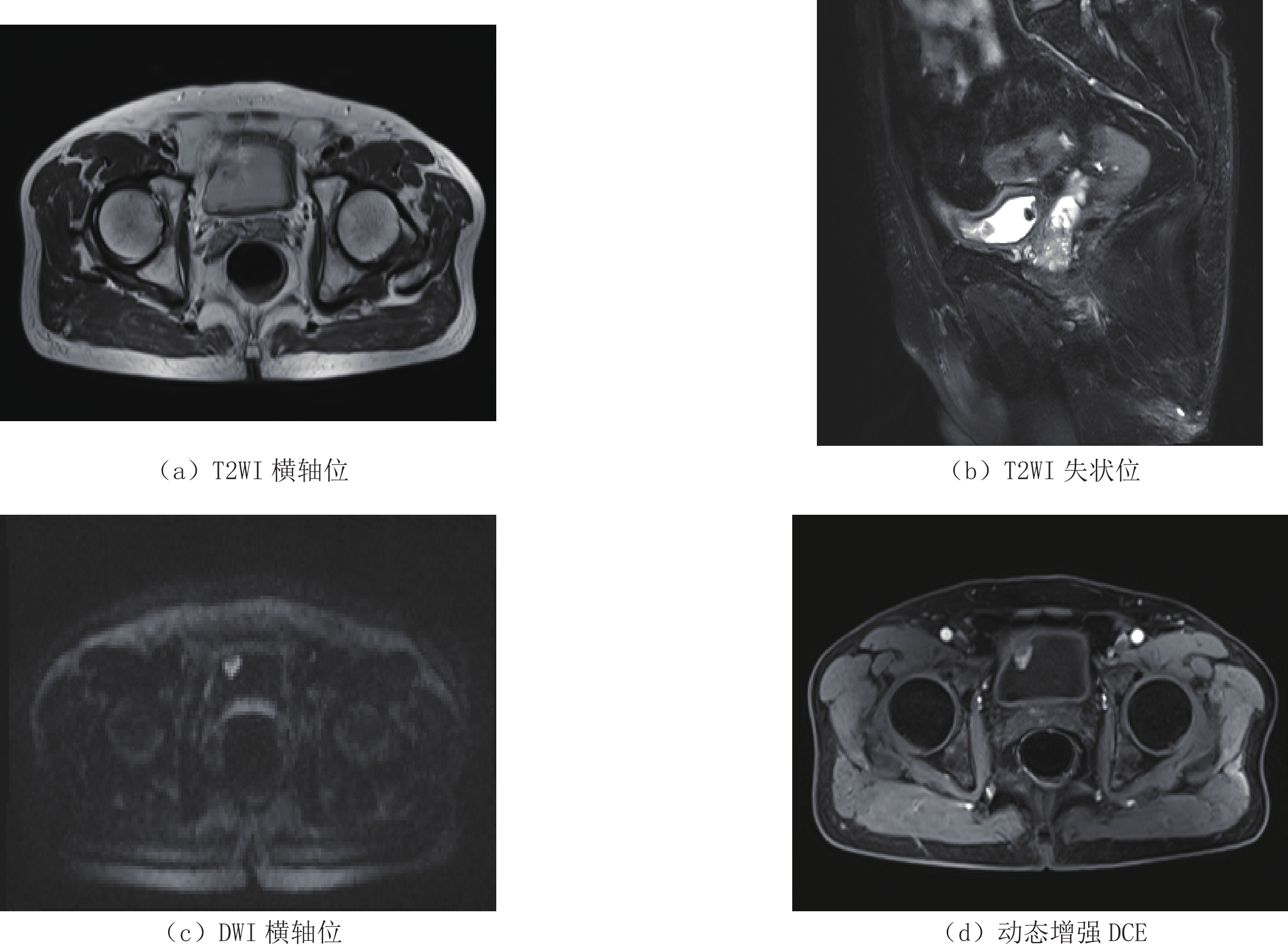
摘要: 目的:探讨两种膀胱准备方法对膀胱癌磁共振成像(MRI)的图像质量及术前鉴别膀胱癌肌层浸润诊断效能的影响。方法:76例膀胱癌患者行术前磁共振检查,分为A组和B组,分别行两种不同的膀胱准备方式,对比两种方法在高分辨率T2WI上图像质量有无差别,并结合弥散加权成像(DWI)及动态增强成像(DCE)序列,判断两组病例磁共振图像对判断膀胱癌病灶肌层浸润情况的诊断效能有无影响。结果:两位阅片者之间一致性良好,A组(禁食4~6 h后于检查前排空膀胱并通过导尿管向膀胱内灌注约100 mL生理盐水后半小时内进行检查),与B组(检查前2 h排空膀胱内尿液后禁食禁水)相比,前者具有更优的膀胱充盈度评分;在诊断效能上,A组病例诊断肌层浸润性膀胱癌的敏感性及准确度均高于B组,但其差异无统计学意义。结论:检查前通过导尿管向膀胱内灌注适当容量(本研究为100 mL)生理盐水可作为一种辅助膀胱准备方法,帮助膀胱癌患者的膀胱适度充盈,以提高磁共振图像质量,进而提高膀胱磁共振诊断效能。Abstract: Purpose: To investigate the effects of two preparation methods on the image quality of bladder cancer magnetic resonance imaging (MRI) and the diagnostic performance of differentiation of muscular invasion of bladder cancer. Methods: 76 cases of bladder cancer patients underwent preoperative MRI. They were divided into two groups and underwent two different bladder preparation methods respectively. The image quality of the two methods on high-resolution T2WI combined with diffusion-weighted imaging (DWI) and dynamic contrast-enhanced imaging (DCE) was compared. To determine whether the MR images of the two groups of patients have any influence on the diagnostic performance of the bladder cancer lesions in muscle invasion. Results: The consistency between the two readers was good. Compared with group B (urinate 2 hours prior to bladder MRI with no drinking), group A (after 4 to 6 hours of fasting, inject about 100 ml of physiological saline into the bladder before the examination through the catheter) had a better bladder filling degree. In terms of diagnostic efficiency, the sensitivity and accuracy of group A in diagnosing muscle-invasive bladder cancer were higher than those of group B, the difference has no statistically significant. Conclusion: Injecting an appropriate volume of physiological saline into the bladder before MR examination can be used as a clinical assistant bladder preparation method which help patients with bladder cancer properly fill the bladder, improve the quality of magnetic resonance images, and help improve the diagnosis of bladder cancer with magnetic resonance.
-
Keywords:
- magnetic resonance image /
- bladder neoplasms /
- diagnostic quality
-
-
表 1 1.5T磁共振膀胱扫描序列参数表
Table 1 The scanning parameters of 1.5T MRI
扫描参数 TR/ms TE/ms FOV/mm 矩阵 层厚/层间距/mm 平均次数 采集时间/s 激发角度 b值/(s/mm2) T2WI Tra 6190 85 180 256×256 7.0/2.1 3 192 150 — Sag 6900 85 180 256×256 4.0/1.2 3 214 150 Cor 6000 85 180 256×256 4.0/0.8 3 114 150 DWI 4400 100 400 — 6 6 120 — 0,1500 DCE 7.97 2.9 250 — 3 1 10 —  下载: 导出CSV
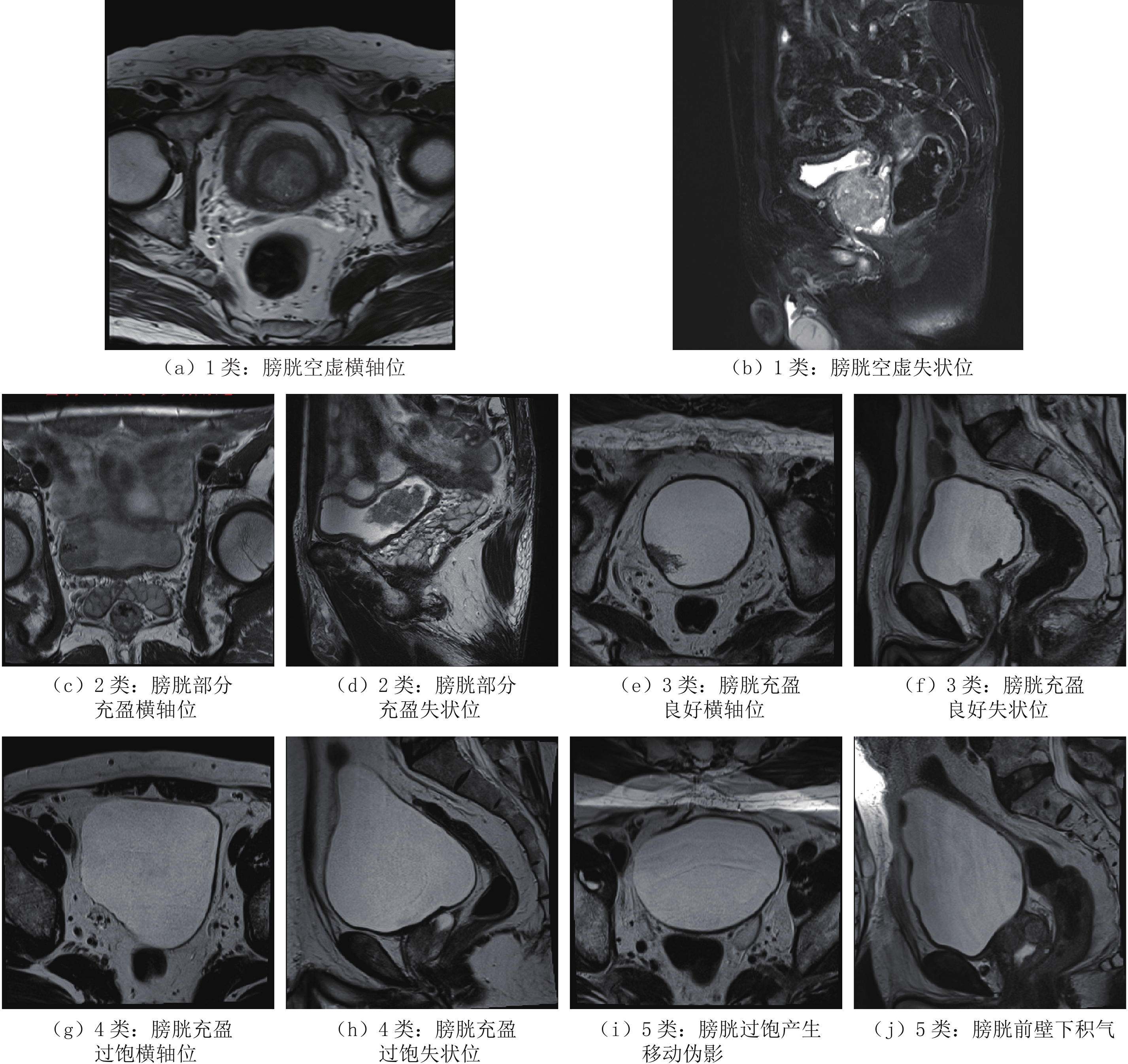
表 2 两组病例膀胱充盈情况
Table 2 Filling of bladder of two groups
组别 膀胱空虚/
例(%)膀胱部分充盈/
例(%)膀胱充盈良好/
例(%)膀胱充盈过饱/
例(%)膀胱内移动伪影/
例(%)合计/例 A组 0(0.0) 2(6.5) 26(83.9) 1(3.2) 2(2.5) 31 B组 3(6.7) 14(31.1) 21(46.7) 2(4.4) 5(11.1) 45 合计 3 16 47 3 7 76
下载: 导出CSV
表 3 两组病例膀胱充盈度评分
Table 3 Bladder filling score of two groups
组别 1分/例(%) 2分/例(%) 3分/例(%) 合计/例 A组 2(6.5) 3(9.7) 26(83.9) 31 B组 8(17.8) 16(35.6) 21(46.7) 45 合计 10 19 57 76
下载: 导出CSV
表 4 病例肌层浸润情况
Table 4 Diagnostic effectiveness of
病理结果 影像评估有无肌层浸润 无(A组/B组) 有(A组/B组) 合计(A组/B组)/例 无 19/27 1/1 20/28 有 0/3 3/11 3/14 合计 19/30 4/12 23/42
下载: 导出CSV
-
[1] BARCHETTI G, SIMONE G, CERAVOLO I, et al. Multiparametric MRI of the bladder: Inter-observer agreement and accuracy with the vesical imaging-reporting and data system (VI-RADS) at a single reference center[J]. European Radiology, 2019, 29(10): 5498−5506. doi: 10.1007/s00330-019-06117-8
[2] 赵秋枫, 华佳. 十大恶性肿瘤影像分级检查推荐方案(1.0版)之膀胱癌[J]. 中国医学计算机成像杂志, 2019,25(5): 478−481. ZHAO Q F, HUA J. Recommendation scheme for image grading examination of ten malignant tumors (version 1.0)[J]. Chinese Computed Medical Imaging, 2019, 25(5): 478−481. (in Chinese).
[3] KLAASSEN Z, KAMAT A M, KASSOUF W, et al. Treatment strategy for newly diagnosed T1 high-grade bladder urothelial carcinoma: New insights and updated recommendations[J]. European Urology, 2018, 74: 597−608. doi: 10.1016/j.eururo.2018.06.024
[4] SUSHENTSEV N, TANNER J, SLOUGH R A, et al. The effect of different drinking and voiding preparations on magnetic resonance imaging bladder distention in normal volunteers and patients[J]. Canadian Association of Radiologists Journal, 2018, 69: 383−389. doi: 10.1016/j.carj.2018.07.001
[5] BABJUK M, BÖHLE A, BURGER M, et al. EAU guidelines on non-muscle-invasive urothelial carcinoma of the bladder: Update 2016[J]. European Urology, 2017, 71: 447−461. doi: 10.1016/j.eururo.2016.05.041
[6] JACOBS B L, LEE C T. Bladder cancer in 2010: How far have we come?[J]. CA: A Cancer Journal for Clinicians, 2010, 60(4): 244−272. doi: 10.3322/caac.20077
[7] GENDY R, DELPRADO W, BRENNER P, et al. Repeat transurethral resection for non-muscle-invasive bladder cancer: A contemporary series[J]. BJU International, 2016, 117: 54−59. doi: 10.1111/bju.13265
[8] BARENTSZ J, RUIJS S H, STRIJK S P. The role of MR imaging in carcinoma of the urinary bladder[J]. American Journal of Roentgenology, 1993, 160: 937−947. doi: 10.2214/ajr.160.5.8470608
[9] 翟凤仪, 王焕军, 蔡华崧, 等. 检查前准备及扫描体位对膀胱壁磁共振成像图像质量的影响[J]. 中华腔镜泌尿外科杂志(电子版), 2015,9(5): 38−41. ZHAI F Y, WANG H J, CAI H S, et al. Bladder mnucle wall detection on 3.0T MR: Evaluation for subjects preparation and scanning position effect[J]. Chinese Journal of Endourology (Electronic Version), 2015, 9(5): 38−41. (in Chinese).
[10] SIROKY M B. The aging bladder[J]. Nature Reviews Urology, 2004, 6(S1): S3-7.
[11] FUJIOKA C, ISHII K, YAMANAGA T, et al. Optimal bladder volume at treatment planning for prostate cancer patients receiving volumetric modulated arc therapy[J]. Practical Radiation Oncology, 2016, 6: 395−401. doi: 10.1016/j.prro.2016.05.007
[12] PANEBIANCO V, NARUMI Y, ALTUN E, et al. Multiparametric magnetic resonance imaging for bladder cancer: Development of VI-RADS (vesical imaging-reporting and data system)[J]. European Urology, 2018, 74: 294−306. doi: 10.1016/j.eururo.2018.04.029
[13] 张添辉, 古志聪, 姚纯, 等. 多参数磁共振成像VI-RADS评分对膀胱癌肌层浸润诊断价值的初步研究[J]. 中国临床医学影像杂志, 2019,30(8): 569−573. ZHANG T H, GU Z C, YAO C, et al. Diagnostic value of multiparametric MRI using vesical imaging reporting and datd system in detecting muscle-invasiveness of bladder cancer[J]. Journal of China Clinic Medical Imaging, 2019, 30(8): 569−573. (in Chinese).
[14] THOENY H C, BELLIN M F, COMPERAT E M, et al. Vesical imaging-reporting and data system (VI-RADS): Added value for management of bladder cancer patients?[J]. European Urology, 2018, 74(3): 307−308. doi: 10.1016/j.eururo.2018.06.017
-
期刊类型引用(2)
1. 李洋森,王伟,李炳颖,毛云新,刘晓晖. 分频AVO技术在西湖凹陷中深层薄储层评价中的应用. CT理论与应用研究(中英文). 2025(03): 409-418 .  百度学术
百度学术
2. 朱焱辉. 基于压缩感知的地震频带拓宽方法——以珠江口盆地东部惠州地区为例. 中外能源. 2023(06): 44-52 . 百度学术
其他类型引用(0)

计量
- 文章访问数: 255
- HTML全文浏览量: 120
- PDF下载量: 16
- 被引次数: 2


