
Effect of Computed Tomography Window Technique on the Results of Artificial Intelligence Classification of Lung Lesions
-
摘要: 目的:应用3种不同的3D CNN的算法及5种CT窗口设置,探讨CT窗技术对人工智能分类肺部病变结果的影响。方法:回顾性分析172例周围型肺癌及185例局灶性肺炎的胸部CT影像资料,选择ResNet、ResNext以及DenseNet 3种不同的3D CNN的算法将病变分为两组,并在每1种3D CNN算法处理过程中应用5种不同的CT窗口设置,包括肺窗(1500,-600),纵隔窗(350,40),自定义窗口1(SW1)(1000,40),自定义窗口2(SW2)(1000,-100),全窗(4096,1024),分别计算分类准确率及AUC结果,并进行ROC曲线的两两对比。结果:ResNet的平均分类准确率最低为纵隔窗85.732%,AUC值为0.871;平均分类准确率最高为全窗,达91.596%,AUC值为0.946。ResNext的平均分类准确率最低为纵隔窗81.528%,AUC值为0.814;平均分类准确率最高为全窗,达86.568%,AUC值为0.882。DenseNet的平均分类准确率最低为纵隔窗87.954%,AUC值为0.906;平均分类准确率最高为SW2,达93.274%,AUC值为0.951。应用medcalc将3种3D CNN的5种窗口下的ROC曲线进行了两两对比发现,纵隔窗与肺窗、纵隔窗与SW1、纵隔窗与SW2之间的AUC值比较均有统计学意义。结论:3种3D CNN的分类诊断效能差别不大;CT窗口设置对CNN分类肺部病变结果有影响,在纵隔窗设置下以上3种人工智能算法对该两类肺部病变的诊断效能最差。Abstract: Objective: To use three different 3D CNN algorithms and five different computed tomography (CT) window settings to study the effect on the results of artificial intelligence classification of lung lesions in different CT window techniques. Method: A total of 172 cases of peripheral lung cancer and 185 of focal pneumonia who underwent chest CT were analyzed. Three different 3D CNN algorithms were selected (ResNet, ResNext, and DenseNet) to divide the lesions into two groups. Five different CT window settings, including lung window (1500, 600), mediastinal window (350, 40), custom window 1 (SW1) (1000, 40), and custom window 2 (SW2) (1000, 100), were used retrospectively. We calculated classification accuracy, receiver operating characteristic (ROC) curve, and area under the curve (AUC). The ROC curve was compared in pairs. Results: The average classification accuracy of ResNet was the lowest in the mediastinal window (85.732%; AUC value: 0.871) and the highest in the full window (91.596%; AUC value: 0.946). The average classification accuracy of ResNext was the lowest in the mediastinal window (81.528%; AUC value: 0.814) and the highest in the full window (86.568%; AUC value: 0.882). The average classification accuracy of DenseNet was the lowest in the mediastinal window (87.954%; AUC value: 0.906) and the highest in the SW2 window (93.274%; AUC value: 0.951). Medcalc was used to compare ROC curves under five windows of three 3D CNN. The AUC values between mediastinal window and lung window, mediastinal window and SW1, and mediastinal window and SW2 were statistically significant. Conclusion: There is little difference in the diagnostic efficacy of the three 3D CNN. Different CT window settings have an influence on the results of CNN classification of the lung lesions, and the diagnostic efficiency of the three 3D CNN is the worst under the mediastinal window.
-
Keywords:
- artificial intelligence /
- CT window technique /
- 3D CNN /
- lung lesions
-
窗技术的合理应用在CT诊断中非常重要,特别在胸部CT。由于肺内大部分病变,如肿块、炎症、间质病变等病变及周围结构的复杂性,对临床放射医师来说,除了常规选择肺窗、纵隔窗,应用更多的窗口设置能更好的观察、诊断病变。
人工智能当前在胸部CT的应用越来越多,包括图像分割、病变检测、病变分类等领域[1],如肺结节的检测、良性和恶性肺结节的分类、肺癌分期等。卷积神经网络(convolutional neural networks,CNN)是深度学习的一种代表性算法,它能很好的提取和组合空间特征以形成高级特征[2-5]。
目前文献中报道的人工智能对CT数据的处理一般是运用CT机扫描时的原始数据或选择其中一种窗宽窗位(常为肺窗),较少考虑到窗口设置对人工智能结果的影响。因此本研究与中国科学院计算技术研究所合作,选择了3种不同的3D CNN的算法:ResNet、ResNext以及DenseNet,并在每1种3D CNN算法中应用不同的CT窗口设置,探讨CT窗技术对3D CNN分类肺部病变结果的影响。
1. 资料与方法
1.1 一般资料
回顾性分析2013年3月至2018年3月在首都医科大学附属北京友谊医院诊断为周围型肺癌的172例患者及诊断为局灶性肺炎的185例的患者胸部CT薄层影像,分为两组:周围型肺癌组和局灶性肺炎组。其中周围型肺癌组(94名男性和78名女性,年龄范围为32~82岁;平均年龄为63岁)均经手术后病理证实(病理类型包括腺癌、鳞状细胞癌和小细胞癌);局灶性肺炎组(85名男性和100名女性,年龄范围为8~96岁;平均年龄为44岁)均经抗炎治疗后复查实变病变吸收。
所有患者选择胸部CT平扫图像(非增强检查),CT扫描参数:CT扫描仪为GE Discovery CT 750 HD,扫描仪参数为120 kVp,5 mm准直采集,螺距为0.984~1,重建厚度为1.25 mm,或飞利浦iCT,扫描仪参数为120 kVp,5 mm准直采集,螺距为0.915,重建厚度为1.25 mm。每幅图像的像素分辨率为512×512。
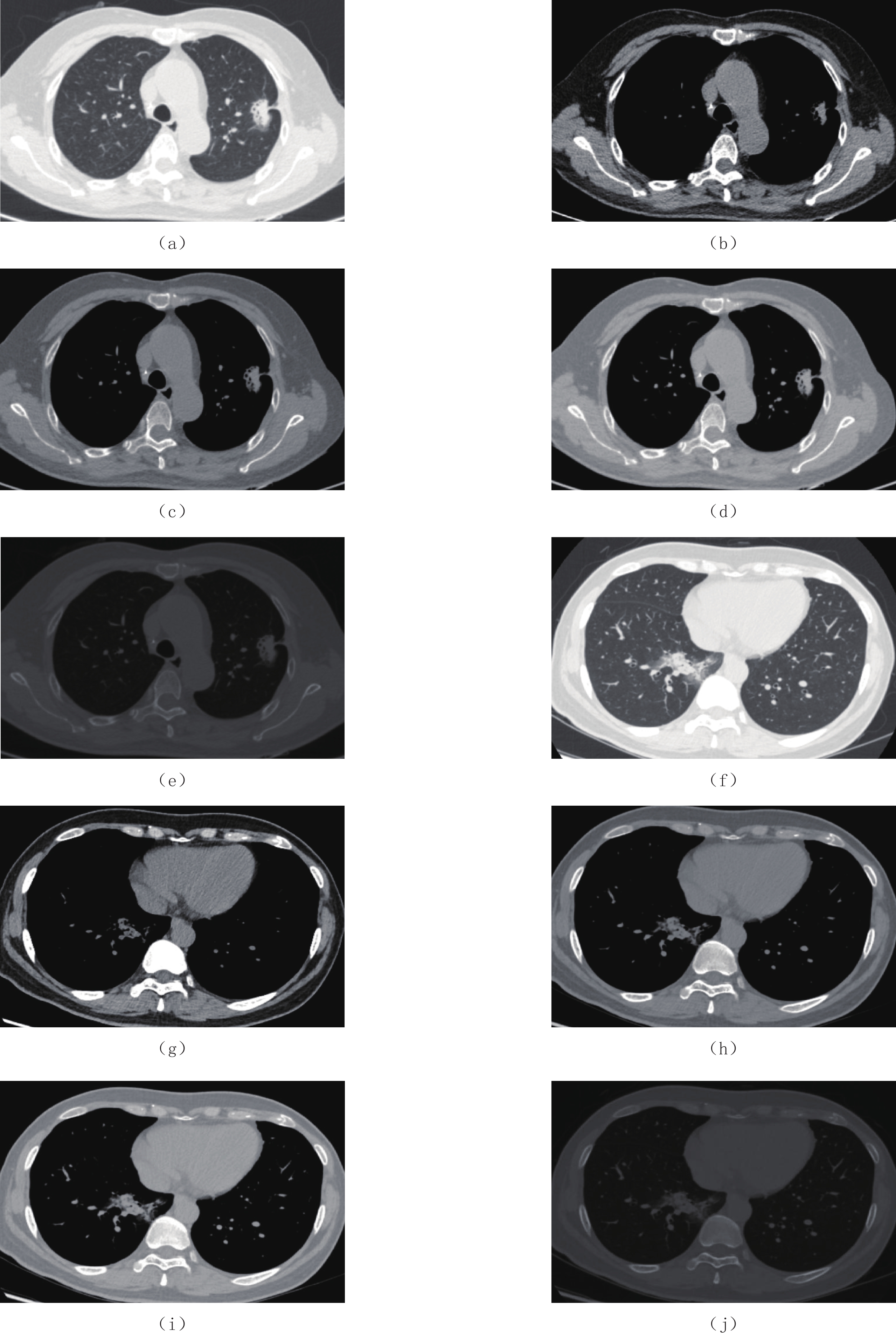
5种窗口设置(窗宽,窗位):肺窗(1500,-600);纵隔窗(350,40);自定义窗口1(SW1)(1000,40);自定义窗口2(SW2)(1000,-100);全窗(4096,1024)。自定义窗口1(SW1)是选择和纵隔窗相同的窗位,然后扩大窗宽为1000;自定义窗口2(SW2)是选择和SWI相同的窗宽1000,窗位定为 -100;全窗是将CT扫描及DICOM图像存储的原始数据CT值范围为-1024~3071 HU换算成的窗宽窗位(4096,1024)。5种窗口设置下的病变显示见图1。
![]() 图 1 不同窗口设置时对病灶的显示不同:(a)~(e)左肺上叶周围型肺癌(腺癌);(f)~(j)右肺下叶局灶性肺炎(a)和(f)肺窗;(b)和(g)纵隔窗;(c)和(h)SW1;(d)和(i)SW2;(e)和(j)全窗Figure 1. Different display of lesions in different windows: (a)~(e) is the same patient, diagnosed as peripheral lung cancer in the upper lobe of the left lung (pathological examination shows adenocarcinoma); (f)~(j) is the same patient, diagnosed as focal pneumonia in the lower lobe of the right lung
图 1 不同窗口设置时对病灶的显示不同:(a)~(e)左肺上叶周围型肺癌(腺癌);(f)~(j)右肺下叶局灶性肺炎(a)和(f)肺窗;(b)和(g)纵隔窗;(c)和(h)SW1;(d)和(i)SW2;(e)和(j)全窗Figure 1. Different display of lesions in different windows: (a)~(e) is the same patient, diagnosed as peripheral lung cancer in the upper lobe of the left lung (pathological examination shows adenocarcinoma); (f)~(j) is the same patient, diagnosed as focal pneumonia in the lower lobe of the right lung1.2 3D CNN的设置
本研究与中国科学院计算技术研究所合作,对图像进行预处理,并选择3种不同的3D CNN的算法:ResNet、ResNext以及DenseNet。
本研究的图像预处理是在三维图像空间中进行,步骤包括:①高斯去噪,应用sigma为0.5的高斯滤波器,将图像的背景噪声去除;②肺分割,使用基于阈值的方法进行粗略肺分割;③对齐及缩放,根据肺实质区域的分割结果得到肺实质区域的边界框,将输入大小统一设置为(96,64,96)。
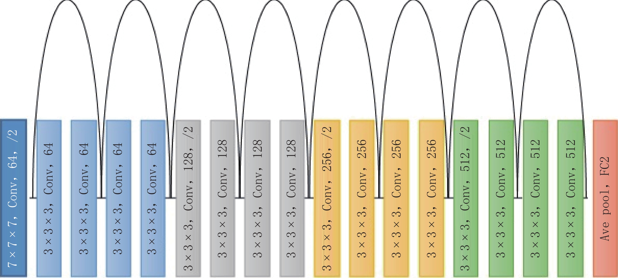
ResNet网络的超参数:使用Adam优化器,初始学习率为0.0001,β1=0.9,β2=0.999,权值衰减为0.001。所有实验都经过200个epochs的训练。计算机配备Intel Core i7-8700 CPU和NVIDIA GTX1080 ti GPU,深度学习平台为PyTorch。网络构建:主干使用18层的3D ResNet网络(图2),最后,利用一个全局池化层和一个具有两个输出的全连接层,将病例分为两类:周围型肺癌与局灶性肺炎。为了避免训练数据的过度拟合,在最后一个全连接层之前使用一个比率为0.5的dropout层[6]。
![]() 图 2 3D ResNet网络构建主干使用18层的3D ResNet网络,在每个卷积块中,有1个卷积层(其参数在图中列出)、1个批标准化(Batch Normalization)层和1个ReLu激活层,图中的第1个参数“3×3×3”表示3D内核大小,第3个参数“64、128、256、512”表示通道数,最后1个参数“/2”表示步长为2的池层。Figure 2. 3D ResNet network design
图 2 3D ResNet网络构建主干使用18层的3D ResNet网络,在每个卷积块中,有1个卷积层(其参数在图中列出)、1个批标准化(Batch Normalization)层和1个ReLu激活层,图中的第1个参数“3×3×3”表示3D内核大小,第3个参数“64、128、256、512”表示通道数,最后1个参数“/2”表示步长为2的池层。Figure 2. 3D ResNet network designResNext网络及DenseNet分别为50层的主干及121层的主干,并保持其他训练参数与ResNet的参数相同。
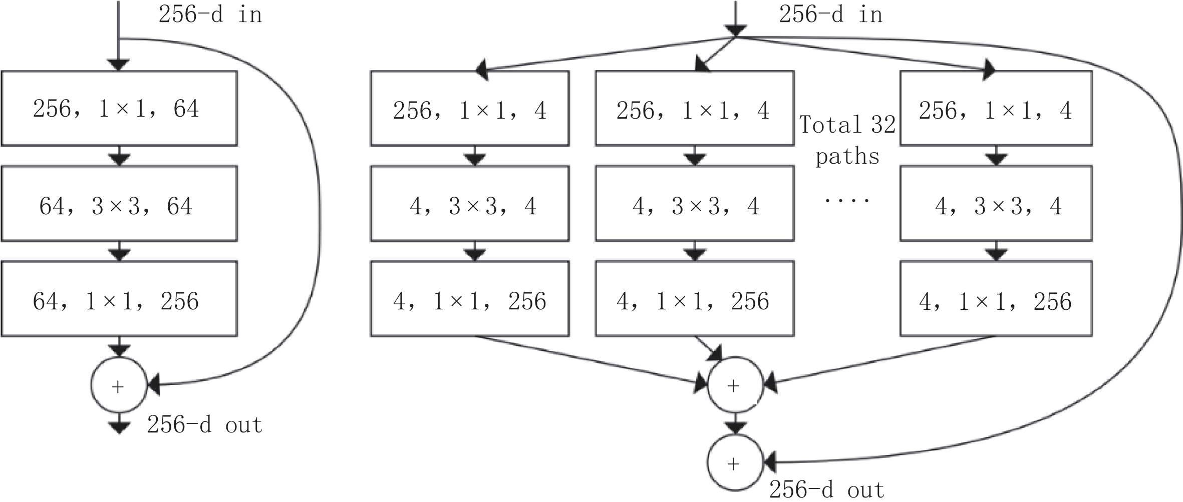
ResNext对于ResNet中的每一个模块(Block)进行多次重复之后再对结果求和,该构建块的不同重复单元具有相同的网络拓扑结构以减轻网络结构设计负担。与ResNet相比,它增加了重复次数这一新维度,即基数(Cardinality),作为深度和宽度维度之外的重要因素[7](图3)。
![]()
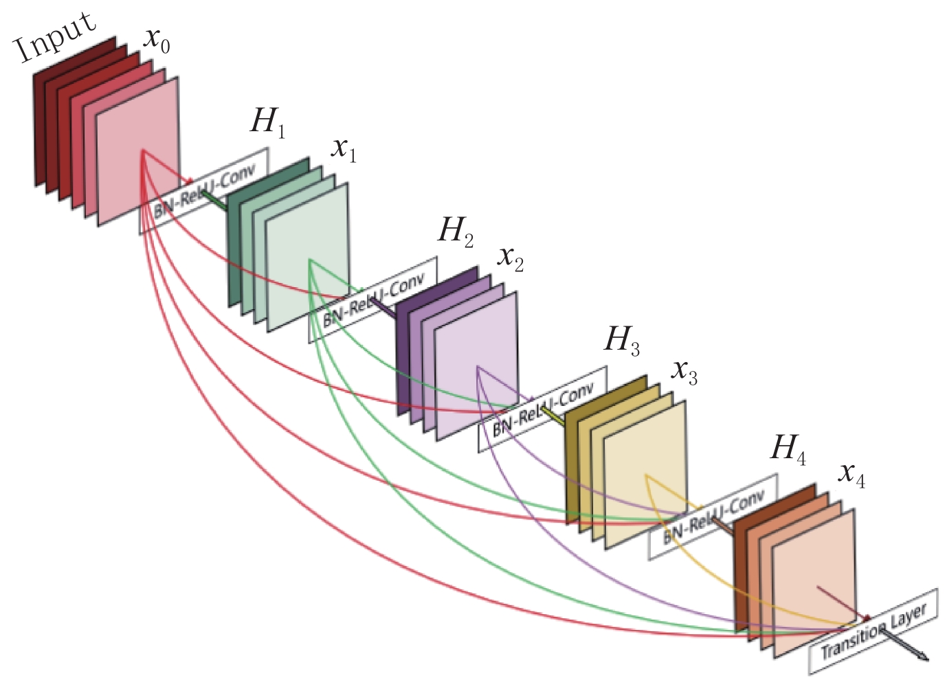
相比ResNet,DenseNet引入密集连接机制。每个层都会将前面所有层作为其输入。图4表示DenseNet的密集连接机制。网络的每一层都会与前面所有层在通道维度连接在一起,并作为下一层的输入[8]。
1.3 统计分析
使用IBM SPSS 19进行数据分析,进行5倍交叉验证,计算分类准确率,绘制受试者工作特征曲线(receiver operating characteristic curve,ROC),统计曲线下面积(area under curve,AUC)。使用medcalc 19.1版本Delong测试进行ROC曲线的两两对比,统计Z值及P值,并以P<0.05为差异具有统计学意义。
2. 结果
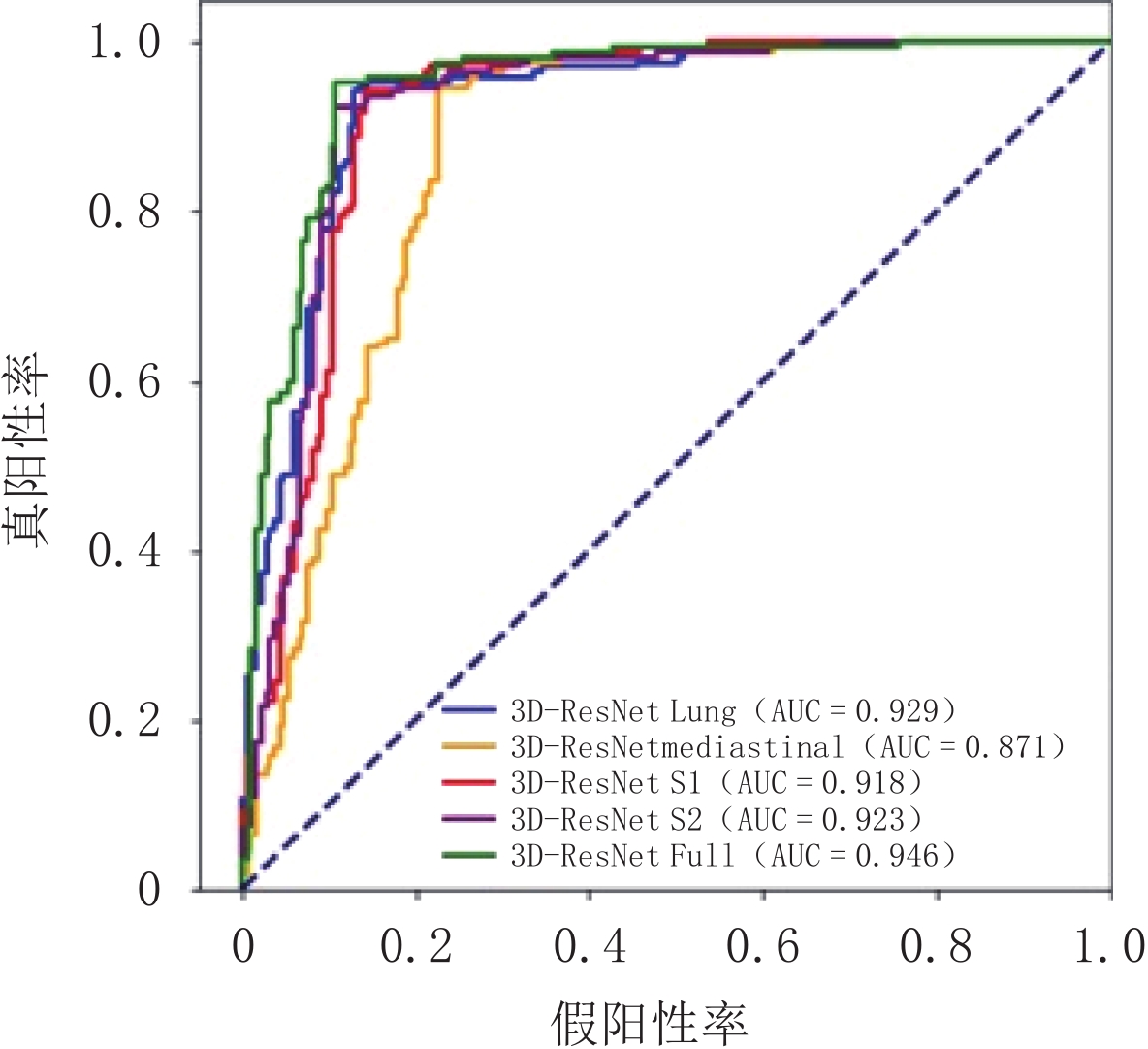
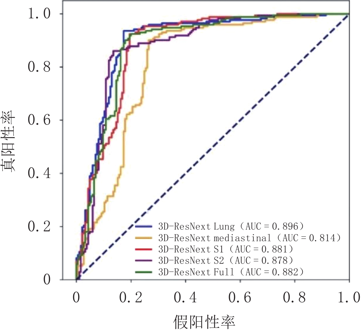
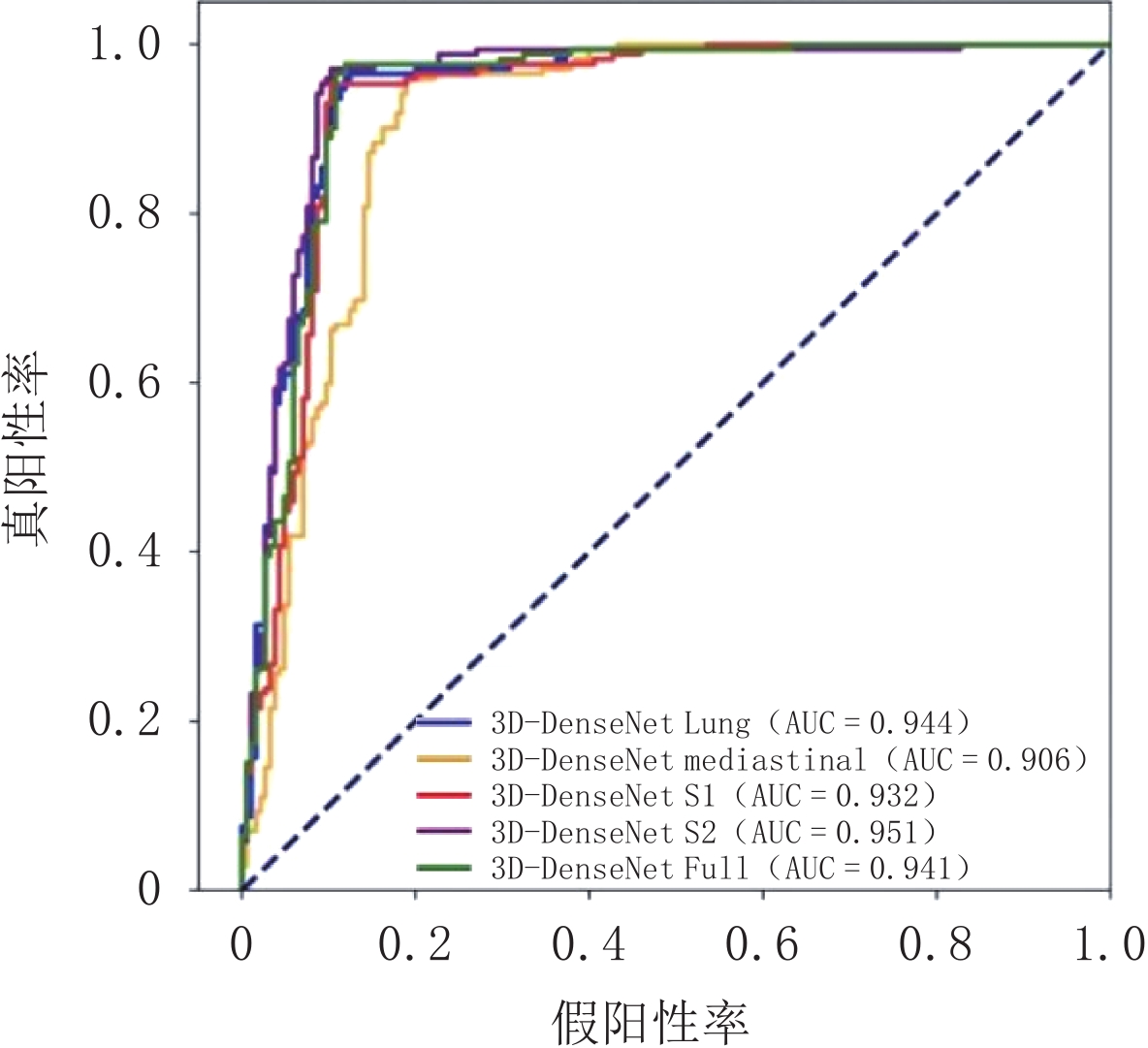
本研究将整个数据集随机分成5个部分,进行5倍交叉验证,结果表明3种神经网络之间的分类诊断效能没有太大差异,总体来说ResNext的性能稍差,DenseNet的性能稍好。在窗口设置方面,ResNet的平均分类准确率最低为纵隔窗;平均分类准确率最高为全窗。ResNext的平均分类准确率最低为纵隔窗;平均分类准确率最高为全窗。DenseNet的平均分类准确率最低为纵隔窗;平均分类准确率最高为自定义窗口2(表1~表3)ROC曲线图见图5~图7。
表 1 ResNet 5个窗口设置的5倍交叉验证的分类准确率(%)及AUC结果Table 1. Classification accuracy (%) and AUC results of 5-fold cross validation of 5 window settings in ResNet窗口 (WW,WL) Fold 1 Fold 2 Fold 3 Fold 4 Fold 5 平均分类准确率/% AUC 肺窗 (1500,−600) 87.50 88.73 91.55 91.55 93.06 90.478 0.929 纵隔窗 (350,40) 80.56 87.32 90.14 85.92 84.72 85.732 0.871 SW1 (1000,40) 86.11 92.96 88.73 91.55 90.28 89.926 0.918 SW2 (1000,−100) 87.50 92.96 90.14 91.55 90.28 90.486 0.923 Full (4096,1024) 87.50 90.14 90.14 94.37 95.83 91.596 0.946 表 2 ResNext 5个窗口设置的5倍交叉验证的分类准确率(%)及AUC结果Table 2. Classification accuracy (%) and AUC results of 5-fold cross validation of 5 window settings in ResNext窗口 (WW,WL) Fold 1 Fold 2 Fold 3 Fold 4 Fold 5 平均分类准确率/% AUC 肺窗 (1500,−600) 84.72 80.14 87.32 88.73 87.50 85.682 0.896 纵隔窗 (350,40) 77.78 83.10 81.69 84.51 80.56 81.528 0.814 SW1 (1000,40) 86.11 85.92 88.73 85.92 84.72 86.280 0.881 SW2 (1000,−100) 84.72 88.73 83.10 85.92 87.50 85.994 0.878 Full (4096,1024) 83.33 91.55 85.92 87.32 84.72 86.568 0.882 表 3 DenseNet 5个窗口设置的5倍交叉验证的分类准确率(%)及AUC结果Table 3. Classification accuracy (%) and AUC results of 5-fold cross validation of 5 window settings in DenseNet窗口 (WW,WL) Fold 1 Fold 2 Fold 3 Fold 4 Fold 5 平均分类准确率/% AUC 肺窗 (1500,−600) 87.50 91.55 92.96 94.37 93.06 91.888 0.944 纵隔窗 (350,40) 88.89 87.32 91.55 84.51 87.50 87.954 0.906 SW1 (1000,40) 94.44 94.37 90.14 90.14 93.06 92.430 0.932 SW2 (1000,−100) 93.06 95.77 91.55 91.55 94.44 93.274 0.951 Full (4096,1024) 91.67 92.96 90.14 94.37 94.44 92.716 0.941 将ResNet网络的5种窗口下的ROC曲线进行了两两对比,结果如下:肺窗与纵隔窗AUC的值比较有统计意义(Z=3.099,P=0.002);肺窗与SW1之间(Z=0.800,P=0.424)、肺窗与SW2之间(Z=0.478,P=0.633)、肺窗与全窗之间(Z=1.811,P=0.072)的AUC值比较均无统计学意义。纵隔窗与SW1(Z=2.884,P=0.004)、纵隔窗与SW2(Z=2.869,P=0.004)、纵隔窗与全窗(Z=4.115,P=0.000)之间的AUC值比较均有统计学意义。SW1与SW2之间无统计学意义(Z=0.376,P=0.707)。SW1与全窗之间有统计学意义(Z=2.043,P=0.041),SW2与全窗之间无统计学意义(Z=1.705,P=0.088)。
将ResNext网络的5种窗口下的ROC曲线进行了两两对比,结果如下:肺窗与纵隔窗AUC的值比较有统计意义(Z=4.161,P=0.000);肺窗与SW1之间(Z=0.918,P=0.358)、肺窗与SW2之间(Z=1.210,P=0.226)、肺窗与全窗之间(Z=1.079,P=0.2806)的AUC值比较均无统计学意义。纵隔窗与SW1(Z=3.652,P=0.000)、纵隔窗与SW2(Z=3.265,P=0.000)、纵隔窗与全窗(Z=3.336,P=0.001)之间的AUC值比较均有统计学意义。SW1与SW2之间无统计学意义(Z=0.177,P=0.860)。SW1与全窗之间有统计学意义(Z=0.0338,P=0.969,SW2与全窗之间无统计学意义(Z=0.210,P=0.834)。
将DensNet网络的5种窗口下的ROC曲线进行了两两对比,结果如下:肺窗与纵隔窗AUC的值比较有统计意义(Z=2.332,P=0.020);肺窗与SW1之间(Z=0.962,P=0.336)、肺窗与SW2之间(Z=0.659,P=0.510)、肺窗与全窗之间(Z=294,P=0.769)的AUC值比较均无统计学意义。纵隔窗与SW1(Z=2.193,P=>0.028)、纵隔窗与SW2(Z=2.854,P=0.004)之间的AUC值比较均有统计学意义。纵隔窗与全窗(Z=1.951,P=0.051)的AUC值比较无统计学意义。SW1与SW2之间无统计学意义(Z=1.583,P=0.116)。SW1与全窗之间有统计学意义(Z=0.617,P=0.537),SW2与全窗之间无统计学意义(Z=0.866,P=0.389)。
3. 讨论
本研究应用了3种不同的3D CNN的算法及5种CT窗口设置,初步探讨CT窗技术对人工智能分类肺部病变结果的影响。
由于人体不同组织间CT值有差异,而人眼仅能识别16灰阶,因此合理应用窗技术在CT检查中非常重要。特别在胸部CT,既要观察低密度的含气肺组织(CT值约 -700至 -1000 HU),又要观察中等密度的软组织(CT值约30~80 HU),以及高密度的骨组织(CT值约150~1000 HU以上)。在临床胸部影像读片过程中,至少要采用肺窗、纵隔窗来分别观察肺野和纵隔。其中肺窗能清楚显示病变边缘及周围肺组织,但对病变内部结构显示不佳,纵隔窗对病变内部结构及纵隔结构显示清晰,但对病变的边缘及周围肺组织显示不佳。如图1所示,不同的窗口设置对病变的显示范围不同,在肺窗下病变的边缘、毛刺、磨玻璃等结构显示比纵隔窗要清晰,但对病变内部结构显示不如纵隔窗[9-11]。
本研究选用了5种不同的窗宽窗位:考虑到CT机扫描时及DICOM图像存储的CT值范围为-1024~3071 HU,换算成窗宽窗位为(4096,1024)(本研究称之为全窗),除了选择全窗和常规的肺窗(1500,-600)、纵隔窗(350,40)以外,还自定义了两个窗宽窗位:自定义窗口1(SW1)和自定义窗口2(SW2)。其中SW1由纵隔窗转换而来,本研究选择和纵隔窗相同的窗位(40),然后扩大窗宽为1000(在临床影像读片过程中,我们发现SW1在一定程度上可以兼顾观察到病灶的边缘和病变密度的差异)。SW2是SW1的类似转变,窗宽仍为1000,然后将窗位定为 -100(在临床影像读片过程中,我们发现肺内实性病变的CT值比软组织密度要低,因此将SW2的窗位定为 -100 HU,本研究认为SW2与SW1一样,可以在一定程度上兼顾观察到病灶的边缘和病变密度的差异)。
卷积神经网络是深度学习技术的一种模型,卷积神经网络的关键思想在于多层堆叠、局部连接、权值共享和池化,将单层的卷积神经网络进行多次堆叠,前一层的输出作为后一层的输入,便构成卷积神经网络;CNN中的层与层之间不再是全连接的,而是局部连接的,这样可以大大简化模型的复杂度,减少参数的数量[12-13]。CNN目前可以应用于胸部CT多个方面,包括肺组织分割、肺结节检测、肺部病变的分类等,网络结构也趋于3D化。本研究选择了3种3D CNN网络模型:ResNet、ResNext以及DenseNet,这3种网络均为深度学习常用网络结构。
本研究选择的3种神经网络之间的分类准确率及诊断效能没有太大差异,总体来说ResNext的性能稍差,DenseNet的性能稍好。在窗口设置方面,ResNet的平均分类准确率最低为纵隔窗85.732%,AUC值为0.871;平均分类准确率最高为全窗,达91.596%,AUC值为0.946。ResNext的平均分类准确率最低为纵隔窗81.528%,AUC值为0.814;平均分类准确率最高为全窗,达86.568%,AUC值为0.882。DenseNet的平均分类准确率最低为纵隔窗87.954%,AUC值为0.906;平均分类准确率最高为自定义窗口2,达93.274%,AUC值为0.951。本研究还将3种3D CNN的5种窗口下的ROC曲线进行了两两对比发现,纵隔窗与肺窗、纵隔窗与SW1、纵隔窗与SW2之间的AUC值比较均有统计学意义,而其余4种窗口设置之间结果大多没有显著差异,也就是说纵隔窗设置下的AUC值结果最差。根据公式,CT图像显示的强度范围为“窗位+1/2窗宽~窗位 -1/2窗宽”,可以计算出5个窗口设置下对应的的图像强度:肺窗[-1350,150];纵隔窗[-135,215];自定义窗口1(SW1)[-460,540];自定义窗口2(SW2)[-600,400];全窗图像的强度将保持原始范围[-1024,3071]。由于肺实性病变CT值约 -300~50 HU,磨玻璃病变CT值约 -700~-300 HU[14-15],由此可以发现全窗、肺窗及SW2这3种窗口设置能比较好的覆盖住肺实质以及病变的CT值范围,而纵隔窗和SW1这2种窗口设置并没有完全覆盖住如磨玻璃病变的CT值范围。而本研究结果也表明在纵隔窗下3D CNN的分类效果最差,我们认为其原因可能是在纵隔窗设置下,所覆盖的CT图像显示的强度范围窄,可能会去除一部分有价值的信息,而其他4种窗口下所覆盖的CT图像显示的强度范围相对来说能提供足够的信息。
然而由于本研究的样本量不大,窗口设置的选择以参考文献和临床经验为主,并且数据采集选用了不同机器,参数设置会有一定差异,此外还有待多中心的研究,进一步提高结果的可靠性。
总之,CT窗口设置对人工智能分类肺部病变结果有影响,不同的窗口设置下人工智能对肺部病变的分类准确率及诊断效能有差异,本研究认为在未来人工智能研究中应该适当加入CT窗技术的研究。
-
![]()
图 1 不同窗口设置时对病灶的显示不同:(a)~(e)左肺上叶周围型肺癌(腺癌);(f)~(j)右肺下叶局灶性肺炎
(a)和(f)肺窗;(b)和(g)纵隔窗;(c)和(h)SW1;(d)和(i)SW2;(e)和(j)全窗
Figure 1. Different display of lesions in different windows: (a)~(e) is the same patient, diagnosed as peripheral lung cancer in the upper lobe of the left lung (pathological examination shows adenocarcinoma); (f)~(j) is the same patient, diagnosed as focal pneumonia in the lower lobe of the right lung
![]()
图 2 3D ResNet网络构建
主干使用18层的3D ResNet网络,在每个卷积块中,有1个卷积层(其参数在图中列出)、1个批标准化(Batch Normalization)层和1个ReLu激活层,图中的第1个参数“3×3×3”表示3D内核大小,第3个参数“64、128、256、512”表示通道数,最后1个参数“/2”表示步长为2的池层。
Figure 2. 3D ResNet network design
![]()
表 1 ResNet 5个窗口设置的5倍交叉验证的分类准确率(%)及AUC结果
Table 1 Classification accuracy (%) and AUC results of 5-fold cross validation of 5 window settings in ResNet
窗口 (WW,WL) Fold 1 Fold 2 Fold 3 Fold 4 Fold 5 平均分类准确率/% AUC 肺窗 (1500,−600) 87.50 88.73 91.55 91.55 93.06 90.478 0.929 纵隔窗 (350,40) 80.56 87.32 90.14 85.92 84.72 85.732 0.871 SW1 (1000,40) 86.11 92.96 88.73 91.55 90.28 89.926 0.918 SW2 (1000,−100) 87.50 92.96 90.14 91.55 90.28 90.486 0.923 Full (4096,1024) 87.50 90.14 90.14 94.37 95.83 91.596 0.946  下载: 导出CSV
表 2 ResNext 5个窗口设置的5倍交叉验证的分类准确率(%)及AUC结果
Table 2 Classification accuracy (%) and AUC results of 5-fold cross validation of 5 window settings in ResNext
窗口 (WW,WL) Fold 1 Fold 2 Fold 3 Fold 4 Fold 5 平均分类准确率/% AUC 肺窗 (1500,−600) 84.72 80.14 87.32 88.73 87.50 85.682 0.896 纵隔窗 (350,40) 77.78 83.10 81.69 84.51 80.56 81.528 0.814 SW1 (1000,40) 86.11 85.92 88.73 85.92 84.72 86.280 0.881 SW2 (1000,−100) 84.72 88.73 83.10 85.92 87.50 85.994 0.878 Full (4096,1024) 83.33 91.55 85.92 87.32 84.72 86.568 0.882
下载: 导出CSV
表 3 DenseNet 5个窗口设置的5倍交叉验证的分类准确率(%)及AUC结果
Table 3 Classification accuracy (%) and AUC results of 5-fold cross validation of 5 window settings in DenseNet
窗口 (WW,WL) Fold 1 Fold 2 Fold 3 Fold 4 Fold 5 平均分类准确率/% AUC 肺窗 (1500,−600) 87.50 91.55 92.96 94.37 93.06 91.888 0.944 纵隔窗 (350,40) 88.89 87.32 91.55 84.51 87.50 87.954 0.906 SW1 (1000,40) 94.44 94.37 90.14 90.14 93.06 92.430 0.932 SW2 (1000,−100) 93.06 95.77 91.55 91.55 94.44 93.274 0.951 Full (4096,1024) 91.67 92.96 90.14 94.37 94.44 92.716 0.941
下载: 导出CSV
-
[1] Van GINNEKEN B. Fifty years of computer analysis in chest imaging: Rule-based, machine learning, deep learning[J]. Radiological Physics and Technology, 2017, 10(1): 23−32. doi: 10.1007/s12194-017-0394-5
[2] YU X, LU S, GUO L, et al. ResGNet-C: A graph convolutional neural network for detection of COVID-19[J]. Neurocomputing, 2021, 452: 592−605. doi: 10.1016/j.neucom.2020.07.144
[3] LEE S M, SEO J B, YUN J, et al. Deep learning applications in chest radiography and computed tomography: Current state of the art[J]. Journal of Thoracic Imaging, 2019, 34(2): 75−85. doi: 10.1097/RTI.0000000000000387
[4] SHIN H C, ROTH H R, GAO M, et al. Deep convolutional neural networks for computer-aided detection: CNN architectures, dataset characteristics and transfer learning[J]. IEEE Transactions on Medical Imaging, 2016, 35(5): 1285−1298.
[5] KANG G, LIU K, HOU B, et al. 3D multi-view convolutional neural networks for lung nodule classification[J]. Plos One, 2017, 12(11): 12−22.
[6] CHENG X, WEN H, YOU H, et al. Recognition of peripheral lung cancer and focal pneumonia on chest computed tomography images based on convolutional neural network[J]. Technology in Cancer Research & Treatment, 2022, 21: 1-12.
[7] XIE S, GIRSHICK R, DOLLÁR P, et al. Aggregated residual transformations for deep neural networks[C]//In Proceedings of the IEEE Conference on Computer Vision and Pattern Recognition, 2017: 1492-1500.
[8] HUANG G, LIU Z, Van der MAATEN L, et al. Densely connected convolutional networks[C]//In Proceedings of the IEEE Conference on Computer Vision and Pattern Recognition, 2017: 4700-4708.
[9] 田培林, 徐覃莎, 唐利荣, 等. 胸部CT检查的肺窗技术[J]. 生物医学工程与临床, 2002,6(4): 209−213. doi: 10.3969/j.issn.1009-7090.2002.04.010 TIAN P L, XU Q S, TANG L R, et al. Window techniques for pulmonary image in chest CT[J]. Biomedical Engineering and Clinical Medicine, 2002, 6(4): 209−213. (in Chinese). doi: 10.3969/j.issn.1009-7090.2002.04.010
[10] 张胜超, 陈浩, 秦宣, 等. CT窗口技术在肺磨玻璃结节诊断中的临床意义[J]. 中国现代医学杂志, 2019,29(14): 106−109. ZHANG S C, CHEN H, QIN X, et al. Clinical significance of CT window technique in the diagnosis and treatment of pulmonary ground-glass nodules[J]. China Journal of Modern Medicine, 2019, 29(14): 106−109. (in Chinese).
[11] KITAMI A, SANO F, HAYASHI S, et al. Correlation between histological invasiveness and the computed tomography value in pure ground-glass nodules[J]. Surgery Today, 2016, 46(5): 593−598. doi: 10.1007/s00595-015-1208-1
[12] 张鹏, 徐欣楠, 王洪伟, 等. 基于深度学习的计算机辅助肺癌诊断方法[J]. 计算机辅助设计与图形学学报, 2018,30(1): 90−99. ZHANG P, XU X N, WANG H W, et al. Computer-aided lung cancer diagnosis approaches base on deep learning[J]. Journal of Computer-Aided Design & Computer Graphics, 2018, 30(1): 90−99. (in Chinese).
[13] 尹柯, 张久权, 伍建林, 等. 对比卷积神经网络分类模型与放射科医师鉴别浸润性肺腺癌的效能[J]. 中国医学影像技术, 2021,37(9): 1338−1342. YIN K, ZHANG J Q, WU J L, et al. Comparison on convolutional neural network classification model and radiologists in differentiating invasive lung adenocarcinoma[J]. Chinese Journal of Medical Imaging Technology, 2021, 37(9): 1338−1342. (in Chinese).
[14] 戴书华, 刘国芳, 向东生. 肺磨玻璃结节CT值测量在早期癌症诊断中的意义[J]. 中华肺部疾病杂志(电子版), 2019,12(6): 770−771. DAI S H, LIU G F, XIANG D S. The significance of CT value measurement of pulmonary ground glass nodule in early cancer diagnosis[J]. Chinese Journal of Lung Diseases Electronic Edition, 2019, 12(6): 770−771. (in Chinese).
[15] HE S, CHEN C, WANG Z, et al. The use of the mean computed tomography value to predict the invasiveness of ground-glass nodules: A meta-analysis[J]. Asian Journal of Surgery, 2022, 18: 1-6.
-
期刊类型引用(1)
1. 李梦雨,段诗苗,张雷,周咏春. 提高肺SBRT精度的多窗位动态组合诊疗手段. 中国CT和MRI杂志. 2024(04): 63-65 .  百度学术
百度学术
其他类型引用(1)
计量
- 文章访问数: 453
- HTML全文浏览量: 213
- PDF下载量: 30
- 被引次数: 2


