
Diagnostic Value of Dual-source CT Dual-energy Virtual Non-contrasting Extramural Vascular Invasions of Rectal Cancer
-
摘要:
目的:探讨双源CT双能量虚拟平扫(VNC)对直肠癌壁外血管侵犯(EMVI)的术前诊断价值。方法:选取2019年11月至2021年12月间拟手术治疗的150例直肠癌患者术前行双源CT双能量虚拟平扫,女性64例(42.7%)、男性86例(57.3%),平均年龄为(62.3±11.8)岁。两名高年资影像科医师独立分析术前影像资料(包括ctEMVI情况及有无局部淋巴结浸润和远处转移),判断是否存在术前EMVI。以手术病理结果为金标准,分别评价VNC诊断EMVI的敏感性、特异性、准确性、阳性预测值和阴性预测值并采用受试者操作特征(ROC)曲线下面积评价其诊断效能。结果:150例直肠癌患者中,术后病理证实56例(37.3%)为EMVI阳性,94例(62.7%)为EMVI阴性。医师1对EMVI评价准确性、敏感性、特异性、阳性预测值、阴性预测值分别为86%、80.36%、89.36%、81.82%、88.42%,AUC为0.831(95%CI 0.756~0.905);医师2的准确性、敏感性、特异性、阳性预测值、阴性预测值分别为88.67%、80.36%、93.62%、88.24%、88.89%,AUC为0.870(95%CI 0.802~0.938),医师1与医师2的评估结果一致性较高(k=0.943)。结论:双源CT双能量虚拟平扫对直肠癌EMVI术前评估具有一定的诊断价值。
Abstract:Objective: To investigate the value of dual-source CT dual-energy virtual plain scan in the preoperative diagnosis of extramural vascular invasion (EMVI) in rectal cancer. Methods: A total of 150 patients with rectal cancer (64 females (42.7%) and 86 males (57.3%), with an average age of (62.3±11.8) years) who were scheduled for surgical treatment in our hospital from November 2019 to December 2021 were selected for the preoperative dual-source CT dual-energy virtual plain scan;. Two senior radiologists independently analyzed preoperative imaging data (including ctEMVI status, local lymph node infiltration, and distant metastasis) to determine the existence of preoperative EMVI. With pathological results as the gold standard, the sensitivity, specificity, accuracy, positive predictive value, and negative predictive value of VNC in the diagnosis of EMVI were evaluated, and the area under the receiver operating characteristic curve (ROC) was used to evaluate the diagnostic efficiency. Results: Among 150 patients with rectal cancer, 56 (37.3%) were positive for EMVI and 94 (62.7%) were negative for EMVI. The accuracy, sensitivity, specificity, positive predictive value, and negative predictive value of EMVI evaluation by physician 1 were 86%, 80.36%, 89.36%, 81.82%, and 88.42%, respectively, and the AUC was 0.831 (95%CI, 0.756~0.905). The accuracy, sensitivity, specificity, positive predictive value, and negative predictive value of physician 2 were 88.67%, 80.36%, 93.62%, 88.24%, and 88.89%, respectively, and the AUC was 0.870 (95%CI, 0.802~0.938). The consistency of the evaluation results between physician 1 and physician 2 was high (k=0.943). Conclusion: Dual-source CT dual-energy virtual plain scan has a certain diagnostic value in the preoperative evaluation of EMVI of rectal cancer.
-
据2020年全球癌症流行病学调查结果[1]显示,结直肠癌的发病率及死亡率均位于前5位,高发病率、高死亡率严重威胁全球人类的生命健康。研究发现17%~52% 结直肠癌患者存在直肠癌壁外血管侵犯(extramural vascular invasion,EMVI)阳性[2],已有大量研究证实[2-4]直肠癌EMVI是直肠癌重要的预后因素,可作为预测局部复发、远处转移以及总生存时间减少的独立危险因素,也是是否需要进行新辅助治疗的重要依据,因此,术前影像学精准评价直肠癌EMVI对下一步诊疗策略以及改善患者预后具有重要的临床意义。
有研究[5]得出结论:术前MRI诊断EMVI(mrEMVI)可为患者的治疗决策提供强有力依据,但也有文献报道[6]mrEMVI的敏感性和特异性差异较大,相对于MRI检查而言,CT具有伪影少、风险低、速度快、依从性好等优点,且在NCCN及UICC《结肠癌临床实践指南》中CT被纳入高危人群结肠癌筛查、术前影像学评估及辅助化疗复查的常规检查项目[7]。
目前,国内外将CT虚拟平扫这一技术应用于直肠癌EMVI的报道甚少,本文旨在探讨双源CT双能量虚拟平扫技术对直肠癌EMVI术前评估的诊断效能。
1. 资料与方法
1.1 研究对象
收集内蒙古医科大学附属医院2019年11月至2021年12月直肠癌拟手术的患者169例行双源CT双能量虚拟平扫,纳入标准:①检查后 1周内进行手术且经过病理学检查确诊为直肠癌;②双源 CT双能量虚拟平扫成像前未进行化疗、放疗及靶向治疗等;③患者无 CT成像相关的禁忌症,无碘对比剂及相关过敏史临床、影像资料完整。
排除标准:①合并心、肝、肾功能不全或其他直肠肿瘤;②图像质量较差,无法完成影像评估;③合并严重精神疾病或认知障碍,19例因图像质量不佳、临床资料不全排除,最终纳入150例,其中男性86例,女性64例,年龄43.0~77.0岁,平均年龄(62.3±11.8)岁。本研究通过内蒙古医科大学附属医院伦理委员会同意,并在检查前获取患者签署的书面知情同意书。
1.2 检查方法
检查前1~2日进流食,检查当日禁食6~8 h,检查前2~3 h饮用纯净水500~1000 mL用以充盈膀胱。采用西门子(dual source CT,DSCT)配以高压注射器由膈顶扫描至耻骨联合下缘。扫描参数:管电压100 kV,开启实时动态曝光剂量调节CARE Dose 4D,准直器宽度128×0.6 mm,层厚和层间距5 mm,重建层厚0.75 mm、间隔0.5 mm,矩阵512×512。碘海醇(300 mgI/mL)70 mL,注射速率3 mL/s,扫描结束后利用Liver VNC软件处理得到动脉期、门静脉期融合图像及虚拟平扫图像,将所有图像传送至pacs系统并在该系统进行图像重建及分析。
1.3 图像分析
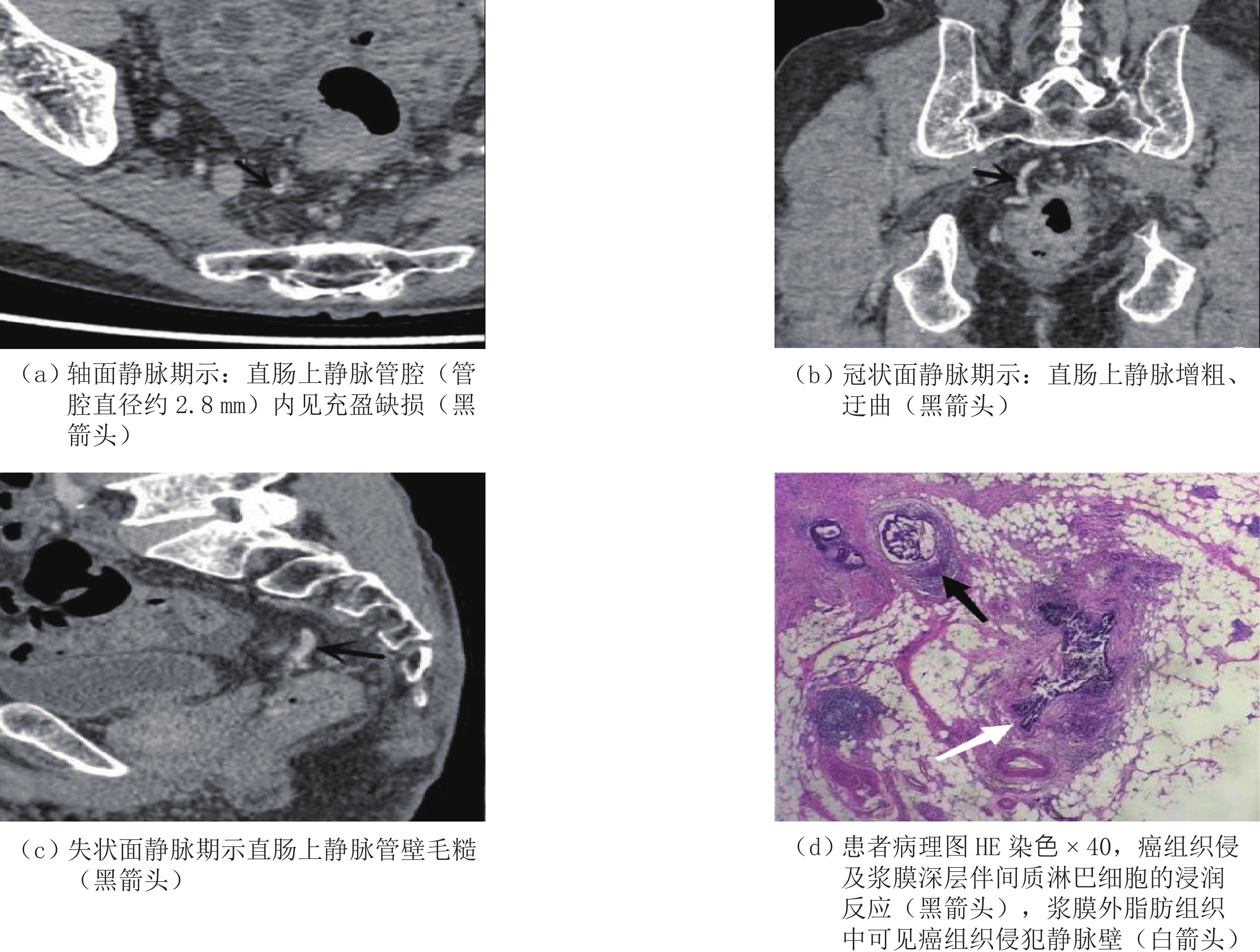
由两名高年资影像科医师采用独立双盲法进行壁外血管侵犯、局部淋巴结浸润、远处转移评估。ctEMVI阳性定义为肿瘤附近可见迂曲血管在静脉期明显强化(图1(b))、或周围系膜血管内有充盈缺损(图1(a)),无以上影像学特征则为ctEMVI阴性[2]。
![]() 图 1 患者男79岁,直肠癌壁外血管侵犯Figure 1. A 79-year-old male patient with extravascular invasion of rectal cancer
图 1 患者男79岁,直肠癌壁外血管侵犯Figure 1. A 79-year-old male patient with extravascular invasion of rectal cancer1.4 统计学分析
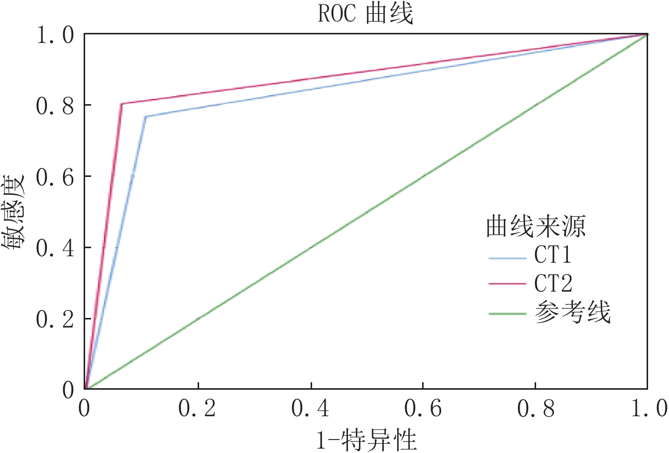
以手术病理结果为金标准,分别计算双源CT双能量虚拟平扫技术诊断EMVI的准确性、敏感性、特异性以及阳性预测值和阴性预测值,绘制ROC曲线(receiver operating characteristic curve),评价其对直肠癌EMVI诊断效能。
计数资料采用
$\chi^2$ 检验确切概率法比较,计量资料采用t检验。采用Kappa一致性检验分析两位医师评价结果、CT评估与病理评估结果。统计软件采用SPSS 17.0,P<0.05认为差异有统计学意义。2. 结果
2.1 患者一般资料
150例病例中,EMVI阳性56例(37.3%),阴性94例(62.7%),组间年龄、性别对EMVI阴阳性无统计学意义,局部淋巴结浸润及远处转移对EMVI阴阳性有统计学意义(表1),且本研究影像资料中ctEMVI表现结合局部淋巴结浸润和/或远处转移较单纯ctEMVI表现诊断准确率有所提高(表2)。
表 1 150例直肠癌患者的一般资料Table 1. General data of 150 patients with rectal cancer项目 ctEMVI P 阳性(n=56) 阴性(n=94) 年龄/岁 52.56 58.47 0.342 性别 男 35 51 0.478 女 21 43 伴局部淋巴结浸润 阳性 37(66.1%) 58(61.7%) 0.000 阴性 19(33.9%) 36(38.3%) 伴远处转移 阳性 35(62.5%) 14(14.9%) 0.031 阴性 21(37.5%) 80(85.1%) 注:局部淋巴结浸润、远处淋巴结转移对EMVI阴阳性诊断具有统计学意义,P<0.05。 表 2 单纯ctEMVI与ctEMVI结合局部淋巴结浸润、远处转移诊断EMVI准确率比较Table 2. Comparison of the accuracy of ctEMVI alone and ctEMVI combined with local lymph node invasion and distant metastasis in the diagnosis of EMVI双源CT双能量虚拟平扫诊断结果 病理学诊断结果 合计 EMVI阳性(n=56) EMVI阴性(n=94) 单纯ctEMVI诊断例数 41 109 150 ctEMVI结合局部淋巴结浸润、远处转移诊断例数 45 105 150 注:当ctEMVI表现结合局部淋巴结浸润和/或远处转移较单纯ctEMVI表现诊断准确率有所提高。 2.2 双源双能量CT虚拟平扫评估直肠癌EMVI的诊断效能
以病理结果为金标准,医师1对ctEMVI评价准确性、敏感性、特异性、阳性预测值、阴性预测值分别为86%、80.36%、89.36%、81.82%和88.42%,AUC为0.831(95%CI 0.756~0.905);医师2对EMVI评价准确性、敏感性、特异性、阳性预测值、阴性预测值分别为88.67%、80.36%、93.62%、88.24%和88.89%,AUC为0.870(95%CI 0.802~0.938),采用Kappa一致性检验分析CT评估结果与病理评估结果的一致性好(表3和图2)。
表 3 两位影像医师评估ctEMVI情况记录表Table 3. Two radiologists evaluated the ctEMVI records双源CT双能量虚拟
平扫诊断结果医师1 医师2 统计检验 例数 百分比/% 例数 百分比/% k P ctEMVI阴性 95 63.33 99 66.00 0.912 0.000 ctEMVI阳性 55 36.67 51 34.00 2.3 辐射剂量
本研究采用双源CT双能量虚拟平扫技术,患者平均辐射剂量为(6.99±2.24)mSv,较常规平扫+三期增强CT扫描辐射剂量(8.98±2.52)mSv降低约22.2%。
3. 讨论
鉴于病理学诊断存在一定的滞后性,影像学评估已成直肠癌的术前个性化诊疗方案选择、术后疗效评估及生存期预测的重要依据,尤其是EMVI状态作为重要的预后相关独立危险因素,是术前治疗方案的重要决策依据[8]。MRI作为国内外指南[9-10]推荐的检查项目,其对EMVI的诊断价值已获得普遍认可[11]。
随着CT新技术的快速发展、更新,双源CT作为辅助检查手段对直肠癌患者术前选择精准治疗方案具有重要意义[12],其临床应用范围也获得进一步推广。因CT平扫对直肠癌EMVI诊断价值有限,本研究采用双能量CT虚拟平扫替代常规平扫,结果表明,运用双能量CT虚拟平扫可在减少检查时间的同时,有效降低辐射剂量。
本研究是参照直肠癌MRI所定义的EMVI评分标准判断ctEMVI状态,采用ctEMVI联合局部淋巴结浸润、远处转移等影像信息整体评估直肠癌EMVI准确率>86%,与文献报道一致[13],且ctEMVI联合局部淋巴结浸润、远处转移较单纯评估ctEMVI的准确性要高,两位影像医师术前诊断ctEMVI具有良好的一致性较强,差异有统计学意义(总体吻合系数k=0.912,P=0.000),此方法未引起评估者间较大的判别误差,具有可行性。
有研究[14]证实发生远处转移的概率与受侵血管管径的大小有关,较大血管(3 mm)发生转移的风险显著高于小血管(<3 mm),分别为57.6% 和20.5%,然而当直肠癌EMVI发生于管腔直径<3 mm的小血管时,由于MRI设备空间分辨率有限,容易产生假阴性的结果[15],且部分患者存在检查禁忌,依从性差等情况,因此采用CT作为辅助检查极为必要,CT可显著缩短扫描时间,提升Z轴分辨率有效减少运动伪影,尤其高质量的三维后处理功明显提高了微小病灶检出率,可以跟踪EMVI的走行方向,了解病变程度和范围,高分辨CT对于肠壁突出的小结节、曲张的血管,毛糙的血管壁诊断效率不亚于MRI[16],被广泛应用于直肠癌术前评估,并取得了较好的效果。
虞云杰等[17]通过常规 CT对直肠癌EMVI的状态进行了系统的评价,其研究发现常规CT与病理检查具有高度符合率,对于直肠癌EMVI状态的评价具有较好的灵敏度和特异度,本研究的灵敏度和特异度高达80%,与该文献报道一致。
本研究结果显示ctEMVI状态与患者的性别、年龄无关,与局部淋巴结侵润、远处转移密切相关,大量研究[14,18]结果发现经病理学检查证实为直肠癌EMVI阳性患者无复发生存率仅为34%,而阴性患者高达73.7%,另一项对447例患者的研究发现直肠癌EMVI阳性患者发生远处转移的概率为42.9%,而阴性则降低至10.6%。本研究中EMVI阳性患者远处转移发生率62.5%(35/56),EMVI阴性患者远处转移发生率为14.9%(14/94)与文献报道非常接近[19],该学者同时得出结论:直肠癌EMVI阳性患者和阴性患者远处转移的发生率相差较大,分别为52.0% 和12.0%。
本研究的局限性:①样本量少且为单中心研究,存在一定的数据偏倚,有待于进一步多中心大数据及多学科综合治疗(multidisciplinary treatment,MDT)的方式进一步研究探讨和论证;②本研究建立模型时仅限于CT征象,未纳入更全面的临床及病理指标(如T分期);③由于病理取材的不确定性,因此本研究无法计算ctEMVI评估pEMVI的真实诊断性能。
综上所述,双源双能量CT虚拟平扫对直肠癌EMVI评估具有较高准确率,可为直肠癌诊断、治疗和预后评估提供有效参考信息,可能成为继TNM分期之后术前直肠癌危险度分层的有效指标,指导临床治疗方案的制定。
-
![]()
图 1 患者男79岁,直肠癌壁外血管侵犯
Figure 1. A 79-year-old male patient with extravascular invasion of rectal cancer
表 1 150例直肠癌患者的一般资料
Table 1 General data of 150 patients with rectal cancer
项目 ctEMVI P 阳性(n=56) 阴性(n=94) 年龄/岁 52.56 58.47 0.342 性别 男 35 51 0.478 女 21 43 伴局部淋巴结浸润 阳性 37(66.1%) 58(61.7%) 0.000 阴性 19(33.9%) 36(38.3%) 伴远处转移 阳性 35(62.5%) 14(14.9%) 0.031 阴性 21(37.5%) 80(85.1%) 注:局部淋巴结浸润、远处淋巴结转移对EMVI阴阳性诊断具有统计学意义,P<0.05。  下载: 导出CSV
表 2 单纯ctEMVI与ctEMVI结合局部淋巴结浸润、远处转移诊断EMVI准确率比较
Table 2 Comparison of the accuracy of ctEMVI alone and ctEMVI combined with local lymph node invasion and distant metastasis in the diagnosis of EMVI
双源CT双能量虚拟平扫诊断结果 病理学诊断结果 合计 EMVI阳性(n=56) EMVI阴性(n=94) 单纯ctEMVI诊断例数 41 109 150 ctEMVI结合局部淋巴结浸润、远处转移诊断例数 45 105 150 注:当ctEMVI表现结合局部淋巴结浸润和/或远处转移较单纯ctEMVI表现诊断准确率有所提高。
下载: 导出CSV
表 3 两位影像医师评估ctEMVI情况记录表
Table 3 Two radiologists evaluated the ctEMVI records
双源CT双能量虚拟
平扫诊断结果医师1 医师2 统计检验 例数 百分比/% 例数 百分比/% k P ctEMVI阴性 95 63.33 99 66.00 0.912 0.000 ctEMVI阳性 55 36.67 51 34.00
下载: 导出CSV
-
[1] SUNG H, FERLAY J, SIEGEL R L, et al. Global cancer statistics 2020: GLOBOCAN estimates of incidence and mortality worldwide for 36 cancers in 185 countries[J]. CA-A Cancer Journal for Clinicians, 2021, 71(3): 209−249. DOI: 10.3322/caac.21660.
[2] CHER H T, RAGHUNANDAN V, PIYAPORN B, et al. Extramural venous invasion by gastrointestinal malignancies: CT appearances[J]. Abdominal Imaging, 2011, 36(5): 491−502. DOI: 10.1007/s00261-010-9667-8.
[3] CHAND M, BHODAY J, BHOME R, et al. mrEMVI status should be used in addition to pEMVI for treatment decision making in rectal cancer to prevent under-reporting of extramural venous invasion[J]. European Journal of Surgical Oncology, 2013, 39(2): 66−79.
[4] PRAMPOLINIF, TASCHINIS, PECCHI, et al. Magnetic resonance imaging performed before and after preoperative chemoradiotherapy chemoradiotherapy in rectal cancer: Predictive factors of recurrence and prognostic significance of MR-detected extramural venous invasion[J]. Abdominal Radiology, 2020, 45(10): 2941−2949. DOI: 10.1007/s00261-018-1838-z.
[5] MASSUCCO P, FONTANA A P, BALBO M A, et al. MRI-detected extramural vascular invasion (mrEMVI) as the best predictive factor to identify candidates to total neoadjuvant therapy in locally advanced rectal cancer[J]. Journal of Surgical Oncology, 2022, 125(6): 1024−1031. DOI: 10.1002/jso.26818.
[6] TAE H K, SUNGMIN W, SANGWON H, et al. The diagnostic performance of MRI for detection of extramural venous invasion in colorectal cancer: A systematic review and meta-analysis of the literature[J]. American Journal of Roentgenology, 2019, 213(3): 575−585. DOI: 10.2214/AJR.19.21112.
[7] TRIVEDI H, CHAMARTHY U, DICARLO L, et al. Prognostic factors of overall survival for patients with stage Ⅱ colon cancer[J]. Acta Medica Academiae Scientiarum Hungaricae, 2014, 43(2): 134−143. DOI: 10.5644/ama2006-124.112.
[8] 白晨. 基于高分辨率MRI影像组学方法直肠癌EMVI诊断模型的研究[D]. 长春: 吉林大学, 2022. BAI C. Study of extramural venous invasion diagnostic model for rectal cancer based on high-resolution MRI radiomics method[D]. Changchun: Jilin University, 2022. DOI:10.27162/d.cnki.gjlin.2022.007006. (in Chinese).
[9] 中华人民共和国国家卫生健康委员会医政医管局, 中华医学会肿瘤学分会. 中国结直肠癌诊疗规范(2020年版)[J]. 中国实用外科杂志, 2020, 40(6): 601-625. Hospital Authority of National Health Commission of the People's Republic of China, Chinese Society of Oncology, Chinese Medical Association. Chinese protocol of diagnosis and treatment of colorectal cancer (2020 edition)[J]. Chinese Journal of Practical Surgery, 2020, 40(6): 601-625. DOI:10.19538/j.cjps.issn1005-2208.2020.06.01. (in Chinese).
[10] BENSON A B, VENOOK A P, AL-HAWARY M M, et al. Rectal cancer, version 2.2018 clinical practice guidelines in oncology[J]. Journal of the National Comprehensive Cancer Network, 2018, 16(7): 874−901. DOI: 10.6004/jnccn.2018.0061.
[11] 唐雪, 江长思, 张世派, 等. 直肠MR造影对直肠癌壁外血管侵犯的诊断价值[J]. 医学影像学杂志, 2022,32(4): 638−642. DOI: 1006-9011(2022)04-0638-05. TANG X, JIANG C S, ZHANG S P, et al. Diagnosis value of MR rectography in assessment for extravasational vascular invasion of rectal cancers[J]. Journal of Medical Imaging, 2022, 32(4): 638−642. DOI: 1006-9011(2022)04-0638-05. (in Chinese).
[12] 路凯, 兰国宾, 戴士林. 双源CT双能量虚拟平扫对结直肠良恶性肿瘤的鉴别诊断价值研究[J]. 中国CT和MRI杂志, 2021,19(9): 131−134. DOI: 10.3969/j.issn.1672-5131.2021.09.042. LU K, LAN G B, DAI S L. Differential diagnosis value of dual-source CT dual-energy virtual non-contrast on benign and malignant colorectal tumors[J]. Chinese Journal of CT and MRI, 2021, 19(9): 131−134. DOI: 10.3969/j.issn.1672-5131.2021.09.042. (in Chinese).
[13] ZHEN G, ZHANG X Y, LI X T, et al. Correlation and prognostic value of CT-detected extramural venous invasion and pathological lymph-vascular invasion in colon cancer[J]. Abdominal Radiology, 2022, 47(4): 1232−1243. DOI: 10.1007/s00261-022-03414-7.
[14] TRIPATHI P, GUO W, RAO S, et al. Additional value of MRI-detected EMVI scoring system in rectal cancer: Applicability in predicting synchronous metastasis[J]. Tumori Journal, 2020, 106(4): 286−294. DOI: 10.1177/0300891620901745.
[15] TRIPATHI P, RAO S X, ZENG M S. Clinical value of MRI-detected extramural venous invasion in rectal cancer[J]. Journal of Digestive Diseases, 2017, 18(1): 2−12. DOI: 10.1111/1751-2980.12439.
[16] BRUCE M, JENNIFER S. Somatostatin receptor scintigraphy of neuroendocrine tumors of the abdomen and pelvis[J]. Seminars in Roentgenology, 2016, 51(2): 112−122. DOI: 10.1053/j.ro.2016.02.008.
[17] 虞云杰, 陈孝娟, 李鹏, 等. 多层螺旋CT在直肠癌术前诊断及⾎管侵犯评估中的应⽤价值[J]. 中国CT和MRI杂志, 2020,18(2): 117−120. DOI: 10.3969/j.issn.1672-5131.2020.02.035. YU Y J, CHEN X J, LI P, et al. Application value of multi-slice spiral CT in the preoperative diagnosis of rectal cancer and assessment of vascular invasion[J]. Chinese Journal of CT and MRI, 2020, 18(2): 117−120. DOI: 10.3969/j.issn.1672-5131.2020.02.035. (in Chinese).
[18] SOHN B, LIM J S, KIM H, et al. MRI-detected extramural vascular invasion is an independent prognostic factor for synchronous metastasis in patients with rectal cancer[J]. European Radiology, 2015, 25(5): 1347−1355. DOI: 10.1007/s00330-014-3527-9.
[19] CHAND M, EVANS J, SWIFT R I, et al. The prognostic significance of postchemoradiotherapy high-resolution MRI and histopathology detected extramural venous invasion in rectal cancer[J]. Annals of Surgery, 2015, 261(3): 473−479. DOI: 10.1097/SLA.0000000000000848.

计量
- 文章访问数: 280
- HTML全文浏览量: 135
- PDF下载量: 37


