
Primary Undifferentiated Small Round Cell Sarcoma of Sellar Region: A Case Report and Literature Review
-
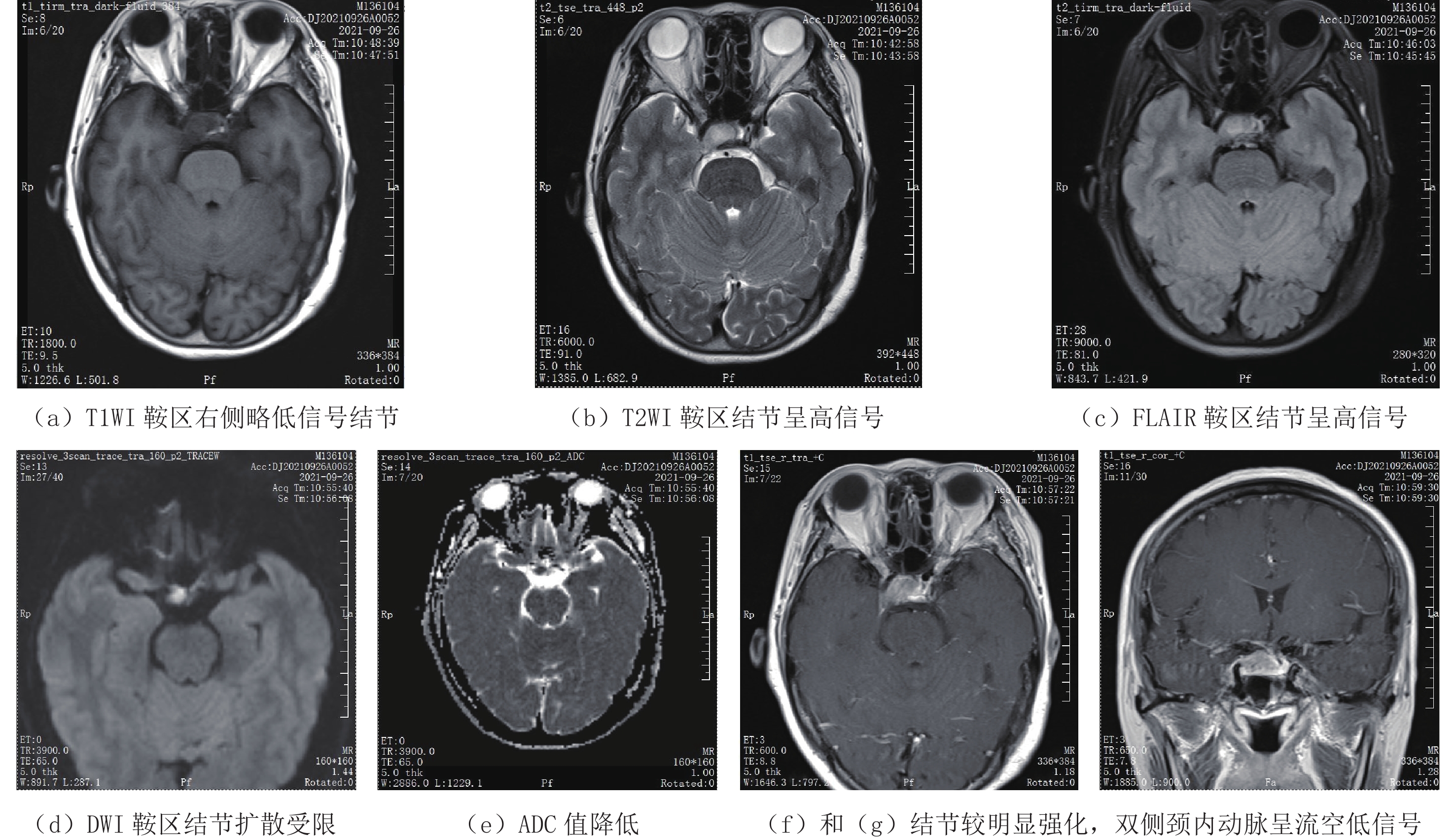
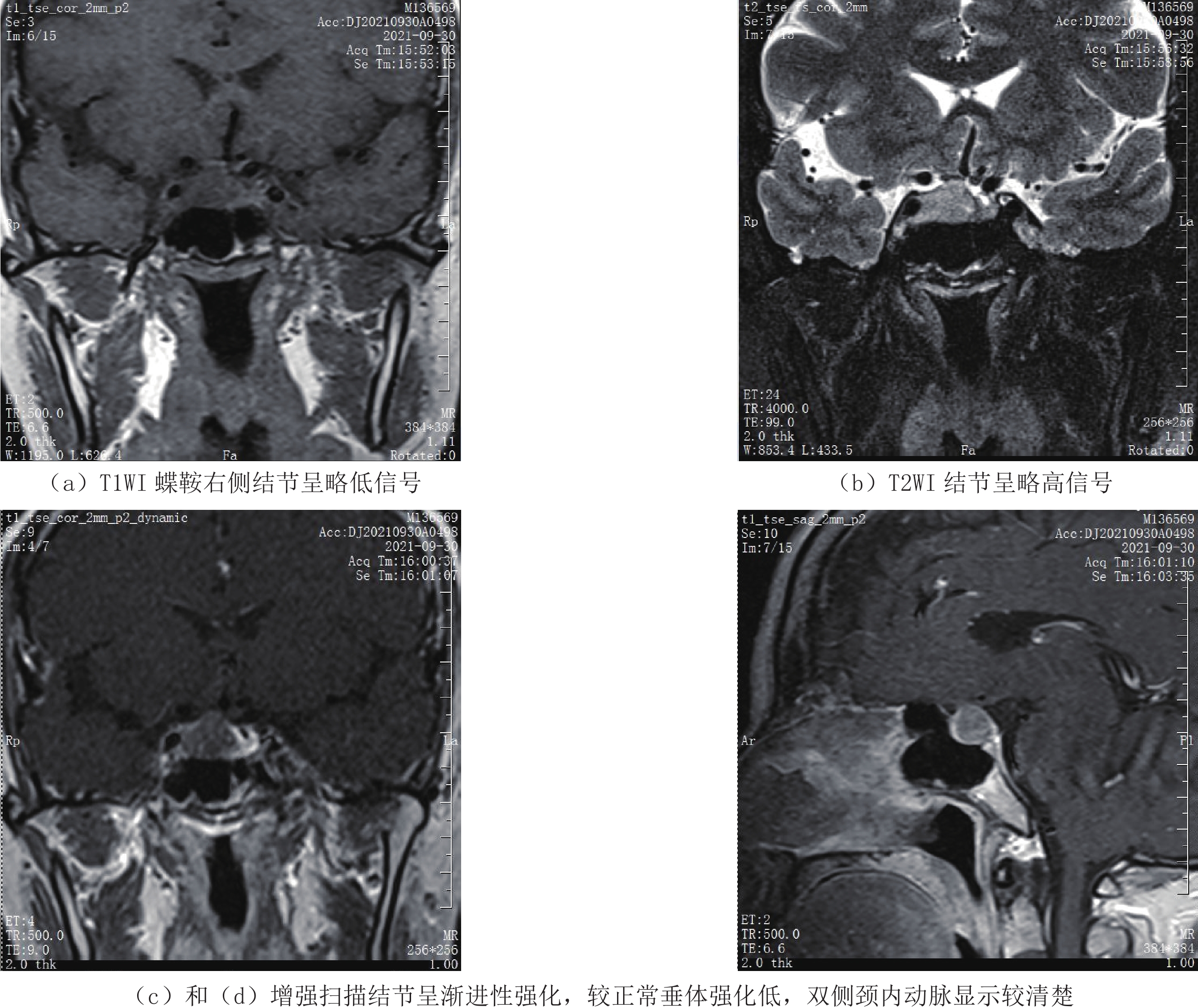
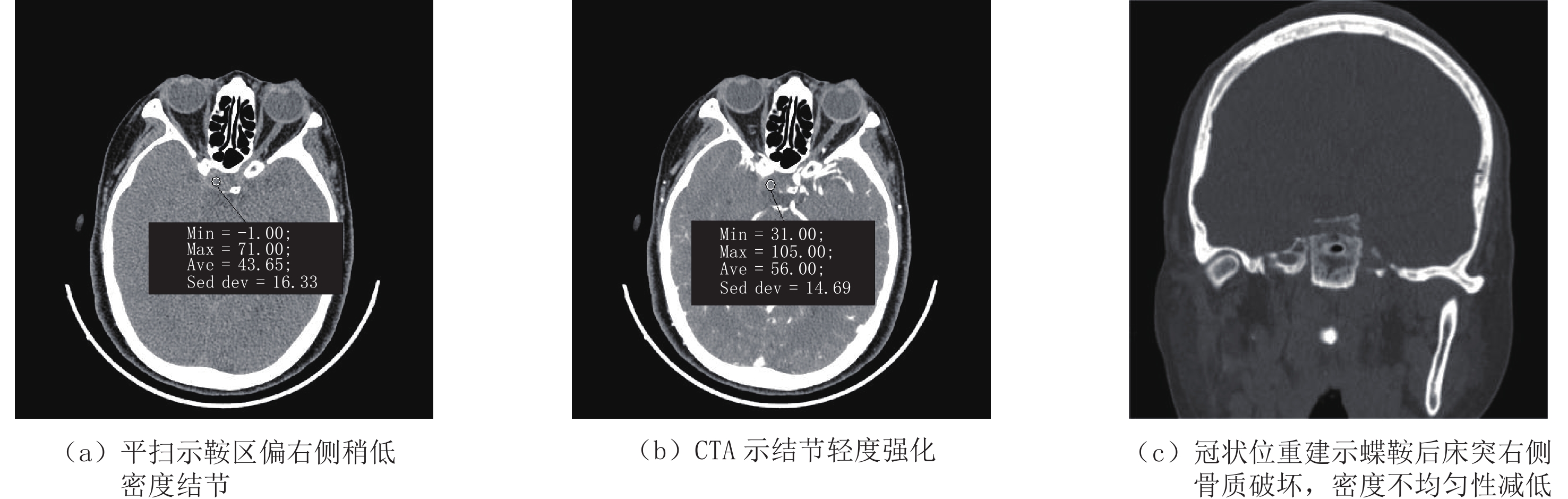
摘要: 目的:探讨鞍区原发性小圆细胞性未分化肉瘤的临床表现、病理特征及影像特点,以提高对该病的影像诊断及鉴别诊断能力。方法:回顾性分析1例经手术病理证实为鞍区原发性小圆细胞性未分化肉瘤的临床病理特征、CT及MR表现,并复习文献。结果:CT平扫示鞍区偏右侧呈稍低密度影,增强欠均匀强化,并累及邻近颅骨;MRI示T1WI呈低信号,T2WI、液体衰减反转恢复(FLAIR)呈略高信号,扩散加权成像(DWI)序列上呈高信号,信号欠均匀;增强病灶较明显强化,边缘较光整,邻近蝶鞍后床突可见骨质破坏。结论:鞍区原发性小圆细胞未分化肉瘤罕见,其临床和影像表现无明显特异性,确诊仍需依靠病理组织学检查。
-
关键词:
- CT诊断 /
- 鞍区 /
- 未分化/未分类软组织肉瘤
Abstract: Objective: This study aimed to investigate the clinical, pathological, and imaging features of primary undifferentiated small round cell sarcoma of the sellar region to improve the ability of imaging and differential diagnosis. Methods: The clinicopathological features and computed tomography (CT) and magnetic resonance imaging (MRI) manifestations of a case with primary undifferentiated small round cell sarcoma of the sellar region confirmed by surgery and pathology were retrospectively analyzed, and the literature was reviewed and summarized. Results: CT showed a node in the right sellar region, reinforcement under-uniform reinforcement and affects adjacent skull; MRI showed homogeneous hypointensity on the T1-weighted image, hyperintense on the T2-weighted image, fluid-attenuated inversion recovery, and diffusion-weighted imaging; and significantly reinforcement with smooth margin on reinforcement scanning. Bone destruction was seen near the posterior clinoid process of the sella turcica. Conclusion: Primary undifferentiated small round cell sarcoma of the sellar region is rare, and its clinical and imaging findings are not significantly specific. Therefore, the diagnosis requires histopathological examination.-
Keywords:
- CT /
- sellar region /
- undifferentiated/unclassified soft tissue sarcoma
-
新型冠状病毒肺炎(COVID-19)是一种有别于其他呼吸道病毒、具有较强传染性和侵袭性的肺部传染性疾病,因其累及部位、范围、病理和程度等差异而呈现明显多样化的影像学表现,所以早期发现和精准诊断对临床治疗和患者预后极为重要[1-4]。
胸部薄层平扫CT诊断早期肺部病变具有较高敏感性和特异性,现总结北京世纪坛医院感染科确诊的153例发病2周内CT表现阳性的COVID-19患者,就与患者年龄和发病时间相关的早期CT表现特征进行单中心回顾性分析,旨在提高临床和影像医师对该病的认识。
1. 材料与方法
1.1 研究人群
收集2022年11月16日至2022年12月16日期间在北京世纪坛医院感染科确诊为COVID-19且2周内初次胸部CT表现阳性的患者153例,男81例(52.9%),女72例(47.1%),年龄26~95岁,平均(68±15)岁;其中≤60岁48例(31.2%)、>60者105例(68.1%)。发病与首次CT检查的时间间隔为1~14天,其中≤7天者104例(68%)、>7天者49例(32%)。临床症状包括咳嗽、发热、气憋、肌肉酸痛、纳差和咯血等;基础性疾病者43例(27.9%),主要为CT表现为肺气肿、间质纤维化、结核性机化或钙化灶、支气管扩张、肿瘤性病变或胸膜钙化的患者。
因为时间较短,本研究只分析COVID-19患者发病后1~14天的首次CT表现,未将患者的临床分期、肺功能、血氧饱和度、血化验指标、治疗方法、患者预后和复查CT的动态演变等指标纳入本研究的统计范畴。
1.2 CT扫描技术
CT扫描仪为32排的北京赛诺威盛Insitum-CT 338机型,扫描参数:探测器宽度16 cm,螺距1.0,电压120 kV,电流150 mAs,重建层厚为肺窗1.5 mm和纵隔窗5 mm,矩阵是512×512,FOV 380~450。肺窗图像的窗宽和窗位为1600和 -600,纵隔窗的窗宽和窗位为400和40;并行冠状位和矢状位肺窗(1×5 mm)和纵隔窗(5×5 mm)重建;放射剂量DLP500~600 mGy·cm。
1.3 影像分析
由两名初或中级医师独立完成,结果有分歧时由高级医师评定。具体指标包括:①累及部位:肺脏、气道、血管、胸膜和纵隔,单叶、单肺和双肺;②病变分布:周围胸膜内和胸膜下、中央血管束周和混合,对称和非对称,叶段和非叶段;③病变大小:长径≤10 mm、10~30 mm和>30 mm;④病变数量:单发和多发,多发又分≤5个和>5个;⑤病变占肺叶体积百分比(半定量分析),即≤10%、11%~30%、31%~50% 和>50%(白肺);⑥病变形状:小结节状(直径≤10 mm)、斑片状(10~30 mm)、大片状(>30 mm)、束带状或混合型;⑦病变密度:蜂窝状、磨玻璃样、网格状、实性(实变或肉芽肿样)和混合型;⑧肺脏背景:肺气肿样、磨玻璃影、网格影和正常等;⑨伴随病变:机化或纤维化、血管壁增厚、胸膜增厚、胸腔积液和胸膜下线等。⑩特殊征象:不规则煎蛋征、铺路石样征、支气管充气征、反晕征、胸膜下黑带、拱廊征等。
1.4 统计学分析
采用SPSS 21.0软件,根据年龄和发病与CT检查时间间隔将患者分别分为两组:≤60岁和>60岁、≤7天和>7天,比较两组患者相关的影像学表现特征。其中正态分布的计量资料以
$\bar x\pm s $ 表示,组间计量和计数资料统计采用连续变量的t检验和分类变量的卡方用检验或 Fisher精确检验。P<0.05表示差异有统计学意义。2. 结果
153例COVID-19患者中,年龄组间的对比显示病变数量、部位、大小、容积和束带影的差异有统计学意义;CT检查时间分组患者之间,病变的形态、密度、机化和纤维化以及胸膜受累等差异有统计学意义。影像学指标对照详见表1和表2。病变形态、密度表型及特征见图1~图3。
表 1 不同年龄和CT检查时间患者CT显示病灶数量、部位和病变体积百分比等情况Table 1. Number and location of lesions and percentage of lesion volume revealed by CT in patients of different ages and CT examination times分布特征 年齡 统计检验 CT检查时间 统计检验 ≤60岁(n=48)
例(%)>60岁
(n=105)
例(%)χ2 P ≤7天
(n=104)
例(%)>7天
(n=49)
例(%)χ2 P 病变数量 单发 5(10.4) 1(0.95) — 0.012 4(3.9) 2(4.1) — 1.000 多发 43(89.6) 104(99.1) — 0.012 100(96.2) 47(95.9) 0.005 0.944 累及部位 单叶 14(29.1) 3(2.8) 23.087 0.000 11(10.6) 6(12.2) 0.094 0.759 单肺 1(2.1) 6(5.8) — 0.434 3(2.9) 4(8.2) — 0.211 双肺 33(68.8) 96(91.4) 12.810 0.000 90(86.5) 39(79.6) 1.215 0.270 病变百分比 ≦10 37(77.1) 45(42.9) 15.516 0.000 59(56.7) 24(49) 0.806 0.369 ≦30 6(12.5) 22(21) 1.574 0.210 20(19.2) 8(16.3) 0.188 0.665 ≦50 2(4.2) 23(21.9) 7.582 0.006 15(14.4) 10(20.4) 0.873 0.350 >50 3(6.3) 15(14.3) 2.049 0.152 12(11.5) 7(14.3) 0.631 0.231 表 2 不同年龄和CT检查时间患者组病灶各类征象占比情况Table 2. Percentage of various types of signs of lesions in the patient group by age and time of CT examination影像学征象 年齡 统计检验 CT检查时间 统计检验 ≤60岁(n=48)
例(%)>60岁(n=105)
例(%)χ2 P ≤7天
(n=104)
例(%)>7天(n=49)
例(%)χ2 P 病变分布 胸膜下 27(56.3) 83(79.0) 8.473 0.004 78(75.0) 32(65.3) 1.549 0.213 胸膜内 41(85.4) 99(94.3) 2.290 0.130 96(92.3) 44(89.8) 0.044 0.834 血管周 38(79.2) 90(85.7) 1.033 0.039 87(83.7) 41(83.7) 0.000 0.998 混合性 33(68.7) 89(84.8) 5.227 0.022 83(79.8) 39(79.6) 0.001 0.975 对称性 22(45.8) 58(55.2) 25.734 0.000 56(53.8) 24(49.0) 0.316 0.574 非叶段性 42(87.5) 93(88.6) 0.036 0.849 89(85.6) 43(87.8) 0.133 0.715 病变大小 ≦10 mm 42(87.5) 46(43.8) 25.734 0.000 86(82.7) 40(81.6) 0.026 0.873 10~30 mm 36(75.0) 22(21.0) 40.881 0.000 89(85.6) 39(79.6) 0.873 0.350 >30 mm 20(41.7) 16(15.2) 12.787 0.000 69(66.3) 29(59.2) 0.742 0.389 混合 38(79.2) 84(80.0) 0.014 0.905 86(82.7) 36(73.5) 1.754 0.185 病变形态 小结节 40(83.3) 84(80.0) 0.238 0.625 85(81.7) 39(79.6) 0.099 0.753 斑片状 38(79.2) 89(84.8) 0.731 0.393 89(85.6) 38(77.6) 1.521 0.217 大片状 16(33.3) 68(64.8) 13.141 0.000 59(56.7) 25(51.0) 0.439 0.508 束带状 6(12.5) 50(47.6) 17.508 0.000 38(36.5) 18(36.7) 0.001 0.981 混合性 39(81.3) 98(93.3) 5.136 0.023 94(90.4) 44(89.8) 0.000 1.000 病变密度/例 GGO 41(85.4) 99(94.3) 2.290 0.130 96(92.3) 44(89.8) 0.044 0.834 实变 25(52.1) 45(42.9) 1.130 0.288 33(31.7) 35(75.5) 21.258 0.000 网格影 31(64.6) 91(86.7) 9.943 0.002 77(74.0) 45(91.8) 6.531 0.011 蜂窝影 1(2.1) 10(9.5) 1.732 0.188 3(2.9) 8(16.3) 7.117 0.008 病变边缘/例 模糊 32(66.7) 51(48.6) 4.346 0.037 56(53.8) 27(55.1) 0.021 0.884 不规则 26(54.2) 47(44.8) 1.168 0.280 53(50) 29(59.2) 0.905 0.341 毛刺 10(20.8) 21(20.0) 0.014 0.905 19(18.3) 12(24.5) 0.798 0.372 伴随病变/例 机化灶 29(60.4) 59(56.2) 0.241 0.624 56(53.8) 32(65.3) 1.790 0.181 间质纤维化 27(56.3) 72(68.6) 2.190 0.139 58(55.8) 41(83.7) 11.356 0.001 气道异常 39(81.3) 74(70.5) 1.980 0.159 77(74.0) 36(73.5) 0.006 0.940 血管增粗 45(93.8) 99(94.3) 0.000 1.000 99(95.2) 45(91.8) 0.207 0.649 胸膜增厚 24(50.0) 84(80.0) 14.280 0.000 70(67.3) 38(77.6) 1.683 0.194 胸水形成 1(2.1) 4(3.8) - 1.000 3(2.9) 2(4.1) - 0.656 3. 讨论
3.1 COVID-19的病理与临床
肺部病毒性感染(pulmonary viral infection,PVI)是一种多由上呼吸道RNA病毒感染引起的气道和/或肺的急性或慢性炎症,常见致病源包括流感或副流感病毒、SARS病毒、COVID-19、巨细胞病毒、腺病毒、鼻病毒、呼吸道合胞病毒和EB病毒等。PVI的诊断标准主要依靠病原学检查,而致病原阴性者,则需结合流行病学史、临床表现、实验室检查、胸部影像学和排除其他疾病表现等综合评判[5-8]。而研究显示[9-10],COVID-19是一种基因组结构不同于其他呼吸道病毒且侵袭性和传播性较强的β属特殊毒株,通过S蛋白与人血管紧张素转化酶-2(ACE2)互相作用感染人呼吸道黏膜上皮细胞、Ⅱ型肺泡上皮细胞和肺间质以及微血管血栓形成和多系统脏器受累等改变。其病理学改变[11-14]主要以肺泡为单元的弥漫性多样化损伤,即肺泡壁内渗出性炎、肉质变、间质炎和纤维化,肺泡腔内充满大量炎性渗出物(浆液或纤维素)、脱落的Ⅱ型肺泡上皮细胞、单核细胞和巨噬细胞等,并沿肺泡腔内侧形成较厚的透明膜。并伴有微血管内透明血栓形成、灶性出血或梗死、肺泡腔渗出物机化或间质纤维化形成以及细支气管炎导致的管壁增厚和管腔狭窄等。这些改变与病程早期出现的严重血管反应及大量炎性趋化因子的影响密切相关。
临床上,COVID-19多见于成人或老年人,男女无差异[13-15],本研究结果与文献一致。COVID-19的诊断金标准是核酸或抗原检测,但存在假阴性、时间较长、无法评判分期和病变程度等不足。临床表现和实验室检查均为非典型或非特异性改变;胸部平片对早期较小病变发现率较低、假阴性率或漏诊及误诊率较高;而胸部增强CT有对比剂过敏、操作复杂且无助于微血管栓子显示等,所以胸部平扫CT成为COVID-19早期筛查、精准诊断和评估程度及预后的最佳影像学方法,尤其高分辨率薄层图像清晰显示病变的有无和细微特征。
3.2 COVID-19的CT表现
COVID-19的CT表现与其累及部位和病理改变密切相关,其CT表现和特征文献上多有报道[1-6,9-12,16-20],对该疾病的专家共识是以病变多发、斑片或大片状、周围或血管束分布、边缘不清的均质或不均质的磨玻璃影为主(图1),极少伴有胸腔积液和纵隔异常等。本研究资料均来自我院感染科发病14天内初次薄层CT表现阳性的患者,统计结果显示多数指标与文献一致,但部分指标与文献存在差异。年龄组间的对比,病变数量、部位、大小、容积和束带影的差异有统计学意义;发病与CT检查时间间隔分组患者之间,病变的形态、密度、机化和纤维化以及胸膜受累等差异有统计学意义。
![]() 图 1 COVID-19肺内病变的CT形态表型Figure 1. CT morphological phenotype of COVID-19 pulmonary lesions
图 1 COVID-19肺内病变的CT形态表型Figure 1. CT morphological phenotype of COVID-19 pulmonary lesions病变的数量、大小和容积是定量诊断、临床分期和明确白肺(病变/肺脏容积百分比>50%)的重要指标,薄层CT图像可清晰显示并进行半定量或AI定量分析。病变分布方面,本研究显示绝大多数病变为周围胸膜内、混合型和非叶段分布,文献报道较少,提示COVID-19在病理机制上是一种气道吸入性或血管源性或继发性间质性病变,有助于该病与特发性间质性肺炎(idiopathic interstitial pneumonia,IIP)、淋巴增生性疾病和叶段性肺泡充盈性病变的鉴别。
病变的CT表型(形态、密度和边缘)因其病理特征和病变程度的不同多呈现不均质的磨玻璃影、实变影、铺路石样、网格影和混合密度影(图2),少数为肿块样、机化或肉芽肿样影、索条影和蜂窝影。本研究结果显示COVID-19的早期病变即出现形态的不规则、密度的不均质和类型的混杂性,尤其实性机化灶、网线状纤维化和病变的皱缩改变是病变机化和纤维化的特征性表现,文献报道较少。这些改变与病程早期出现的大量炎性趋化因子和严重血管反应以及肺泡为单元的弥漫性多样化损伤引起的纤维母细胞增生、胶原化肉芽组织和间质纤维化等密切相关。
COVID-19的伴随病变以往文献报道较少或发生率较低[4-6,16-20],本研究结果显示多数患者(70%~93.5%)合并胸膜、气道和血管的异常以及机化和纤维化改变(图3)。其中胸膜异常多为胸膜或叶间裂局限性线样增厚、胸膜下线和胸膜下栅栏,少有胸水(3%);气道异常为病毒性支气管细支气管炎引起的小气道壁增厚、牵拉支扩和小气道黏液栓等,是导致患者反复咳嗽、少痰、气憋和胸痛的主要原因;血管异常为血管壁模糊和血管增厚,血管/气道外径比大于1.5,其病理机制是病变直接浸润、小血管炎或血管周炎尚有待研究;机化和纤维化表现为变形移位的网格影、纤维索条、皱缩性实变、不规则煎蛋征、拱廊征和反晕征等。
综上所述,COVID-19的CT表现以多部位受累、多样化表型、以肺泡为单元的弥漫性间质性病变,常伴有早期机化或纤维化。胸部薄层平扫CT对早期发现、精准诊断和鉴别、评判临床分期和疗效预后等可提供重要的影像依据。
本研究的局限性包括:①未纳入相关的临床指标、实验室指标、肺功能或血氧饱和度、治疗方法和患者预后等进行综合或对比研究;②所有病例没有病理性诊断资料,多数病例未行核酸和抗原检测;③未行 CT表型和临床分型的对照研究;④本研究以 COVID-19患者初始发病(1~14天)的首次CT检查的影像表型为主,其动态变化规律尚需进一步对照研究。
-
[1] SAREEN P, CHHABRA L, TRIVEDI N. Primary undifferentiated spindlecell sarcoma of sella turcica: Successful treatment with adjuvant temozolomide[J]. BMJ Case Reports, 2013, 2013. pii: bcr201300934.
[2] 王坚, 朱雄增. 软组织肿瘤病理学[M]. 2版. 北京: 人民卫生出版社, 2017: 1380-1381. WANG J, ZHU X Z. Soft-tissue tumor pathology[M]. The 2 ed. Beijing: People's Health Publishing House, 2017: 1380-1381. (in Chinese).
[3] FLETCHER C D M, BRIDGE J A, HOGENDOORN P C W, eds. WHO classification of soft tissue and bone tumours[M]. 4ed, Lyon: IARC Press; 2013: 236-238.
[4] HUANG B Y, CASTILLO M. Nonadenomatous tumors of the pituitary and sella turcica[J]. Topics in Magnetic Resonance Imaging, 2005, 16(4): 289−299. doi: 10.1097/01.rmr.0000224685.83629.18
[5] LOPES M B, LANZINO G, CLOFT H J, et a1. Primary fibrosarcoma of the sella unrelated to previous radiation therapy[J]. Modern Pathology, 1998, ll(6): 579−584.
[6] SCHULTZ A B, BRAT D J, OYESIKU N M, et a1. Intrasellar pituicytoma in a patient with other endocrine neoplasms[J]. Archives of Pathology & Laboratory Medicine, 200 l, 125(4): 527-530.
[7] SHINOJIMA N, OHTA K, YANO S, et a1. Myofibroblastoma in the suprasellar region: Case report[J]. Journal of Neurosurgery, 2002, 97(5): 1203−1207. doi: 10.3171/jns.2002.97.5.1203
[8] MENA H, RIBAS J L, PEZESHKPOUR G H, et a1. Hemangiopericytoma of the central nervous system: A review of 94 cases[J]. Human Pathology, 1991, 22(1): 84−91. doi: 10.1016/0046-8177(91)90067-Y
[9] MANORANJAN B, SYRO L V, SCHEITHAUER B W, et a1. Undifferentiated sarcoma of the sellar region[J]. Endocrine Pathology, 2011, 22(3): 159−164. doi: 10.1007/s12022-011-9166-7
[10] ALPERT T E, HAHN S S, CHUNG C T, et a1. Successful treatment of spindle cell sarcoma of the sella turcica: Case report[J]. Journal of Neurosurgery, 2002, 97(S5): 438−440.
[11] ZHONG J, LI S T, YAO X H, et a1. An intrasellar rhabdomyosarcoma misdiagnosed as pituitary adenoma[J]. Surgical Neurology, 2007, 68(S2): S29−33.
[12] 张广健, 常建勇, 谢英亮, 等. 神经内镜下切除鞍区原发性未分化肉瘤一例并文献复习[J]. 中华神经外科杂志, 2017,33(10): 1070−1071. ZHANG G J, CHANG J Y, XIE Y L, et al. Neuroendoscopic resection of primary undifferentiated sarcoma in the sellar region: A case report and literature review[J]. Chinese Journal of Neurosurgery, 2017, 33(10): 1070−1071. (in Chinese).
-
期刊类型引用(13)
1. 王仁贵. 新型冠状病毒感染的临床、病理与影像表现. CT理论与应用研究. 2023(03): 297-302+296 .  本站查看
本站查看
2. 霍萌,李玲,孙莹,张明霞,孙磊,郭佳,杜常月,李兴鹏,郝琪,张妍,段淑红,刘晓燕,刘薇,段永利,张春燕,王仁贵. 不同中性粒细胞与淋巴细胞比值组间COVID-19胸部HRCT表现分析. CT理论与应用研究. 2023(03): 387-394 . 本站查看
3. 廖倩怡,林飞飞,庄义江,孙龙伟,卢宁,李鹏. 儿童新型冠状病毒奥密克戎毒株感染的胸部CT影像特点. CT理论与应用研究. 2023(03): 347-355 . 本站查看
4. 李玲,张明霞,孙莹,段淑红,郭佳,杜常月,刘梦珂,张怡梦,孙磊,霍萌,王仁贵. 基于深度学习的CT定量指标对糖尿病合并新型冠状病毒肺部感染的影像学研究. CT理论与应用研究. 2023(03): 373-379 . 本站查看
5. 赵建华,梁丹艳,吕高星,闫昕,刘宇,王晓兰,柴军. 深度学习定量测量对新型冠状病毒感染预后的分析. CT理论与应用研究. 2023(05): 587-594 . 本站查看
6. 刘晓燕,鲍中英,段淑红,张捷,张明霞,孙莹,李玲,王仁贵. 发热门诊首诊新型冠状病毒感染患者的临床特征和CT表现分析. CT理论与应用研究. 2023(05): 636-644 . 本站查看
7. 张怡梦,刘梦珂,张晓杰,郝琪,张妍,李兴鹏,张明霞,刘晓燕,王仁贵. 重型新型冠状病毒感染的临床与CT特征. CT理论与应用研究. 2023(05): 579-585+578 . 本站查看
8. 赵建华,梁丹艳,王晓兰,柴军. 不同年龄人群新型冠状病毒感染CT表现分析. CT理论与应用研究. 2023(05): 603-611 . 本站查看
9. 刘梦珂,张怡梦,李兴鹏,张晓杰,张妍,郝琪,李玲,杜常月,王仁贵. 不同年龄新冠肺炎患者CT表现及动态分析. CT理论与应用研究. 2023(05): 645-651 . 本站查看
10. 张妍,黄瑞彬,段永利,刘薇,李玲,郝琪,李兴鹏,刘梦珂,张怡梦,孙小丽,刘晓燕,王仁贵. 有无基础病的COVID-19患者CT表现比较分析. CT理论与应用研究. 2023(05): 652-658 . 本站查看
11. 郝琪,刘晓燕,张妍,李兴鹏,张怡梦,刘梦珂,张晓杰,李玲,郭佳,杜常月,孙莹,霍萌,张明霞,刘薇,段永利,段淑红,王仁贵. 胸部薄层CT平扫对于重型新型冠状病毒感染的诊断价值. CT理论与应用研究. 2023(05): 675-683 . 本站查看
12. 李兴鹏,袁辉,杜常月,刘晓燕,李玲,刘梦珂,张怡梦,张妍,郝琪,段淑红,王仁贵. COVID-19相关性血管异常的薄层CT特征分析. CT理论与应用研究. 2023(05): 667-674 . 本站查看
13. 陈熙来,吴宏楷,甘清鑫,曾庆思,刘晋新,张烈光,邓宇,李征途,李时悦,黎毅敏,钟南山. 基于人工智能对广州新型冠状病毒感染住院患者肺部炎症负荷的分层量化分析及临床匹配度研究. 中华生物医学工程杂志. 2023(05): 495-502 . 百度学术
其他类型引用(1)

 下载:
下载:


计量
- 文章访问数: 251
- HTML全文浏览量: 130
- PDF下载量: 180
- 被引次数: 14
