
Primary Undifferentiated Small Round Cell Sarcoma of Sellar Region: A Case Report and Literature Review
-
摘要: 目的:探讨鞍区原发性小圆细胞性未分化肉瘤的临床表现、病理特征及影像特点,以提高对该病的影像诊断及鉴别诊断能力。方法:回顾性分析1例经手术病理证实为鞍区原发性小圆细胞性未分化肉瘤的临床病理特征、CT及MR表现,并复习文献。结果:CT平扫示鞍区偏右侧呈稍低密度影,增强欠均匀强化,并累及邻近颅骨;MRI示T1WI呈低信号,T2WI、液体衰减反转恢复(FLAIR)呈略高信号,扩散加权成像(DWI)序列上呈高信号,信号欠均匀;增强病灶较明显强化,边缘较光整,邻近蝶鞍后床突可见骨质破坏。结论:鞍区原发性小圆细胞未分化肉瘤罕见,其临床和影像表现无明显特异性,确诊仍需依靠病理组织学检查。
-
关键词:
- CT诊断 /
- 鞍区 /
- 未分化/未分类软组织肉瘤
Abstract: Objective: This study aimed to investigate the clinical, pathological, and imaging features of primary undifferentiated small round cell sarcoma of the sellar region to improve the ability of imaging and differential diagnosis. Methods: The clinicopathological features and computed tomography (CT) and magnetic resonance imaging (MRI) manifestations of a case with primary undifferentiated small round cell sarcoma of the sellar region confirmed by surgery and pathology were retrospectively analyzed, and the literature was reviewed and summarized. Results: CT showed a node in the right sellar region, reinforcement under-uniform reinforcement and affects adjacent skull; MRI showed homogeneous hypointensity on the T1-weighted image, hyperintense on the T2-weighted image, fluid-attenuated inversion recovery, and diffusion-weighted imaging; and significantly reinforcement with smooth margin on reinforcement scanning. Bone destruction was seen near the posterior clinoid process of the sella turcica. Conclusion: Primary undifferentiated small round cell sarcoma of the sellar region is rare, and its clinical and imaging findings are not significantly specific. Therefore, the diagnosis requires histopathological examination.-
Keywords:
- CT /
- sellar region /
- undifferentiated/unclassified soft tissue sarcoma
-
1. 临床资料
患者,女,60岁。因阵发性头痛、头晕10 d、右眼疼痛1 d入院。10 d前患者无明显诱因出现头痛,呈阵发性闷痛,伴头昏、头闷不适,伴轻度恶心,无视物模糊,无耳鸣及听力障碍,无意识障碍、大小便失禁,无畏寒发热,无肢体抽搐、麻木及活动障碍,无言语不清、饮水反呛,无咳嗽咳痰,无胸痛胸闷,无腹痛腹泻;1 d前感右眼疼痛,伴右眼睑下垂。患者既往有高血压病史,长期正规使用降压药物,血压维持正常;否认糖尿病、冠心病、脑梗死等病史,否认手术外伤史。
体格检查:体温36.3℃,脉搏71次/min,呼吸17次/min,血压125/70 mmHg。神志清晰,精神较差,步入病房;全身皮肤无黄染,全身浅表淋巴结未见肿大;双肺呼吸音粗,双肺未闻及啰音,心率71次/min,律齐,无杂音。腹软,无压痛反跳痛,肝脾肋下未触及;双下肢无浮肿。
专科检查:神志清楚,言语清晰,高级智能正常。右眼上睑轻度下垂,眼球内转、上转、下转均部分受限,眼位:-15°,余眼球活动正常;右侧瞳孔稍散大,直径4 mm,光反射迟钝,左侧瞳孔直径3 mm,光反射灵敏;视力:右眼0.6、左眼0.15,矫正不应;眼压:右眼17.3 mmhg,左眼17.5 mmhg;双眼眼底未见明显异常。鼻唇沟等深,口角无歪斜,伸舌居中。四肢肌力、肌张力正常,腱反射对称,病理征未引出。脑膜刺激征阴性,共济运动正常,感觉检查正常。
实验室检查:血常规检查显示白细胞5.85×10⁹/L,红细胞4.27×1012/L,血红蛋白124 g/L,CRP小于10 mg/L,血小板275×109/L;BNP及心梗3项、PCT均正常;电解质正常。泌乳素(PRL)45.34 ng/mL升高;皮质醇(8 Am)2.66 ug/dl降低;促肾上腺皮质激素(0 AM)2.46 pg/mL降低,促肾上腺皮质激素(4 PM)4.96 pg/mL降低,促肾上腺皮质激素(8 AM)3.72 pg/mL降低。
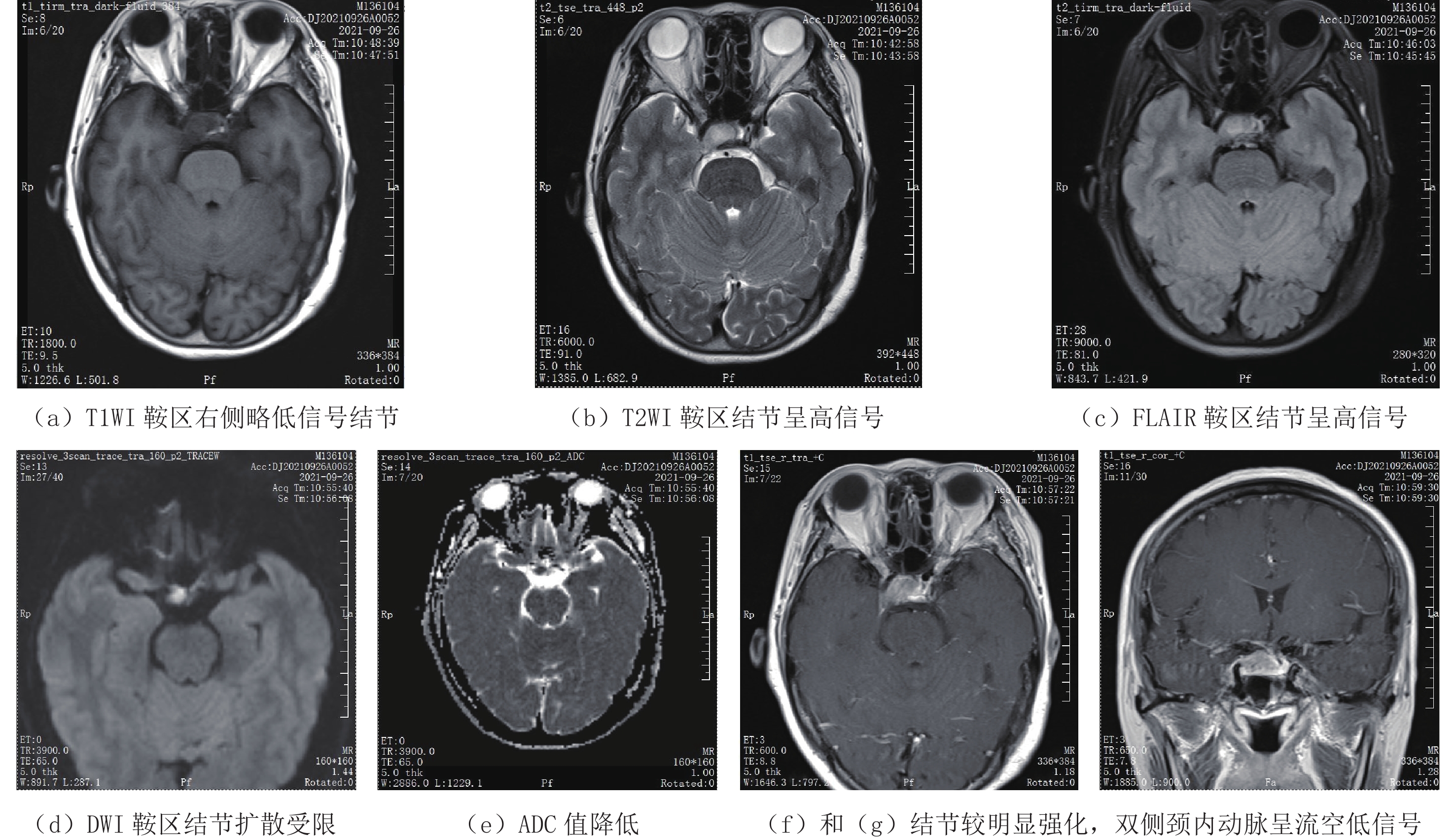
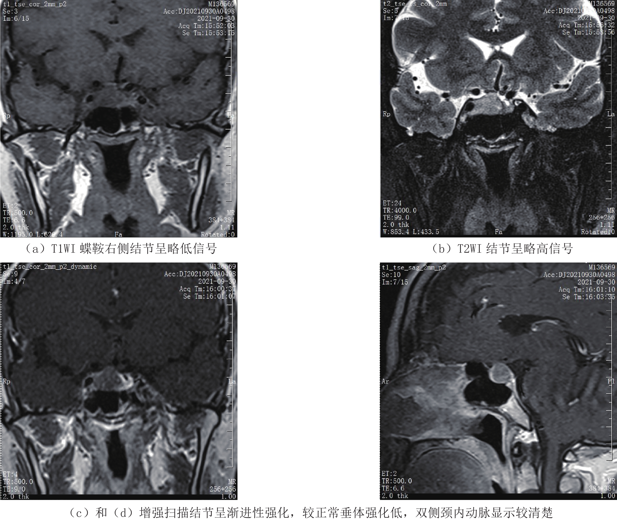
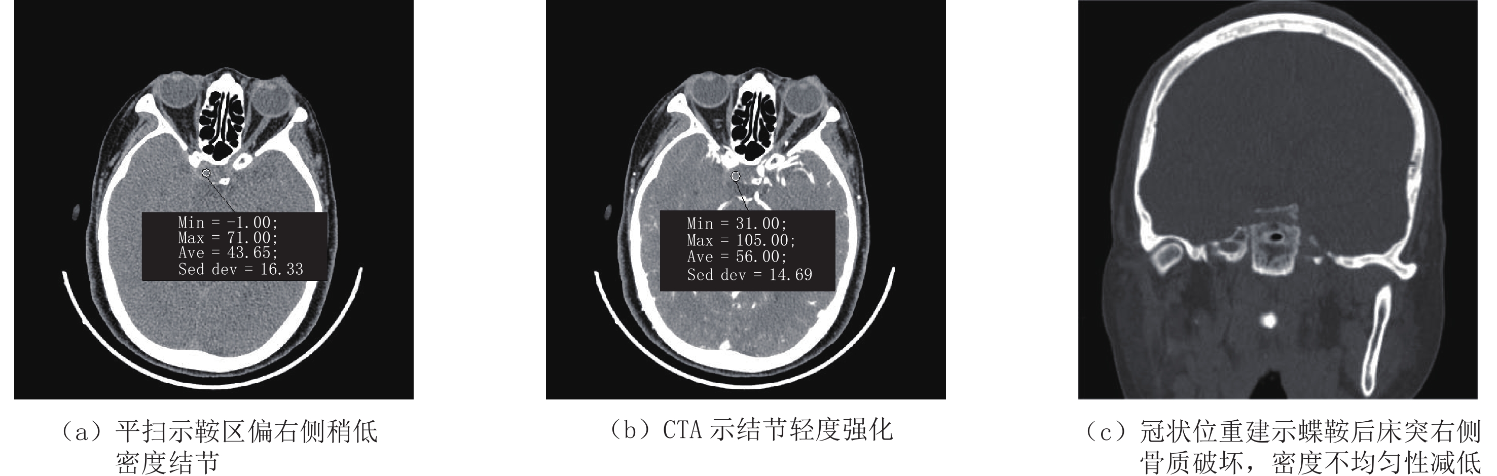
CT、MRI检查:颅脑CT(图1)示鞍区偏右侧稍低密度结节,头颈部CTA增强鞍区偏右侧结节轻度强化,邻近后床突骨质破坏,密度不均匀性减低。颅脑MRI(图2)示鞍区垂体显示不清,垂体窝偏右侧见T1WI呈低信号、T2WI呈高信号结节,FLAIR呈略高信号,且DWI水分子扩散受限呈高信号,增强扫描结节较明显强化,邻近双侧颈内动脉虹吸段呈流空信号。垂体MRI平扫+增强(图3)示垂体窝稍扩大,垂体窝偏右侧见约1.6 cm×1.3 cm×1.3 cm大小软组织信号结节,垂体柄向左侧偏移,增强后呈不均匀强化。双侧颈内动脉海绵窦段位置对称;双侧视交叉上抬;考虑鞍区占位,肿瘤性病变,垂体大腺瘤可能。
临床拟诊垂体瘤,痛性眼肌麻痹,完善术前相关检查,在全麻下行经蝶窦鞍区病损切除术。术中见鞍底突入蝶窦腔,邻近骨质较厚,磨开鞍底骨质即可见肿瘤结节病灶,质软、呈白色,用刮圈刮除鞍区肿瘤组织,冲洗术腔,电凝止血,并在肿瘤残腔填入明胶海绵止血,蝶窦腔填塞明胶海绵止血。退出撑开器,鼻粘膜复位,右侧鼻腔填入膨胀海绵止血结束手术。
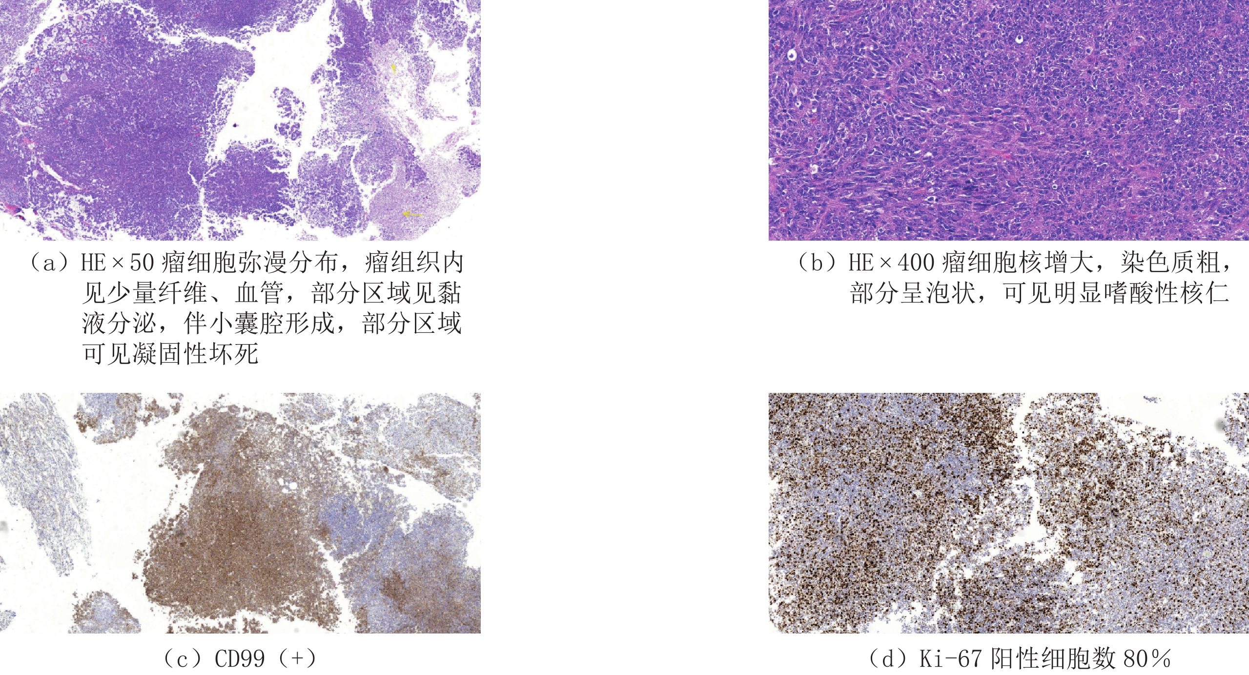
术后病理学检查(图4):(鞍区肿瘤)恶性肿瘤,符合小圆细胞性未分化肉瘤。免疫组化:CK(-),Vim(+),EMA(灶+),NF(灶+),CD99(+),WT-1(灶+),GH(-),PRL(-),ACTH(-),TSH(-),GFAP(-),S-100(-),CK20(-),CD34(-),CgA(-),NSE(-),Syn(-),CD117(-),INI-1(-),LCA(-),Ki67阳性细胞数80%。术后放化疗,5月后复查鞍区MR局部复发,可见新肿块形成(图5)。
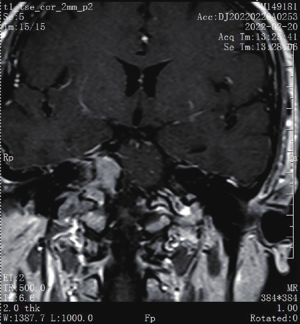
![]() 图 5 术后5月,右侧桥前池明显强化软组织信号结节Figure 5. At 5 months after surgery, the right anterior pontine pool significantly enhanced the soft tissue signal nodules
图 5 术后5月,右侧桥前池明显强化软组织信号结节Figure 5. At 5 months after surgery, the right anterior pontine pool significantly enhanced the soft tissue signal nodules2. 讨论
软组织未分化肉瘤可发生于任何年龄,无明显性别差异。相对来说,梭形细胞未分化肉瘤和多形性未分化肉瘤多发生于50~70岁中老年人,并以男性多见,极少发生于儿童,而小圆细胞性肉瘤多发生于儿童和青少年。垂体腺瘤在成人鞍区肿瘤中最为常见,其他性质的肿瘤仅占10% 左右,有文献报道[1],原发于鞍区小圆细胞性未分化肉瘤非常罕见,且发生在鞍区未分化肉瘤可能与垂体腺瘤患者放疗后相关。
2013年版WHO软组织分类新设立了未分化/未分类软组织肉瘤(undifferentiated/unclassified soft tissue sarcoma,USTS),这一类软组织肉瘤相对比较少见,约占软组织肉瘤的20%[2]。根据瘤细胞的组织学形态,USTS可分为梭形细胞未分化肉瘤、多形性未分化肉瘤、小圆细胞未分化肉瘤和上皮样未分化肉瘤[3]。鞍区肿瘤中,原发性肉瘤极少见,其中发生率最高的是纤维肉瘤,且主要发生于垂体良性肿瘤术后放疗后[4-5],其中至少25% 放射相关的软组织肉瘤为未分化肉瘤。鞍区良性肿瘤主要包括垂体腺瘤、脑膜瘤、神经鞘瘤、肌纤维母细胞瘤、毛细胞性星形细胞瘤等[6-7];其经放疗后易出现的恶性肿瘤主要包括血管周细胞瘤、纤维肉瘤、骨肉瘤、软骨肉瘤、平滑肌肉瘤、横纹肌肉瘤等峰[5,8],其中横纹肌肉瘤是发生于儿童的最常见的肉瘤[9-10],极少发生小圆细胞未分化肉瘤。
本例小圆细胞未分化肉瘤CT表现为稍低密度结节影,平扫与垂体及邻近海绵窦分界不清楚,邻近蝶鞍后床突可见骨质破坏,可提示恶性肿瘤可能。MRI可见垂体窝扩大,垂体窝偏右侧软组织信号结节影,T1WI呈低信号,T2WI呈高信号,似见与正常垂体有边界,垂体柄受压向左侧偏移,视交叉上抬,增强结节明显强化,强化不均匀,早期明显强化与正常垂体可见分界,强化方式不一致。但本例患者结节位于垂体窝内,信号差异不明显,增强扫描呈渐进性强化,早期虽较正常垂体强化低,术前误诊考虑垂体大腺瘤可能。术后复习垂体MRI影像,仔细可见肿瘤与垂体似见边界,偏侧可见正常垂体,垂体呈受压改变,应考虑到肿瘤不来源垂体,另外强化以延迟性强化较明显,且强化不均匀,也不是典型垂体腺瘤强化方式,垂体瘤虽可压迫蝶鞍至蝶鞍扩大,但基本上不会累及蝶鞍导致骨质破坏。国外文献[5,10-11]中非辐射暴露下发生的鞍区原发未分化肉瘤迄今仅报道3例,因此病罕见,无大宗病例报道,因此对其临床、影像表现和治疗都缺乏经验。
复习文献并结合该病例,CT鞍区稍低密度,MR T1WI呈低信号,T2WI呈高信号软组织结节,DWI扩散受限,ADC值降低,可累及邻近颅底骨质,甚至骨质破坏,在排除鞍区其他常见肿瘤后,应考虑到小圆细胞未分化肉瘤等罕少见恶性肿瘤可能。Sateen等[1]报道1例鞍区原发未分化肉瘤患者,术后行放疗、化疗,术后半年未见肿瘤复发。但对其总体预后难以评估,未分化肉瘤恶性程度极高,即使术后及时行放化疗或伽玛刀等综合治疗,其总体预后应较差[12],本例术后半年复发。
未分化肉瘤因缺少大多数特征性的免疫标识,影像学表现不典型,术前诊断与鉴别诊断均较困难。需注意与垂体瘤、颅咽管瘤、鞍区脑膜瘤鉴别。
垂体瘤:肿瘤向上生长,突破鞍膈则可以见到鞍上池变形,甚至有一些情况下会出现大部分的闭塞,其中可以看到有等密度或者稍高密度的肿块。肿瘤当中可以见到有坏死或者囊性的低密度区。如果是向下生长,会在蝶窦内出现圆形的软组织影。CT表现为稍高密度影;MRI提示病变区T1WI为等信号,T2WI为高信号,明显均匀强化。垂体瘤通常DWI信号不高,而小圆细胞未分化肉瘤DWI呈高信号具有一定特征性。
颅咽管瘤:CT表现提示病灶位于鞍上或鞍内,多数呈均匀低密度,部分呈均匀等密度或低等混杂密度灶。肿瘤钙化率较高,约半数沿肿瘤边缘分布,呈壳状,其余为大小不等、多少不一的块状或点状钙化,也可几种钙化形态同时存在。增强后,囊性病灶可见囊壁呈薄环状或壳状增强,少数可显示薄壁分房状强化。实质性肿瘤呈不均匀或均匀强化。颅咽管瘤通常有钙化,且大多位于鞍上区,可资鉴别。
鞍区脑膜瘤:肿瘤多呈圆形或椭圆形,边界较清楚,多以鞍结节或床突为附着点,可向各个方向生长。病灶密度或信号多均匀,CT平扫多呈等密度或稍高密度,其内可见点状或斑片状钙化影;MRI扫描T1WI多呈等信号或稍低信号,T2WI多呈等信号或稍高信号,其内钙化灶在T1WI、T2WI均为低信号;动态增强扫描,肿瘤多呈均匀明显强化,部分病灶在轴位和矢状位上可显示肿瘤邻近脑膜增厚并明显强化,形成典型的“脑膜尾征”。脑膜瘤通常造成邻近骨质增生,具有良性肿瘤的表现,而小圆细胞未分化肉瘤出现骨质破坏,表现出恶性特征。
3. 总结
小圆细胞性未分化肉瘤是一种高度恶性肿瘤,预后较差。本例发生于中老年女性的小圆细胞未分化肉瘤甚少见。软组织肉瘤可发生于任何部位,但绝大多数病例发生于四肢、躯干和头颈部软组织内,发生在鞍区也实属罕见。未分化/未分类软组织肉瘤的临床表现无特异性,主要表现为深部软组织肿块,但本例发生在鞍区表现为软组织密度小结节,病灶小,却引起了邻近器官相应临床表现,并出现垂体相关激素泌乳素(Prolactin,PRL)升高、皮质醇及促肾上腺皮质激素降低,垂体柄和视交叉受压,瞳孔不等大,视物有重影,视物模糊,右眼疼痛,上睑轻度下垂,眼球旋转部分受限。
本例行颅脑CT呈稍低密度结节,邻近骨质可见破坏,MRI可见鞍区结节扩散受限呈高信号,ADC值明显降低,提示恶性肿瘤可能。因此术前阅片不仔细,对鞍区典型肿瘤,如垂体瘤影像表现不熟悉,是本例误诊为垂体瘤的主要因素。但该病例肿瘤病理类型少见,同时发生在罕见部位,术前无论是临床还是影像诊断都困难,最终确诊需术后病理学检查。
-
[1] SAREEN P, CHHABRA L, TRIVEDI N. Primary undifferentiated spindlecell sarcoma of sella turcica: Successful treatment with adjuvant temozolomide[J]. BMJ Case Reports, 2013, 2013. pii: bcr201300934.
[2] 王坚, 朱雄增. 软组织肿瘤病理学[M]. 2版. 北京: 人民卫生出版社, 2017: 1380-1381. WANG J, ZHU X Z. Soft-tissue tumor pathology[M]. The 2 ed. Beijing: People's Health Publishing House, 2017: 1380-1381. (in Chinese).
[3] FLETCHER C D M, BRIDGE J A, HOGENDOORN P C W, eds. WHO classification of soft tissue and bone tumours[M]. 4ed, Lyon: IARC Press; 2013: 236-238.
[4] HUANG B Y, CASTILLO M. Nonadenomatous tumors of the pituitary and sella turcica[J]. Topics in Magnetic Resonance Imaging, 2005, 16(4): 289−299. doi: 10.1097/01.rmr.0000224685.83629.18
[5] LOPES M B, LANZINO G, CLOFT H J, et a1. Primary fibrosarcoma of the sella unrelated to previous radiation therapy[J]. Modern Pathology, 1998, ll(6): 579−584.
[6] SCHULTZ A B, BRAT D J, OYESIKU N M, et a1. Intrasellar pituicytoma in a patient with other endocrine neoplasms[J]. Archives of Pathology & Laboratory Medicine, 200 l, 125(4): 527-530.
[7] SHINOJIMA N, OHTA K, YANO S, et a1. Myofibroblastoma in the suprasellar region: Case report[J]. Journal of Neurosurgery, 2002, 97(5): 1203−1207. doi: 10.3171/jns.2002.97.5.1203
[8] MENA H, RIBAS J L, PEZESHKPOUR G H, et a1. Hemangiopericytoma of the central nervous system: A review of 94 cases[J]. Human Pathology, 1991, 22(1): 84−91. doi: 10.1016/0046-8177(91)90067-Y
[9] MANORANJAN B, SYRO L V, SCHEITHAUER B W, et a1. Undifferentiated sarcoma of the sellar region[J]. Endocrine Pathology, 2011, 22(3): 159−164. doi: 10.1007/s12022-011-9166-7
[10] ALPERT T E, HAHN S S, CHUNG C T, et a1. Successful treatment of spindle cell sarcoma of the sella turcica: Case report[J]. Journal of Neurosurgery, 2002, 97(S5): 438−440.
[11] ZHONG J, LI S T, YAO X H, et a1. An intrasellar rhabdomyosarcoma misdiagnosed as pituitary adenoma[J]. Surgical Neurology, 2007, 68(S2): S29−33.
[12] 张广健, 常建勇, 谢英亮, 等. 神经内镜下切除鞍区原发性未分化肉瘤一例并文献复习[J]. 中华神经外科杂志, 2017,33(10): 1070−1071. ZHANG G J, CHANG J Y, XIE Y L, et al. Neuroendoscopic resection of primary undifferentiated sarcoma in the sellar region: A case report and literature review[J]. Chinese Journal of Neurosurgery, 2017, 33(10): 1070−1071. (in Chinese).
 下载:
下载:
计量
- 文章访问数: 251
- HTML全文浏览量: 130
- PDF下载量: 180


