
Imaging Diagnosis of a Rare Case of Cervical Necrotizing Fasciitis (CNF) in the Nasopharynx: A Clinical Case Analysis
-
摘要: 本文介绍1例罕见的发生于鼻咽部的颈部坏死性筋膜炎(CNF)。该患者以顽固性慢性化脓性中耳炎就诊,进行包括CT、MRI及PET/CT在内的影像学检查,均误诊为鼻咽癌。最终该患者通过鼻咽肿物活检术确诊为CNF。本文通过回顾性分析该患者的影像学表现及误诊原因,总结出CT、MRI、PET/CT在CNF早期诊断中的特征和要点,为其临床精准诊断提供重要的帮助。Abstract: This case report describes a rare case of cervical necrotizing fasciitis (CNF) of the nasopharynx. The patient presented with intractable chronic suppurative otitis media and underwent radiological examination including CT, MRI, and PET/CT. All of the images misdiagnosed this condition as nasopharyngeal carcinoma. Finally, the patient underwent a nasopharyngeal mass biopsy and CNF was diagnosed. A retrospective analysis of the radiological scans was performed and the causes of the misdiagnosis were evaluated. The CT, MRI, and PET/CT characteristics and key features for the early diagnosis of CNF were summarized. Our findings may contribute to current knowledge of the precise diagnosis of CNF.
-
Keywords:
- PET/CT /
- neck /
- necrotizing fasciitis /
- radiology
-
坏死性筋膜炎(necrotizing fasciitis,NF)是危及生命的侵袭性软组织感染,其坏死区域主要累及筋膜及肌肉。这是一种罕见的疾病,常好发于躯干、会阴及四肢,而对于颈部坏死性筋膜炎(cervical necrotizing fasciitis,CNF)来说,由于发病部位的血供丰富,因此其发病率更低,仅占NF的1%~10%[1]。牙源性以及扁桃体感染是常见的发病原因,颌下以及咽旁间隙是最好发的感染部位,发生于鼻咽部较为罕见。
CNF如果不及时诊断以及治疗,病变区域有可能沿着颈深区域向纵隔蔓延,从而导致严重的后果。不受控制的高血糖会导致患者的免疫系统随着年龄的增长越来越虚弱,这往往是该疾病的主要诱发因素[2]。由于CNF在发病初期并无特异性的体征以及实验室指标的异常,因此影像学检查,尤其是CT检查在该病的精准诊断中显得尤为重要。
1. 病史资料
1.1 临床资料
患者男性,58岁,1年余前无明显诱因出现左侧头痛,伴有左耳流脓,无鼻塞、流涕、涕中带血,无耳痛、耳内流血等不适。于当地医院诊断为“慢性化脓性中耳炎”,予以抗炎治疗后头痛稍有减轻。此后患者上述症状持续存在,均予以抗炎治疗,但头痛并未缓解。
10月前患者自觉上述症状加重,并出现左侧面瘫及吞咽困难,之后对症治疗后均未见好转。既往双侧慢性化脓性中耳炎50余年;糖尿病史5年,合并糖尿病性视网膜病史,平时胰岛素控制;高血压病史4年。
1.2 实验室检查
血常规:红细胞3.70×10¹²/L(下降),血红蛋白108 g/L(上升),红细胞压积32.6%(下降),淋巴细胞15.9%(下降),其余正常。血生化:谷丙转氨酶6.3 U/L(下降),谷草转氨酶10.6 U/L(下降),总胆汁酸6.8 umol/L(上升),总蛋白61.3 g/L(下降),白蛋白33.6 g/L(下降),肌酐101 umol/L(上升),葡萄糖4.39 mmol/L,糖化血红蛋白21.9%(上升),其余正常。肿瘤指标:AFP、CEA、CA199、CA125、CA724、PSA、fPSA、fPSA/PSA、CTFRA21-1、SCC、CA50均阴性。
1.3 影像学表现
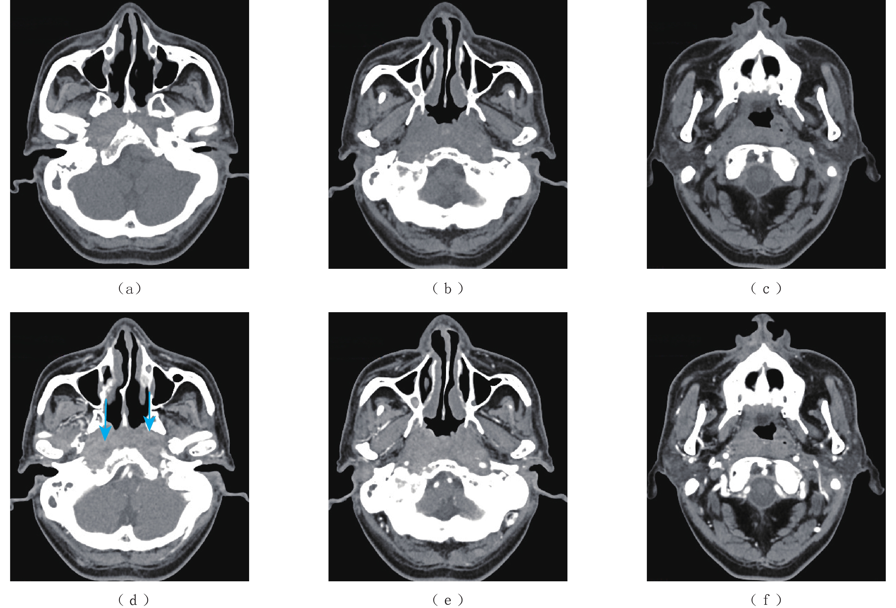
CT图像显示鼻咽腔基本对称,双侧咽隐窝略变浅(图1);两侧咽旁间隙及咽后间隙软组织肿胀,颅底斜坡骨质破坏,蓝色箭头处可见无强化的筋膜征象(图1(d))。
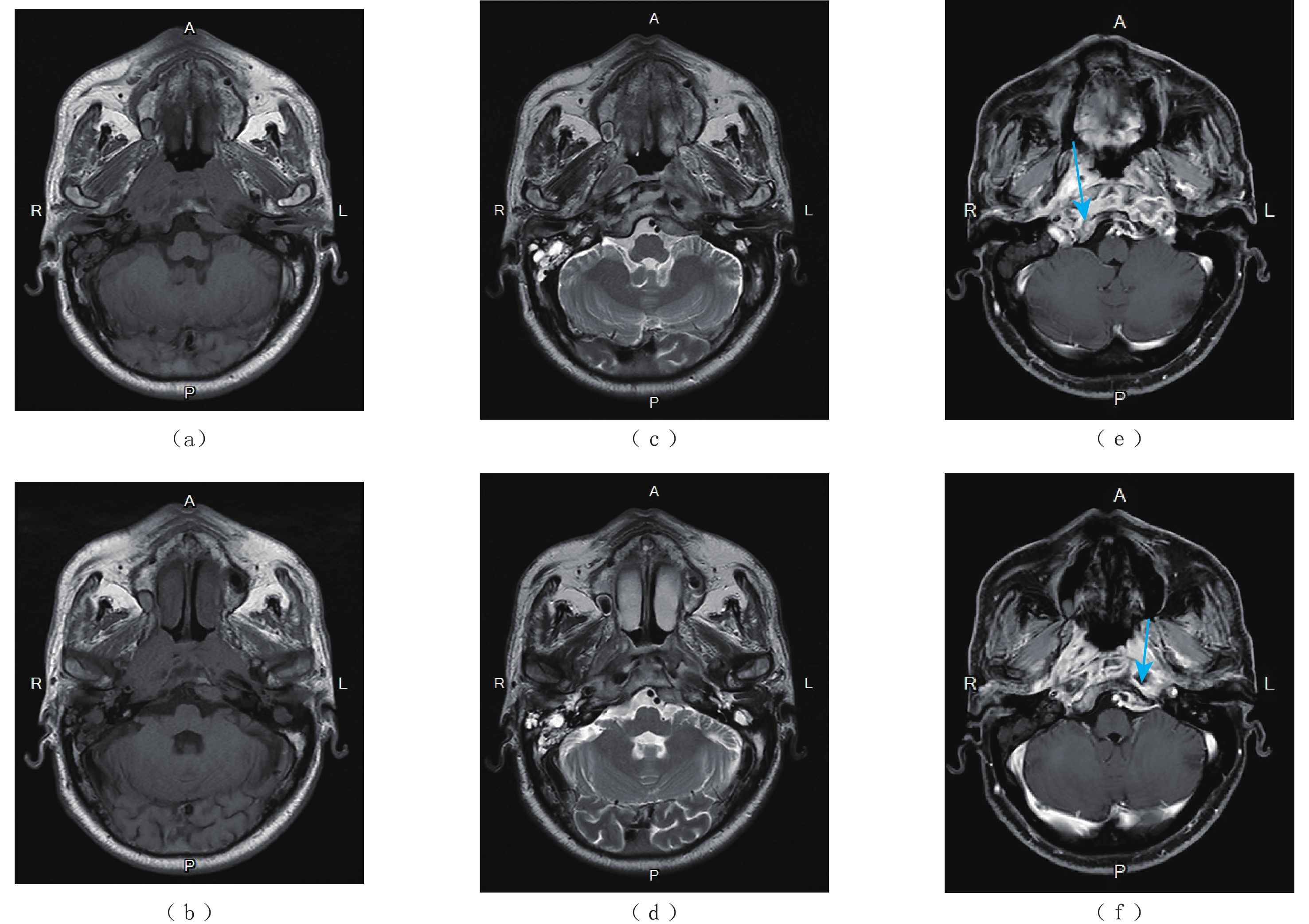
MRI图像示鼻咽腔基本对称,双侧咽隐窝略变浅(图2);两侧咽旁间隙及咽后间隙软组织肿胀,肌间脂肪消失,增强后咽旁间隙可见散乱不均匀的异常强化,病变沿肌间隙走行,鼻咽粘膜未见异常增厚。蓝色箭头见颅骨斜坡右侧及左下可见稍长T1稍短T2信号影,增强后强化不均匀,周围见强化的软组织信号影(图2(e)和图2(f)),两侧乳突气房可见液性信号影。
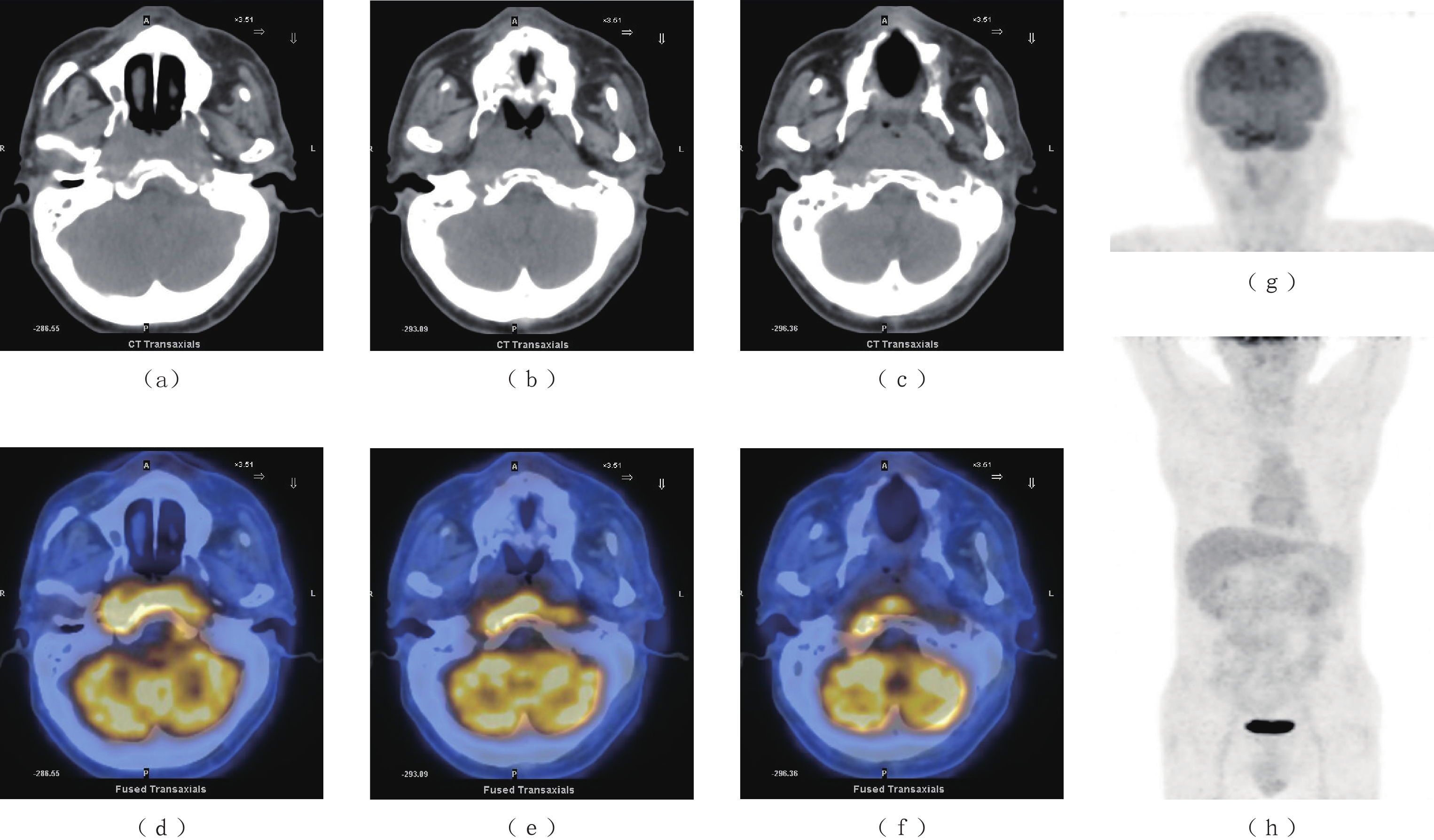
PET/CT图像显示FDG异常代谢区域位于鼻咽深部软组织伴邻近颅底斜坡骨质破坏(图3),鼻咽粘膜未见异常代谢(图3(a)~图3(f));头颈部PET图像显示颈部未见肿大淋巴结影(图3(g))。CT、MRI以及PET/CT检查,均误诊为鼻咽癌。
1.4 诊疗过程
患者行鼻内镜下鼻咽部肿物活检术,于鼻咽部可见包裹性脓腔及坏死组织,取部分病理组织送病理及脓性分泌物送细菌培养,广泛彻底清除病灶至正常黏膜。
病理诊断:局部见炎性渗出,局部纤维结缔组织增生及粘液变性,胶原变性。
1.5 随访
患者出院1月后门诊复查,疼痛及吞咽困难的症状缓解,面瘫有所好转。半年后电话跟踪随访,患者疼痛及吞咽困难的症状消失,面瘫基本缓解,仅左眼睑闭合不佳。
2. 讨论
2.1 病因及发病机制
NF是一种组织坏死并且进展迅速的疾病,病原体入侵软组织并且引起血管血栓,最终导致脂肪组织、筋膜及皮肤坏死[3]。CNF更是一种罕见的累及颈部筋膜的微生物感染,它的易感因素包括糖尿病、不良的口腔卫生、酗酒、肿瘤以及静脉吸毒[4]。
CNF最常见原因是牙源性感染(27.5%),其次是扁桃体疾病(22.5%)、皮肤感染(8.75%)和腮腺感染(6.25%)[5]。最常见的并发症是气道阻塞以及下行性坏死性纵隔炎[6]。纵隔炎的预后非常差,因此它与感染性休克一样都是CNF最为严重的并发症[7-8]。
2.2 临床特征
由于CNF预后不良,及时诊断并且早期干预就显得尤为重要。压痛、发热和皮肤红斑是早期NF的常见体征[9]。回顾本病例,可能是由于发病位置的特殊(鼻咽部),患者并无上诉症状,而是表现为左侧头痛伴左耳流脓。
Wong等[10]为了对包括CNF在内的NF进行早期诊断,提出了坏死性感染实验室风险指标(LRINEC)评分。LRINEC评分是以6项实验室指标的异常进行评分,其中包括血清C反应蛋白(>150 mg/L)、白细胞(WBC)计数(>15000/μL)、血红蛋白(<13.5 g/dL)、血清钠水平(<135 mmol/L)、血清肌酐水平(142 mmol/L)和血清葡萄糖水平(10 mmol/L)。LRINEC评分大于等于8分,发生NSTI的风险为75%。虽然之后一些研究对LRINEC评分进行评估,证实了该评分在NF感染初期诊断中的有效性,但是近期的一些研究又发现该指标的敏感性较差,并不能作为排除NF的有效手段[11,12-13]。通过回顾本例患者的实验室指标也验证了这一结论,患者的LRINEC评分仅为2分,远没有达到诊断NF的水平。
2.3 影像学表现
对于临床体征及实验室指标均不明确的早期NF患者,影像学检查可以发挥重要作用。如怀疑为NF,CT扫描是一个有价值的影像学工具。一项关于坏死性筋膜炎的CT研究发现,CT的敏感性达到了100%,特异性为98%,因此CT阴性结果可以有效的排除坏死性筋膜炎,CT阳性结果对诊断坏死性筋膜炎具有很高的价值[14]。当CT图像中出现脂肪受累、沿着筋膜平面走行的液体以及气体聚集,尤其是增强图像出现无强化的筋膜增厚等征象,需要考虑NF[14]。
而MR则被认为是诊断NF最佳的影像学检查,当T2加权像上出现深筋膜增厚>3 mm并伴有多个肌筋膜室受累,此为诊断NF的重要征象[15]。虽然MRI的表现优于CT,但是MRI在某些紧急情况下难以进行,因此不建议将其作为首选的影像学检查技术[11]。我们回顾该患者CT及MRI图像,图像中虽然出现两侧咽旁间隙及咽后间隙肿胀,伴双侧欠对称,咽隐窝变浅,合并颅底骨破坏等表现,这些都是与鼻咽癌相同的征象,但是图像中另外可见典型的深筋膜增厚的表现,尤其是MR上可见散乱不均匀的异常强化,病变沿肌间隙走行,这些均提示需要与坏死性筋膜炎进行鉴别。
在该患者的PET/CT图像上,虽然鼻咽部肿胀伴有较大范围的FDG代谢增高,同时合并颅底骨的破坏,但是FDG异常代谢的区域主要局限在鼻咽深部区域,粘膜并未见FDG异常代谢,这点从MR中信号正常的鼻咽粘膜中得到了印证(图2(c)和图2(d))。
2.4 治疗及转归
本例患者通过鼻咽肿物活检术发现鼻咽部包裹性浓腔及坏死组织,并且病灶进行广泛彻底地清除,完成对CNF的诊断以及治疗。术后患者症状明显缓解,出院后继续接受头孢曲松抗感染治疗。
2.5 诊断与鉴别诊断
目前NF的诊断主要依赖症状学、实验室指标、影像学以及侵入性诊断。然而早期的NF体征与症状几乎没有特异性,通常难以明确诊断。因此当出现肿胀、发热以及与症状不成比例的剧烈疼痛时,需要高度怀疑NF。既往LRINEC评分曾经作为NF诊断的重要依据,但是该评分诊断的敏感性较低,并不能作为排除NF的有效手段。当NF诊断不明确时,影像学检查可以提供相对有价值的信息。
一项包含23项研究总计纳入5982名患者的META分析评估了体格检查、影像学检查以及LRINEC评分在NF诊断中的准确性[13]。该研究发现影像学检查具有敏感性及特异性,尤其是CT的敏感性为88.5%,特异性为93.3%;而体格检查以及LRINEC评分敏感性较差,均不能应用于排除NF。侵入性诊断——手术探查是诊断NF的金标准,当手指可轻易分离筋膜(手指实验阳性)、组织缺血坏死以及恶臭分泌物,均提示NF的诊断[16]。由于该例患者CNF发生于鼻咽部,需要与以下疾病进行鉴别。
2.5.1 鼻咽癌
鼻咽癌病理类型目前以未分化癌及鳞状细胞癌为主,EB病毒感染与鼻咽癌发病率密切相关。早期鼻咽癌基本无症状,也可能因为咽鼓管阻塞引起一系列症状,其中包括鼻塞、鼻出血、中耳炎、听力下降等。影像学表现:CT及MR表现基本相似,表现为鼻咽部两侧欠对称,局部见软组织肿块突入鼻腔内,咽隐窝变浅或消失;当咽旁间隙受累时,其脂肪间隙消失,再向外可累及翼腭窝及颞下窝,向后可累及颅底;增强扫描多表现为不均匀强化。PET/CT表现为FDG高代谢。
2.5.2 鼻咽淋巴瘤
鼻咽淋巴瘤是仅次于鼻咽癌第2常见的鼻咽部恶性肿瘤,它的发病率与EB病毒感染也密切相关。影像学表现,它好发于鼻咽顶壁咽扁桃体和咽鼓管扁桃体附近粘膜内聚集的淋巴小结,往往表现为一种弥漫对称分布的肿块[17]。同时它通常沿着粘膜或脂肪间隙扩散至口咽部及下咽扁桃体,极少累及深层结构,因此鼻咽淋巴瘤很少累及颅底。增强扫描多为均匀性的强化表现,PET/CT也表现为FDG高代谢。
3. 结论
鼻咽部坏死性筋膜炎缺乏早期诊断的特征性表现,但是影像学检查,尤其是CT对该病的早期诊断具有重要价值。
-
[1] BÖTTGER S, ZECHEL-GRAN S, SCHMERMUND D, et al. Odontogenic cervicofacial necrotizing fasciitis: Microbiological characterization and management of four clinical cases[J]. Pathogens, 2022, 11(1): 78. doi: 10.3390/pathogens11010078
[2] MTENGA A A, KALYANYAMA B M, OWIBINGIRE S S, et al. Cervicofacial necrotizing fasciitis among patients attending the Muhimbili National Hospital, Dar es Salaam, Tanzania[J]. Bmc Infectious Diseases, 2019, 19(1): 642. doi: 10.1186/s12879-019-4267-x
[3] ABDURRAZAQ T O, IBIKUNLE A A, BRAIMAH R O. Cervical necrotizing fasciitis: A potentially fatal disease with varied etiology[J]. Annals of Medical and Health Sciences Research, 2016, 6(4): 251−256. doi: 10.4103/amhsr.amhsr_33_16
[4] CONGEDO M T, NACHIRA D, PENNISI M A, et al. Risk factors associated with post-operative complications in multidisciplinary treatment of descending necrotizing mediastinitis[J]. Journal of Clinical Medicine, 2022, 11(21): 6364. doi: 10.3390/jcm11216364
[5] SUEHARA A B, GONÇALVES A J, ALCADIPANI F A, et al. Deep neck infection: analysis of 80 cases[J]. Brazilian Journal of Otorhinolaryngology, 2008, 74(2): 253−259. doi: 10.1016/S1808-8694(15)31097-1
[6] CHOU P Y, HSIEH Y H, LIN C H. Necrotizing fasciitis of the entire head and neck: Literature review and case report[J]. Biomedical Journal, 2020, 43(1): 94−98. doi: 10.1016/j.bj.2019.08.002
[7] NOUGUÉ H, LE MAHO A L, BOUDIAF M, et al. Clinical and imaging factors associated with severe complications of cervical necrotizing fasciitis[J]. Intensive Care Medicine, 2015, 41(7): 1256−1263. doi: 10.1007/s00134-015-3830-1
[8] 王家晨, 李春香, 王建明. 颈部坏死性筋膜炎的早期诊疗进展[J]. 中国眼耳鼻喉科杂志, 2021,21(1): 69−73. doi: 10.14166/j.issn.1671-2420.2021.01.019 WANG J C, LI C X, WANG J M. Progress in early diagnosis and treatment of cervical necrotizing fasciitis[J]. Chinese Journal of Ophthalmology and Otorhinolaryngology, 2021, 21(1): 69−73. (in Chinese). doi: 10.14166/j.issn.1671-2420.2021.01.019
[9] WANG J M, LIM H K. Necrotizing fasciitis: Eight-year experience and literature review[J]. Brazilian Journal of Infectious Diseases, 2014, 18(2): 137−143. doi: 10.1016/j.bjid.2013.08.003
[10] WONG C H, KHIN L W, HENG K S, et al. The LRINEC (Laboratory Risk Indicator for Necrotizing Fasciitis) score: A tool for distinguishing necrotizing fasciitis from other soft tissue infections[J]. Critical Care Medicine, 2004, 32(7): 1535−1541. doi: 10.1097/01.CCM.0000129486.35458.7D
[11] SARTELLI M, COCCOLINI F, KLUGER Y, et al. WSES/GAIS/WSIS/SIS-E/AAST global clinical pathways for patients with skin and soft tissue infections[J]. World Journal of Emergency Surgery, 2022, 17(1): 3. doi: 10.1186/s13017-022-00406-2
[12] KIM M C, KIM S, CHO E B, et al. Utility of magnetic resonance imaging for differentiating necrotizing fasciitis from severe cellulitis: A magnetic resonance indicator for necrotizing fasciitis (MRINEC) algorithm[J]. Journal of Clinical Medicine, 2020, 9(9): 3040. doi: 10.3390/jcm9093040
[13] FERNANDO S M, TRAN A, CHENG W, et al. Necrotizing soft tissue infection: diagnostic accuracy of physical examination, imaging, and LRINEC score: A systematic review and meta-analysis[J]. Annals of Surgery, 2019, 269(1): 58−65. doi: 10.1097/SLA.0000000000002774
[14] MARTINEZ M, PEPONIS T, HAGE A, et al. The role of computed tomography in the diagnosis of necrotizing soft tissue infections[J]. World Journal of Surgery, 2018, 42(1): 82−87. doi: 10.1007/s00268-017-4145-x
[15] KIM K T, KIM Y J, WON LEE J, et al. Can necrotizing infectious fasciitis be differentiated from nonnecrotizing infectious fasciitis with MR imaging?[J]. Radiology, 2011, 259(3): 816-824.
[16] ANDREASEN T J, GREEN S D, CHILDERS B J. Massive infectious soft-tissue injury: Diagnosis and management of necrotizing fasciitis and purpura fulminans[J]. Plastic and Reconstructive Surgery, 2001, 107(4): 1025−1035. doi: 10.1097/00006534-200104010-00019
[17] KING A D, LEI K I, RICHARDS P S, et al. Non-Hodgkin's lymphoma of the nasopharynx: CT and MR imaging[J]. Clinical Radiology, 2003, 58(8): 621−625. doi: 10.1016/S0009-9260(03)00182-X
-
期刊类型引用(1)
1. 郑晓枫,马新宇,于开锋. 造影剂肾病发病机制及中医药防治研究进展. 辽宁中医药大学学报. 2023(03): 169-172 .  百度学术
百度学术
其他类型引用(0)
 下载:
下载:
计量
- 文章访问数: 297
- HTML全文浏览量: 110
- PDF下载量: 35
- 被引次数: 1


