
Analysis of Thin Slice Computed Tomography Features of Coronavirus Disease 2019 Related Vascular Abnormalities
-
摘要: 目的:探讨胸部薄层CT平扫对新型冠状病毒感染(COVID-19)相关性血管异常的CT特征分析的临床价值。材料与方法:回顾性收集2022年12月5日至2022年12月17日北京世纪坛医院感染科确诊COVID-19且胸部薄层CT平扫图像显示有病变累及血管的患者73例,所有患者有完整的胸部薄层CT平扫资料和有较完整的临床资料。依据年龄>60岁和≤60岁将患者分为老年组和青壮年组,观察所有患者胸部影像学表现,并进行不同年龄组间统计学分析。结果:73例COVID-19患者中,青壮年组和老年组组间对比有统计学意义的影像学指标如下:病变分布中央血管周、病变大小10~30 mm、病变大小>30 mm、病变占肺叶体积百分比≤30%、病变占肺叶体积百分比>50%(白肺)、病变形态大片状、病变优势类型腺泡样、血管扭曲、血管周缘模糊、树芽征、粗大纤维索条。结论:①胸部薄层 CT平扫可明确COVID-19相关性血管异常的病变数量、位置、累及部位、范围、血管异常和病变类型,对COVID-19血管异常的定性诊断和鉴别诊断有一定的意义;②胸部薄层CT平扫检查对于发现临床症状不典型但有COVID-19累及血管的老年患者有重要意义;③COVID-19相关性“血管增粗”既可以是血管本身管径的增粗,也可以由血管周围间质炎性水肿造成。Abstract: Objective: This study aimed to explore the clinical value of thin slice computed tomography (CT) plain scan in the analysis of CT features of vascular abnormalities associated with coronavirus disease 2019 (COVID-19). Materials and methods: A total of 73 patients with COVID-19 confirmed by the Department of Infection of Beijing Shijitan Hospital from December 5, 2022 to December 17, 2022, were included in the study. Chest thin CT plain scan images showed that the lesions involved blood vessels were retrospectively collected. All patients had complete chest thin CT plain scan and relatively complete clinical data. According to age (>60 and ≤60 years), the patients were divided into the young and elderly groups. The chest imaging manifestations of all patients were observed and statistically analyzed between different age groups. Results: Among the 73 patients with COVID-19, the imaging indexes with statistical significance between the young and elderly groups were as follows: the distribution of the lesion around the central blood vessel, size of the lesion (10~30 mm), size of the lesion (>30 mm), percentage of the lesion to the volume of the lung lobe (≤30), percentage of the lesion to the volume of the lung lobe (>50) (white lung), shape of the lesion was large, the dominant type of the lesion was acinar, vascular distortion, vascular margin fuzzy, and tree-bud sign thick fiber rope. Conclusion: (1) Chest thin-slice CT plain scan can identify the number, location, involved location, scope, vascular abnormality, and pathological type of COVID-19-related vascular abnormality, which has certain significance for the qualitative and differential diagnosis of COVID-19 vascular abnormality. (2) The chest thin CT plain scan is of great significance for finding elderly patients with COVID-19 involving blood vessels. (3) COVID-19-related "blood vessel thickening" can be caused by either the diameter of the blood vessel itself or the inflammatory edema of the perivascular interstitium.
-
Keywords:
- tomography /
- X-ray computer /
- novel coronavirus pneumonia /
- high resolution
-
-
表 1 COVID-19不同年龄组的临床指标比较
Table 1 Comparison of clinical indicators of COVID-19 in different age groups
临床指标 组别 统计检验 青壮年组(年龄≤60岁;
n=29)老年组(年龄>60;
n=44)$\chi^2 /t$ P 平均年龄/岁 40.6±12.6 76.9±9.8 -13.806 0.00 性别/例(%) 男 18(62.1) 34(77.3) 1.972 0.16 女 11(37.9) 10(22.7) 1.972 0.16 病程/d 4.6±2.4 4.0±2.9 0.803 0.42 发热/例(%) 29(100.0) 44(100.0) — — 咳嗽/例(%) 19(65.5) 30(68.2) 0.056 0.81 咽痛/例(%) 13(44.8) 14(31.8) 1.269 0.26 胸闷/例(%) 1(3.4) 4(9.1) — 0.64 气憋/例(%) 1(3.4) 4(9.1) — 0.64  下载: 导出CSV
表 2 COVID-19不同年龄组的影像学指标比较
Table 2 Comparison of imaging indicators of COVID-19 in different age groups
影像学指标 组别 统计检验 青壮年组(年龄
≤60岁;n=29)老年组(年龄
>60;n=44)$\chi^2 /t$ P 病变数量/例(%) 单发 2(6.9) 0(0.0) — 0.15 多发 27(93.1) 44(100.0) — 0.15 ≤10 6(20.7) 12(27.3) 0.408 0.52 >10 21(72.4) 32(72.7) 0.001 0.98 病变分布/例(%) 单叶 2(6.9) 1(2.3) — 0.56 单肺 4(13.8) 3(6.8) — 0.43 双肺 23(79.3) 40(90.9) — 0.18 胸膜内 26(89.7) 38(86.4) — 1.00 中央血管束周 18(62.1) 39(88.6) 7.209 0.01 累及部位/例(%) 气道 27(93.1) 42(95.5) — 1.00 血道 29(100.0) 44(100.0) — — 间质 27(93.1) 39(88.6) — 0.70 混合 29(100.0) 44(100.0) — 1.00 病变大小/例(%) ≤10 mm 1(3.4) 1(2.3) — 1.00 10~30 mm 11(38.0) 6(13.6) 5.775 0.02 >30 mm 17(58.6) 37(84.1) 5.890 0.02 病变占肺叶体积百分比
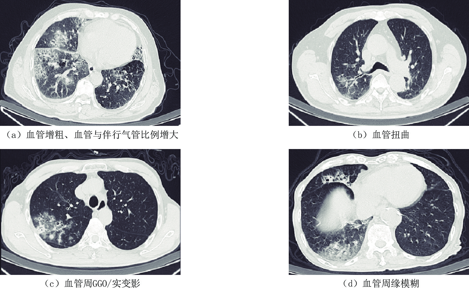
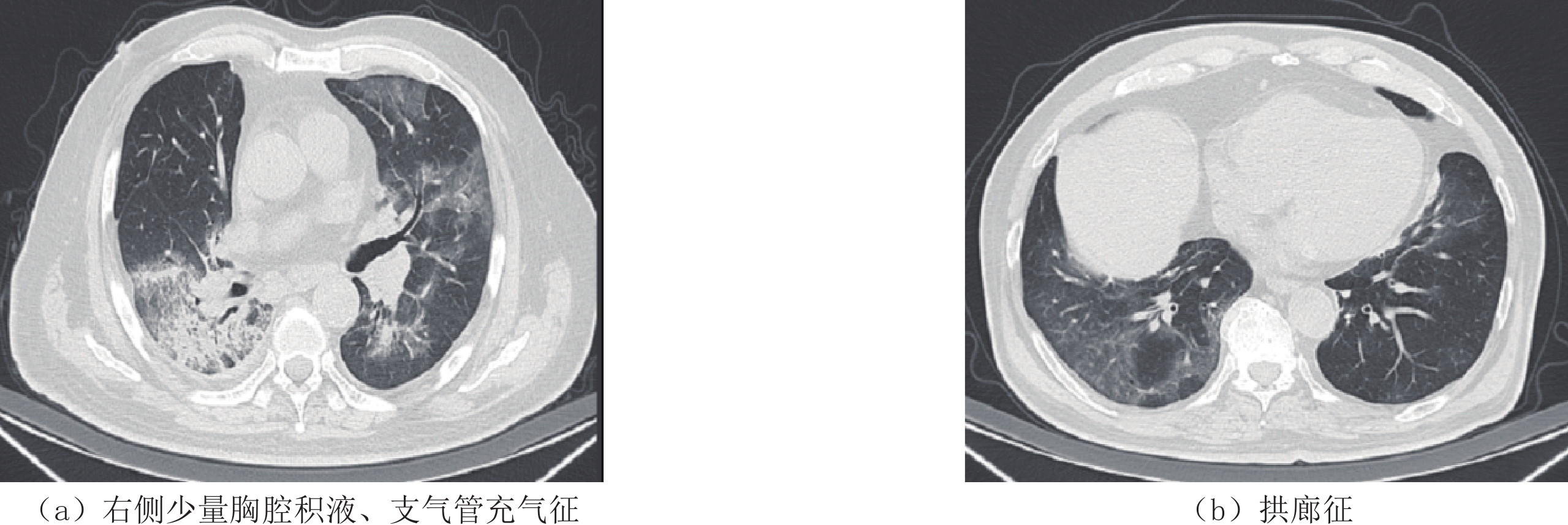
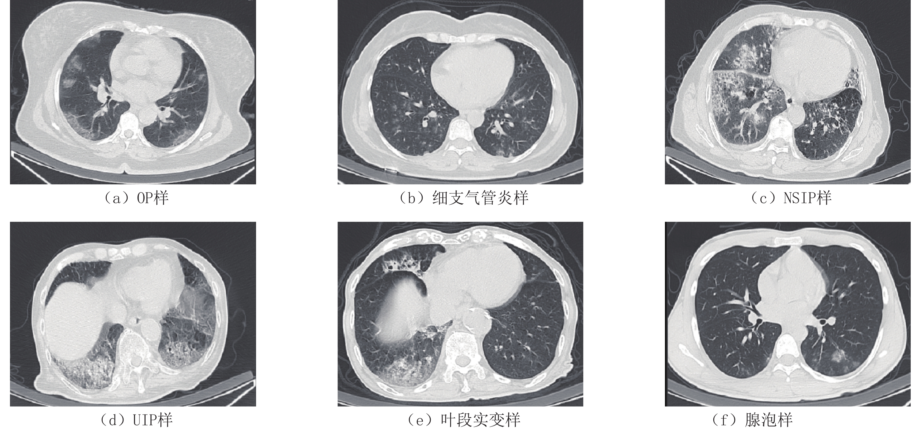
(半定量分析)/例(%)≤10 6(20.7) 8(18.1) 0.071 0.79 ≤30 18(62.1) 16(36.4) 4.642 0.03 ≤50 4(13.8) 9(20.5) 0.530 0.47 >50(白肺) 1(3.4) 11(25.0) — 0.02 病变形态/例(%) 小结节 8(27.6) 7(15.9) 1.460 0.23 斑片状 25(86.2) 33(75.0) 1.345 0.25 大片状 14(48.3) 36(81.8) 5.670 0.02 束带状 4(13.8) 6(13.6) — 1.00 混合型 16(55.2) 30(68.2) 1.269 0.26 病变密度/例(%) GGO 26(89.7) 34(77.3) 1.831 0.18 实变 12(41.4) 20(45.5) 0.118 0.73 网格影 11(37.9) 21(47.7) 0.681 0.41 蜂窝影 0(0.0) 3(6.8) — 0.27 病变优势类型/例(%) OP样 13(44.8) 13(29.5) 1.780 0.18 细支气管炎样 10(34.5) 15(34.1) 0.001 0.97 NSIP样 0(0.0) 5(11.4) — 0.15 UIP样 0(0.0) 3(6.8) — 0.27 叶段实变样 2(6.9) 8(18.2) — 0.30 腺泡样 4(13.8) 0(0.0) — 0.02 病变边缘/例(%) 模糊 25(86.2) 41(93.2) — 0.43 清楚 4(13.8) 3(6.8) — 0.43 不规则 17(58.6) 34(77.3) 2.888 0.09 血管异常/例(%) 血管增粗 28(96.6) 44(100.0) — 0.40 血管扭曲 2(6.9) 16(36.4) 8.170 0.00 血管周GGO/实变 29(100.0) 44(100.0) — — 血管与伴行气管
比例增大17(58.6) 20(45.5) 1.212 0.27 血管周缘模糊 23(79.3) 31(70.5) 7.290 0.01 其他特殊征象/例(%) 胸腔积液 0(0.0) 4(9.1) — 0.15 晕征 14(48.3) 22(50.0) 0.021 0.89 反晕征 5(17.2) 8(18.2) 0.011 0.92 拱廊征 5(17.2) 10(22.7) 0.322 0.57 铺路石征 11(37.9) 18(40.9) 0.065 0.80 胸膜下线 8(27.6) 21(47.7) 2.961 0.09 支气管充气征 9(31.0) 16(36.4) 0.220 0.64 树芽征 9(31.0) 1(2.3) — 0.00 粗大纤维索条 9(31.0) 27(61.4) 6.433 0.01
下载: 导出CSV
-
[1] WIERSINGA W J, RHODES A, CHENG A C, et al. Pathophysiology, transmission, diagnosis, and treatment of coronavirus disease 2019 (COVID-19): A review[J]. The Journal of the American Medical Association, 2020, 324(8): 782−793. doi: 10.1001/jama.2020.12839
[2] MACHNICKI S, PATEL D, SINGH A, et al. The usefulness of chest CT imaging in patients with suspected or diagnosed COVID-19: A review of literature[J]. Chest, 2021, 160(2): 652−670. doi: 10.1016/j.chest.2021.04.004
[3] 严祥虎, 包雨微, 朱桐, 等. COVID-19肺炎: 胸部病灶在恢复期的CT影像演变特征[J]. 放射学实践, 2020,35(4): 428−432. YAN X H, BAO Y W, ZHU T, et al. Evolution characteristics of thoracic lesions on CT of COVID-19 in recovery stage[J]. Radiologic Practic, 2020, 35(4): 428−432. (in Chinese).
[4] 龚晓明, 李航, 宋璐, 等. 新型冠状病毒肺炎(COVID-19) CT表现初步探讨[J]. 放射学实践, 2020,35(3): 261−265. doi: 10.13609/j.cnki.1000-0313.2020.03.003 GONG X M, LI H, SONG L, et al. Preliminary study on CT characteristics of corona virus disease 2019[J]. Radiologic Practic, 2020, 35(3): 261−265. (in Chinese). doi: 10.13609/j.cnki.1000-0313.2020.03.003
[5] 张贺诚, 褚岩, 刘晶, 等. 新型冠状病毒肺炎COVID-19的临床特征与CT表现[J]. CT理论与应用研究, 2020,29(5): 559−565. DOI: 10.15953/j.1004-4140.2020.29.05.06. ZHANG H C, CHU Y, LIU J, et al. The clinical features and CT manifestations of the novel coronavirus pneumonia COVID-19[J]. CT Theory and Applications, 2020, 29(5): 559−565. DOI: 10.15953/j.1004-4140.2020.29.05.06. (in Chinese).
[6] SOLOMON J J, HEYMAN B, KO J P, et al. CT of post-acute lung complications of COVID-19[J]. Radiology, 2021, 301(2): E383-E395.
[7] CHAMS N, CHAMS S, BADRAN R, et al. COVID-19: A multidisciplinary review[J]. Front in Public Health, 2020, 8: 383.
[8] ZHANG H, PENNINGER J M, LI Y, et al. Angiotensin-converting enzyme 2 (ACE2) as a SARS-CoV-2 receptor: Molecular mechanisms and potential therapeutic target[J]. Intensive Care Medicine, 2020, 46(4): 586−590. doi: 10.1007/s00134-020-05985-9
[9] SHUKLA A K, BANERJEE M. Angiotensin-converting-enzyme 2 and renin-angiotensin system inhibitors in COVID-19: An update[J]. High Blood Pressure & Cardiovascular Prevention, 2021, 28(2): 129−139.
[10] 卢虎, 郑义山. 新型冠状病毒肺炎与肾素-血管紧张素系统相关研究进展[J]. 中国临床研究, 2022,35(6): 840−843. LU H, ZHENG Y S. Novel coronavirus disease-2019 and renin-angiotensin system[J]. Chinese Journal of Clinical Research, 2022, 35(6): 840−843. (in Chinese).
[11] 孙莹, 李玲, 刘晓燕, 等. 早期新型冠状病毒肺炎的胸部薄层平扫CT表现特征[J]. CT理论与应用研究, 2023,32(1): 131−138. DOI: 10.15953/j.ctta.2023.006. SUN Y, LI L, LIU X Y, et al. Imaging features of early COVID-19 on chest thin-slice non-enhanced CT[J]. CT Theory and Applications, 2023, 32(1): 131−138. DOI: 10.15953/j.ctta.2023.006. (in Chinese).
[12] 代瑜, 褚志刚. COVID-19胸部CT表现及可能的病理基础[J]. 西南医科大学学报, 2020,43(6): 644−647. DAI Y, CHU Z G. Chest CT findings and possible pathological basis of coronavirus disease 2019[J]. Journal of Southwest Medical University, 2020, 43(6): 644−647. (in Chinese).
[13] 郭颖韵, 张亮, 李瑞雪, 等. 新型冠状病毒肺炎合并胸腔积液及心包积液10例临床特征[J]. 武汉大学学报(医学版), 2021,42(6): 878−883. GUO Y Y, ZHANG L, LI R X, et al. Clinical characteristics of COVID-19 complicated with pleural and pericardial effusion in 10 patients[J]. Medical Journal of Wuhan University, 2021, 42(6): 878−883. (in Chinese).
[14] 刘建, 袁怀平, 焦以庆, 等. 新型冠状病毒肺炎的影像学特征及与常见病毒性肺炎鉴别诊断的研究进展[J]. 心肺血管病杂志, 2020,39(6): 629−634. doi: 10.3969/j.issn.1007-5062.2020.06.001 LIU J, YUAN H P, JIAO Y Q, et al. COVID-19 and other viral pneumonia radiological patterns and differential diagnosis[J]. Journal of Cardiovascular and Pulmonary Diseases, 2020, 39(6): 629−634. (in Chinese). doi: 10.3969/j.issn.1007-5062.2020.06.001
[15] MINAULT Q, KAROL A, VEILLON F, et al. Tree-in-bud sign[J]. Abdominal Radiology, 2018, 43(11): 3188−3189. doi: 10.1007/s00261-018-1562-8
[16] RUARO B, SALTON F, BRAGA L, et al. The history and mystery of alveolar epithelial type II cells: Focus on their physiologic and pathologic role in lung[J]. International Journal of Molecular Sciences, 2021, 22(5): 2566. doi: 10.3390/ijms22052566
[17] PARIMON T, YAO C, STRIPP B R, et al. Alveolar epithelial type II cells as drivers of lung fibrosis in idiopathic pulmonary fibrosis[J]. International Journal of Molecular Sciences, 2020, 21(7): 2269. doi: 10.3390/ijms21072269
[18] 宋兰, 宋伟, 隋昕, 等. 北京协和医院普通型新型冠状病毒肺炎患者的初诊临床特征及CT影像学表现[J]. 中国医学科学院学报, 2020,42(3): 376−382. SONG L, SONG W, SUI X, et al. Preliminary study on clinical features and CT findings of common-type coronavirus disease 2019 patients in peking union medical college hospital[J]. Acta Academiae Medicinae Sinicae, 2020, 42(3): 376−382. (in Chinese).
[19] 张岩, 南成睿, 刘海霞, 等. 新型冠状病毒肺炎CT征象再讨论[J]. 临床荟萃, 2020,35(2): 106−112. doi: 10.3969/j.issn.1004-583X.2020.02.002 ZHANG Y, NAN C R, LIU H X, et al. Re-discussion about CT signs of novel coronavirus pneumonia[J]. Clinical Focus, 2020, 35(2): 106−112. (in Chinese). doi: 10.3969/j.issn.1004-583X.2020.02.002
[20] 史河水, 韩小雨, 樊艳青, 等. 新型冠状病毒(2019-nCoV)感染的肺炎临床特征及影像学表现[J]. 临床放射学杂志, 2020,39(1): 8−11. doi: 10.13437/j.cnki.jcr.20200206.002 SHI H S, HAN X Y, FAN Y Q, et al. Radiologic features of patients with 2019-nCoV infection[J]. Journal of Clinical Radiology, 2020, 39(1): 8−11. (in Chinese). doi: 10.13437/j.cnki.jcr.20200206.002
[21] YE Z, ZHANG Y, WANG Y, et al. Chest CT manifestations of new coronavirus disease 2019 (COVID-19): A pictorial review[J]. European Radiology, 2020, 30(8): 4381−4389. doi: 10.1007/s00330-020-06801-0
-
期刊类型引用(2)
1. 高子晴,卢吴柱,叶大林,林宇红,陈晓波. 床旁超声无创性评估新型冠状病毒感染中型患者肺部病变的临床研究. 中国中西医结合影像学杂志. 2023(04): 428-431 .  百度学术
百度学术
2. 林军,李焕兴,罗槑,陈竹,吴桂辉,曾义岚. 新型冠状病毒肺炎患者肺部超声特征. 中华实验和临床感染病杂志(电子版). 2021(01): 37-45 . 百度学术
其他类型引用(0)
计量
- 文章访问数: 133
- HTML全文浏览量: 69
- PDF下载量: 14
- 被引次数: 2


