
High-resolution Computed Tomography (HRCT) Characteristics of Coronavirus Disease 2019 (COVID-19) in Patients with Diabetes
-
摘要: 目的:探讨糖尿病患者肺部新型冠状病毒感染(COVID-19)HRCT特点。材料与方法:收集2022年12月14日至2023年1月10日确诊COVID-19且胸部CT表现异常的患者584例,男359例、女225例,年龄范围60~99岁,平均年龄(76±9)岁。其中合并糖尿病225例,非糖尿病359例;比较糖尿病患者COVID-19胸部HRCT与非糖尿病患者COVID-19胸部HRCT表现不同;定义发病与CT检查时间间隔<7d为急性期,363例入组患者,分析急性期糖尿病组与非糖尿病组新型冠状病毒肺炎(COVID-19)HRCT特点。结果:糖尿病患者COVID-19胸部感染与非糖尿病患者COVID-19胸部感染两组肺内病变在发病部位、分布、形态及伴随征象差异无统计学意义。两组病变在密度(细网格、病变密度不均匀)及病变边缘(病变边缘模糊)差异有统计学意义。无糖尿病组的肺部影像网格、不均匀和模糊征象显著高于有糖尿病组。其中细网格影:糖尿病组54例(24%),非糖尿病组127例(35.38%);密度不均匀:糖尿病组181例(80.44%),非糖尿病组313例(87.19%);边缘模糊:糖尿病组205例(91.11%),非糖尿病组344(95.82%)。急性期糖尿病组患者肺内网格影明显少于非糖尿病组患者,糖尿病组35例(24.65%),非糖尿病组82例(37.10%),差异有统计学意义。结论:糖尿病患者肺部新型冠状病毒感染(COVID-19)胸部HRCT病变渗出为主、密度均匀、边缘清晰,较非糖尿病组间质改变不明显。Abstract: Objective: To explore the characteristics of high-resolution computed tomography (HRCT) in diabetes complicated with coronavirus disease 2019 (COVID-19)-associated pneumonia. Materials and Methods: This study included 584 patients (359 males and 225 females), aged between 60~99 years old (mean, (76±9) years), with positive chest computed tomography (CT) findings and diagnosed with COVID-19 in our hospital from December 14, 2022, to January 10, 2023. Of these, 225 patients were diabetic and 359 were non-diabetic. The features of the chest HRCT from patients with diabetes mellitus complicated with COVID-19 and those without diabetes mellitus complicated with COVID-19 were compared. Moreover, 363 patients in the acute stage of COVID-19 (defined as the time interval between onset and CT examination <7 days) were selected for subgroup analysis, and the HRCT characteristics of COVID-19 between the diabetes group and the non-diabetic group in the acute stage. Results: The location, distribution, morphology, and concomitant signs of pulmonary lesions between the two groups of patients with COVID-19 did not differ significantly. Conversely, statistically significant differences in density (fine mesh, uneven density) and lesion margin (fuzzy lesion margin) were detected. In particular, the grid, uneven, and fuzzy signs on lung imaging were significantly higher in the non-diabetic group than that in the diabetic group. Additionally, 54 patients (24%) in the diabetic group and 127 patients (35.38%) in the non-diabetic group demonstrated fine mesh shadows. There were 181 patients (80.44%) in the diabetic group and 313 patients (87.19%) in the non-diabetic group with uneven density. Furthermore, 205 patients (91.11%) in the diabetic group and 344 patients (95.82%) in the non-diabetic group had blurred edges. There was significantly less pulmonary grid shadowing in the acute subgroup with diabetes (35, 24.65%) than in the acute subgroup without diabetes (82, 37.10%). Conclusion: The features of chest HRCT in patients with diabetes mellitus and COVID-19 are mainly exudation, uniform density, and a clear edge, while the interstitial changes are not obvious compared with patients in the non-diabetic group.
-
Keywords:
- CT /
- high resolution /
- coronavirus disease 2019 /
- diabetes mellitus
-
新型冠状病毒感染(COVID-19)具有较强传染性[1],根据《新型冠状病毒感染诊疗方案(试行第十版)》,将有心血管疾病(含高血压)、慢性肺疾病病、糖尿病、慢性肝病、肾脏疾病、肿瘤等基础病者定义为重型/危重型高危人群[2]。
本文回顾分析柳州市柳铁中心医院收治COVID-19患者584例,其中糖尿病患者肺部COVID-19 225例;无糖尿病患者感染COVID-19 359例,比较两组COVID-19胸部高分辨CT表现特点;定义发病与CT检查间隔7 d为急性期,分析糖尿病患者合并COVID-19感染急性期肺部影像特点。提高糖尿病者感染COVID-19影像学认识,进一步为临床诊断提供依据并改善预后。
1. 材料与方法
1.1 研究人群
收集2022年12月14日至2023年1月10日期间在柳州市柳铁中心医院以上呼吸道感染就医患者2679例,其中胸部CT表现阳性且抗原或核酸检测阳性的患者1099例,排除图像伪影较重者;无空腹血糖或糖化血红蛋白者;最终纳入584例患者,其中男性359例,女性225例,年龄范围60~99岁,平均年龄(76±9)岁;无糖尿病患者359例;糖尿病患者225例,糖尿病病史不等,本组病例中以病史10年以上者多见,糖尿病病史10年以上102人;225例患者住院期间糖化血红蛋白HbA1c>6.5%。
本研究经柳州市柳铁中心医院伦理委员会批准,同意开展本项研究(意见号KY2023-056-01),且本研究不需要患者知情。
1.2 CT扫描技术
采用西门子drive扫描仪,患者仰卧位,头先进或足先进,吸气后屏气,扫描范围从肺尖到膈顶。扫描参数:管电压120 kV,管电流80~120 mA,层间距0.625 mm,层厚1 mm。图像采用高分辨算法重建。
1.3 影像分析
有两名主治及以上放射科诊断医师独立完成,并由1名主任医师审核,在PACS工作站上分别选择肺窗(窗宽和窗位为1600和 -600),纵隔窗(窗宽和窗位为350和45)观察并分析图像。
具体指标包括:①病变分布:单叶、单肺和双肺;②病变部位:周围(即胸膜下)、中央(沿支气管血管束)及混合;③病变形态:结节(1 cm以内)、斑片(3 cm)、大片(大于3 cm);④病变密度:磨玻璃样、网格状、实变、混合型、病变密度均匀、不均匀;⑤病变边缘:模糊、清晰;⑥伴随病变:膜增厚、胸腔积液。
1.4 统计学分析
采用SPSS 21.0软件,根据患者有无糖尿病将患者分为两组,比较两组患者相关的HRCT表现特征。定义发病与CT检查时间间隔<7 d为急性期,分析急性期糖尿病合并COVID-19与非糖尿病合并COVID-19两组患者HRCT表现特征。
组间计数资料统计采用分类变量的卡方检验,定量数据进行独立样本t检验,P<0.05表示差异有统计学意义。
2. 结果
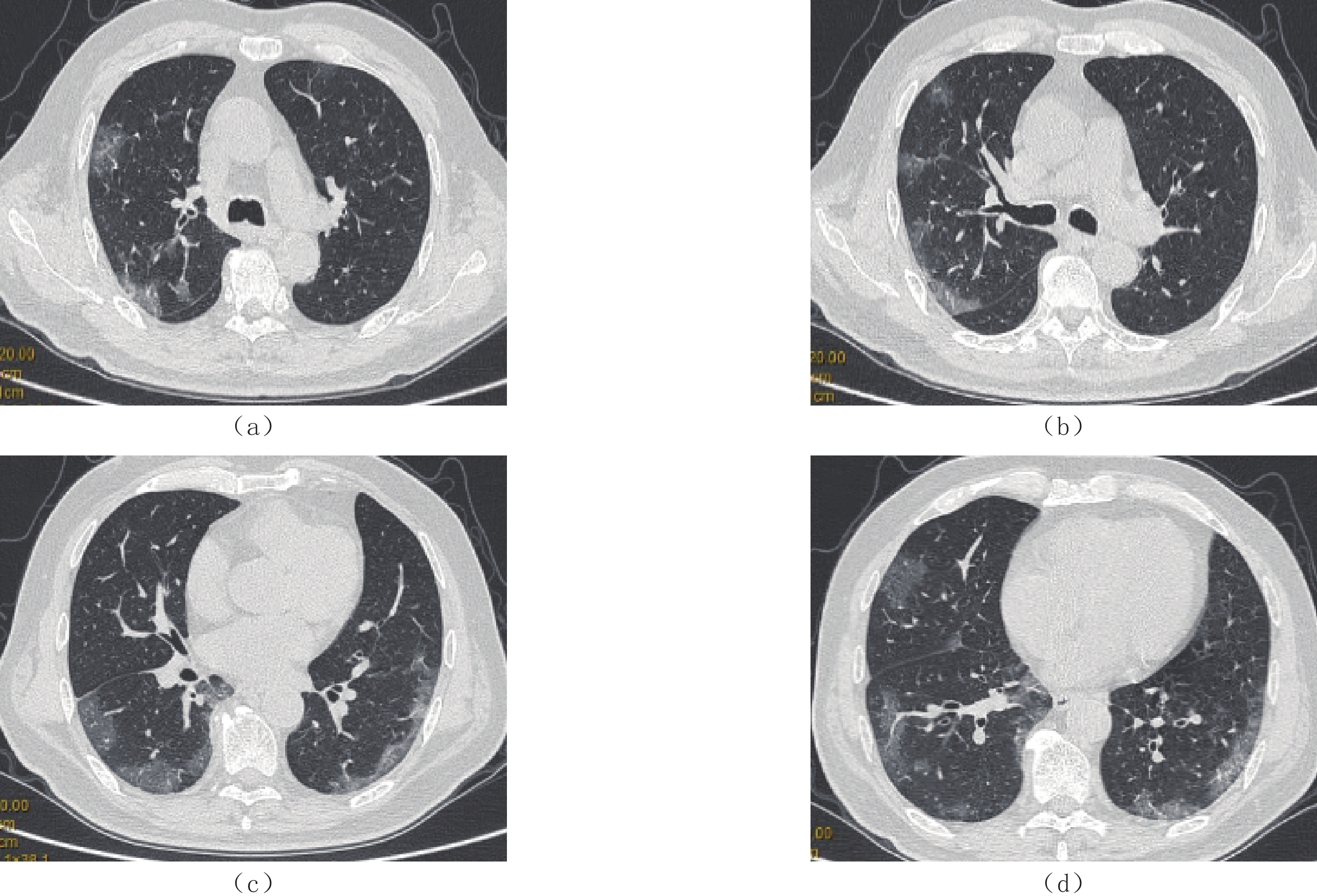
糖尿病患者感染COVID-19与非糖尿病患者感染COVID-19两组患者肺部病变密度(网格、密度不均匀)、边缘(病灶边缘模糊)比较差异均有统计学意义,糖尿病组肺内病灶实变多见,且病灶密度均匀且边缘清晰(图1和图2)。两组病变在发病部位、分布、形态及伴随征象差异无统计学意义;急性期肺内病变在糖尿病患者肺部COVID-19与非糖尿病患者肺部COVID-19两组患者肺部HRCT表现比较,网格影在急性期非糖尿病组患者中更多见。两组具体影像学征象对照及比较结果详见表1、表2和表3。
![]() 图 1 男性,63岁,发热3 d,无糖尿病,HRCT显示双肺外周胸膜下GGO,密度不均匀,部分病灶边缘模糊Figure 1. A 63-year-old man, a patient without diabetes mellitus, presented with a fever for 3 days, HRCT demonstrates peripheral subpleural ground glass opacities (GGO) in both lungs, with some lesions showing uneven density and blurred edges
图 1 男性,63岁,发热3 d,无糖尿病,HRCT显示双肺外周胸膜下GGO,密度不均匀,部分病灶边缘模糊Figure 1. A 63-year-old man, a patient without diabetes mellitus, presented with a fever for 3 days, HRCT demonstrates peripheral subpleural ground glass opacities (GGO) in both lungs, with some lesions showing uneven density and blurred edges![]() 图 2 女,65岁,糖尿病病史9年,餐后血糖控制不佳,咳嗽3 d肺内多发实变样,病变边缘清晰Figure 2. A 65-year-old female with a history of diabetes for 9 years and elevated blood sugar after meals, presented with a cough for 3 days. HRCT shows multiple lung lesions with clear lesion edges表 1 有无糖尿病两组患者的HRCT表现特征一览表Table 1. HRCT features in patients with and without diabetes mellitus
图 2 女,65岁,糖尿病病史9年,餐后血糖控制不佳,咳嗽3 d肺内多发实变样,病变边缘清晰Figure 2. A 65-year-old female with a history of diabetes for 9 years and elevated blood sugar after meals, presented with a cough for 3 days. HRCT shows multiple lung lesions with clear lesion edges表 1 有无糖尿病两组患者的HRCT表现特征一览表Table 1. HRCT features in patients with and without diabetes mellitus项目 组别 统计检验 无糖尿病/例(%) 有糖尿病/例(%) $\chi^2 $ P 病变数量 多发 359(100.00) 224(99.50) 1.590 0.206 累及部位 单叶 3(0.84) 0(0.00) 1.890 0.169 单肺 7(1.95) 2(0.89) 1.026 0.311 双肺 351(97.77) 220(97.78) 0.000 0.996 病变分布 周围(胸膜下) 114(31.75) 83(36.89) 1.631 0.202 中央(血管周) 6(1.67) 6(2.67) 0.681 0.409 混合性 239(66.57) 137(60.89) 1.949 0.163 病变形态 结节(1 cm) 159(44.29) 109(48.44) 0.962 0.327 斑片状(3 cm) 275(76.60) 178(79.11) 0.501 0.479 大片状(>3 cm) 240(66.85) 149(66.22) 0.025 0.875 病变密度 GGO 314(87.47) 189(84.00) 1.390 0.238 实变 41(11.42) 37(16.44) 3.017 0.082 网格影 127(35.38) 54(24.00) 8.369 0.004** 不均匀 313(87.19) 181(80.44) 4.823 0.028** 均匀 77(21.45) 59(26.22) 1.764 0.184 病变边缘 模糊 344(95.82) 205(91.11) 5.448 0.020* 清晰 19(5.29) 18(8.00) 1.709 0.191 伴随病变 血管增粗 26(7.24) 25(11.11) 2.597 0.107 胸膜增厚 203(56.55) 130(57.78) 0.086 0.770 胸水形成 54(15.04) 27(12.00) 1.071 0.301 注:*-P<0.05,**-P<0.01。 表 2 COVID-19急性期糖尿病患者与非糖尿病患者的临床信息Table 2. Clinical information on patients with and without diabetes in the acute phase of COVID-19项目 组别 统计检验 无糖尿病(n=221) 有糖尿病(n=142) t P 发病时间/d 4.74±2.12 4.45±2.28 1.211 0.227 年龄 78.00±9.37 75.85±8.63 2.203 0.028* 注:急性期定义为发病时间<7 d。*-P<0.05。 表 3 COVID-19急性期糖尿病患者与非糖尿病患者的HRCT特征一览表Table 3. HRCT characteristics in patients with and without diabetes in the acute phase of COVID-19项目 特征 组别 统计检验 无糖尿病(n=221)
/
例(%)有糖尿病(n=142)
/
例(%)$\chi^2 $ P 病变数量 多发 221(100.00) 141(99.50) 1.590 0.206 累及部位 单肺 6(2.71) 2(1.41) 0.685 0.408 双肺 214(96.83) 138(97.18) 0.036 0.849 病变分布 周围(胸膜下) 65(29.41) 49(34.51) 1.042 0.307 中央(血管周) 5(2.26) 5(3.52) 0.511 0.475 混合性 153(69.23) 90(63.38) 1.377 0.248 病变形态 结节/树丫(1 cm) 86(38.91) 63(44.37) 1.062 0.303 斑片状(3 cm) 161(72.85) 110(77.46) 0.973 0.324 大片状(>3 cm) 157(71.04) 100(70.42) 0.016 0.899 病变密度 GGO 190(85.97) 121(85.21) 0.041 0.840 实变 30(13.57) 28(19.72) 2.431 0.119 网格影 82(37.10) 35(24.65) 6.141 0.013* 不均匀 188(85.07) 117(82.39) 0.460 0.497 均匀 49(22.17) 38(26.76) 0.999 0.318 病变边缘 模糊 212(95.93) 130(91.55) 3.041 0.081 清晰 11(4.98) 11(7.75) 1.164 0.281 伴随病变 血管增粗 17(7.69) 18(12.68) 2.465 0.116 胸膜增厚 129(58.37) 85(59.86) 0.079 0.779 胸水形成 35(15.84) 20(14.08) 0.207 0.649 注:*-P<0.05。 3. 讨论
COVID-19是一种基因组结构不同于其他呼吸道病毒且侵袭性和传播性较强的β属特殊毒株,通过S蛋白与人血管紧张素转化酶-2(ACE2)互相作用感染人呼吸道黏膜上皮细胞、Ⅱ型肺泡上皮细胞和肺间质以及微血管血栓形成和多系统脏器受累等改变[3-4]。人群普遍易感,重型及危重型多见于合并基础病者如糖尿病。COVID-19感染主要依靠病毒核酸检测确诊,核酸检测存在时间长和假阴性,HRCT有助于提高COVID-19的检出率和诊断准确率[5],部分患者核酸阴性影像却具有典型病毒性肺炎征象,国家卫生健康委员会《新型冠状病毒感染的肺炎诊疗方案(试行第五版)》中[6],将CT表现纳入临床诊断依据。
本研究分析了584例新型冠状病毒感染(COVID-19)HRCT表现阳性的患者,统计结果显示多数影像学征象与文献一致[3-4,7-16]。225例糖尿病患者新型冠状病毒感染后双肺HRCT表现在发病部位、分布、形态及伴随征象与常规人群感染COVID-19无明显差异,均表现为双肺多发、周围(胸膜下)分布为主的结节、斑片及大片磨玻璃及实变影;病变密度均匀与否及病变边缘改变差异有统计学意义;发病与CT检查间隔小于7 d分组,糖尿病组患者COVID-19和非糖尿病组患者COVID-19之间,病变密度网格影改变差异有统计学意义。病变的HRCT征象如密度、形态和边缘因其病理特征和病变程度的不同多呈现不均匀磨玻璃、实变影及网格影,边缘多表现为模糊。
本研究结果显示糖尿病组患者COVID-19肺炎早期病变即出现密度均匀和病变边缘清晰的表现,文献报道较少。尸体解剖证实COVID-19主要是引起深部气道、间质和肺泡损伤为特征的炎性反应[17],糖尿病患者COVID-19感染肺部病变HRCT显示网格影少于非糖尿病组,说明糖尿病患者感染COVID-19时肺泡渗出为主,间质受累少见,有研究显示小叶内间隔增厚是COVID-19肺炎的独立预测因子之一[18-19],而糖尿病合并COVID-19感染,肺内网格影少见,这可能与糖尿病患者自身免疫有关。渗出为主是否是糖尿病患者易发生重症原因,需要进一步研究证实。
总之,糖尿病患者合并COVID-19肺炎,HRCT表现双肺多发,周围分布为主,渗出为主,网格影少见,病变密度均匀、病变边缘相对清晰。合并或者不合并胸膜改变。基础病是COVID-19感染后发生重症的主要原因之一,熟悉糖尿病患者合并COVID-19肺炎HRCT征象,为临床治疗及评估预后提供依据。
本研究的局限在性,两组病例未纳入临床指标及氧饱和度与患者影像改变进行相关性分析;糖尿病组患者病史长短及血糖控制情况感染后机体反应是否存在差异;此外糖尿病往往合并心脑血管病变,并发症有无是否与肺内病灶改变及预后的研究尚需进一步研究。
-
![]()
图 1 男性,63岁,发热3 d,无糖尿病,HRCT显示双肺外周胸膜下GGO,密度不均匀,部分病灶边缘模糊
Figure 1. A 63-year-old man, a patient without diabetes mellitus, presented with a fever for 3 days, HRCT demonstrates peripheral subpleural ground glass opacities (GGO) in both lungs, with some lesions showing uneven density and blurred edges
![]()
图 2 女,65岁,糖尿病病史9年,餐后血糖控制不佳,咳嗽3 d肺内多发实变样,病变边缘清晰
Figure 2. A 65-year-old female with a history of diabetes for 9 years and elevated blood sugar after meals, presented with a cough for 3 days. HRCT shows multiple lung lesions with clear lesion edges
表 1 有无糖尿病两组患者的HRCT表现特征一览表
Table 1 HRCT features in patients with and without diabetes mellitus
项目 组别 统计检验 无糖尿病/例(%) 有糖尿病/例(%) $\chi^2 $ P 病变数量 多发 359(100.00) 224(99.50) 1.590 0.206 累及部位 单叶 3(0.84) 0(0.00) 1.890 0.169 单肺 7(1.95) 2(0.89) 1.026 0.311 双肺 351(97.77) 220(97.78) 0.000 0.996 病变分布 周围(胸膜下) 114(31.75) 83(36.89) 1.631 0.202 中央(血管周) 6(1.67) 6(2.67) 0.681 0.409 混合性 239(66.57) 137(60.89) 1.949 0.163 病变形态 结节(1 cm) 159(44.29) 109(48.44) 0.962 0.327 斑片状(3 cm) 275(76.60) 178(79.11) 0.501 0.479 大片状(>3 cm) 240(66.85) 149(66.22) 0.025 0.875 病变密度 GGO 314(87.47) 189(84.00) 1.390 0.238 实变 41(11.42) 37(16.44) 3.017 0.082 网格影 127(35.38) 54(24.00) 8.369 0.004** 不均匀 313(87.19) 181(80.44) 4.823 0.028** 均匀 77(21.45) 59(26.22) 1.764 0.184 病变边缘 模糊 344(95.82) 205(91.11) 5.448 0.020* 清晰 19(5.29) 18(8.00) 1.709 0.191 伴随病变 血管增粗 26(7.24) 25(11.11) 2.597 0.107 胸膜增厚 203(56.55) 130(57.78) 0.086 0.770 胸水形成 54(15.04) 27(12.00) 1.071 0.301 注:*-P<0.05,**-P<0.01。  下载: 导出CSV
表 2 COVID-19急性期糖尿病患者与非糖尿病患者的临床信息
Table 2 Clinical information on patients with and without diabetes in the acute phase of COVID-19
项目 组别 统计检验 无糖尿病(n=221) 有糖尿病(n=142) t P 发病时间/d 4.74±2.12 4.45±2.28 1.211 0.227 年龄 78.00±9.37 75.85±8.63 2.203 0.028* 注:急性期定义为发病时间<7 d。*-P<0.05。
下载: 导出CSV
表 3 COVID-19急性期糖尿病患者与非糖尿病患者的HRCT特征一览表
Table 3 HRCT characteristics in patients with and without diabetes in the acute phase of COVID-19
项目 特征 组别 统计检验 无糖尿病(n=221)
/
例(%)有糖尿病(n=142)
/
例(%)$\chi^2 $ P 病变数量 多发 221(100.00) 141(99.50) 1.590 0.206 累及部位 单肺 6(2.71) 2(1.41) 0.685 0.408 双肺 214(96.83) 138(97.18) 0.036 0.849 病变分布 周围(胸膜下) 65(29.41) 49(34.51) 1.042 0.307 中央(血管周) 5(2.26) 5(3.52) 0.511 0.475 混合性 153(69.23) 90(63.38) 1.377 0.248 病变形态 结节/树丫(1 cm) 86(38.91) 63(44.37) 1.062 0.303 斑片状(3 cm) 161(72.85) 110(77.46) 0.973 0.324 大片状(>3 cm) 157(71.04) 100(70.42) 0.016 0.899 病变密度 GGO 190(85.97) 121(85.21) 0.041 0.840 实变 30(13.57) 28(19.72) 2.431 0.119 网格影 82(37.10) 35(24.65) 6.141 0.013* 不均匀 188(85.07) 117(82.39) 0.460 0.497 均匀 49(22.17) 38(26.76) 0.999 0.318 病变边缘 模糊 212(95.93) 130(91.55) 3.041 0.081 清晰 11(4.98) 11(7.75) 1.164 0.281 伴随病变 血管增粗 17(7.69) 18(12.68) 2.465 0.116 胸膜增厚 129(58.37) 85(59.86) 0.079 0.779 胸水形成 35(15.84) 20(14.08) 0.207 0.649 注:*-P<0.05。
下载: 导出CSV
-
[1] ZHU N, ZHANG D Y, WANG W L, et al. A novel coronavirus from patients with pneumonia in China, 2019[J]. The New England Journal of Medicine, 2020, 382(8): 727−733. doi: 10.1056/NEJMoa2001017
[2] 国家卫生健康委办公厅, 国家中医药局综合司. 《新型冠状病毒感染诊疗方案(试行第十版)》[EB/OL].(2023-01-05)[2023-01-06]. http://www.nhc.gov.cn/ylyjs/pqt/202301/32de5b2ff9bf4eaa88e75bdf7223a65a/files/02ec13aadff048ffae227593a6363ee8.pdf. [3] WU R, GUAN W, GAO Z, et al. The arch bridge sign: A newly described CT feature of the coronavirus disease-19 (COVID-19) pneumonia[J]. Quantitative Imaging in Medicine And Surgery 2020, 10(7): 1551-1558.
[4] YOON S H, LEE J H, KIM B N. Chest CT findings in hospitalized patients with SARS-CoV-2: Delta versus Omicron variants[J]. Radiology, 2023, 306(1): 252−260. doi: 10.1148/radiol.220676
[5] 郑颖彦, 马昕, 王慧英, 等. 新型冠状病毒肺炎的薄层高分辨率计算机断层扫描征象[J]. 上海医学, 2020,43(5): 261−265. ZHENG Y Y, MA X, WANG H Y, et al. Thin high-resolution computed tomography findings of novel coronavirus pneumonia[J]. Shanghai Medical Science, 2020, 43(5): 261−265. (in Chinese).
[6] 国家卫生健康委办公厅, 国家中医药管理局办公室. 《新型冠状病毒感染的肺炎诊疗方案(试行第五版)》[EB/OL]. (2020-02-05) [2020-02-10]. http://bgs.satcm.gov.cn/zhengcewenjian/2020-02-06/12847.html. [7] LIU Y, ZHOU X, LIU X, et al. Systematic review and meta-analysis of the CT imaging characteristics of infectious pneumonia[J]. Annals of Palliative Medicine, 2021, 10(10): 10414−10424. doi: 10.21037/apm-21-2101
[8] TANG Y, LIAO H, WU Q, et al. Chest CT imaging characteristics and their evolution of 48 patients with COVID-19 in Hengyang, China[J]. American Journal of Translational Research, 2021, 13(9): 9983−9992.
[9] PAKDEMIRLI E, MANDALIA U, MONIB S. Characteristics of chest CT images in patients with COVID-19 pneumonia in London, UK[J]. Cureus, 2020, 12(9): e10289.
[10] MARCHIORI E, NOBRE L F, HOCHHEGGER B, et al. CT characteristics of COVID-19: Reversed halo sign or target sign?[J]. Diagnostic and Interventional Radiology, 2021, 27(2): 306−307. doi: 10.5152/dir.2020.20734
[11] HUANG C, WANG Y, LI X, et al. Clinical features of patients infected with 2019 novel coronavirus in Wuhan, China[J]. Lancet, 2020, 395(10223): 497−506. doi: 10.1016/S0140-6736(20)30183-5
[12] 黄益龙, 张振光, 李翔, 等. CT影像组学联合征象鉴别新型冠状病毒肺炎与其他病毒性肺炎的价值[J]. 中华放射学杂志, 2022,56(1): 36−42. doi: 10.3760/cma.j.cn112149-20201220-01318 HUANG Y L, ZHANG Z G, LI X, et al. Value of CT imaging combined signs in distinguishing novel coronavirus pneumonia from other viral pneumonia[J]. Chinese Journal of Radiology, 2022, 56(1): 36−42. (in Chinese). doi: 10.3760/cma.j.cn112149-20201220-01318
[13] 赵小二, 邓克学, 王朋. 不同阶段新型冠状病毒肺炎的CT影像演变分析[J]. 实用放射学杂志, 2021,37(8): 1254−1257. doi: 10.3969/j.issn.1002-1671.2021.08.008 ZHAO X E, DENG K X, WANG P. CT imaging evolution analysis of novel coronavirus pneumonia at different stages[J]. Journal of Practical Radiology, 2021, 37(8): 1254−1257. (in Chinese). doi: 10.3969/j.issn.1002-1671.2021.08.008
[14] 李声鸿, 曾献军, 鄢海蓝, 等. 新型冠状病毒肺炎薄层CT评价[J]. 实用放射学杂志, 37(7): 1074-1076, 1130. LI S H, ZENG X J, YAN H L, et al. Thin-slice CT evaluation of novel coronavirus pneumonia[J]. Journal of Practical Radiology, 37(7): 1074-1076, 1130. (in Chinese).
[15] 张庆, 熊浩, 彭婕, 等. 胸部CT对新型冠状病毒肺炎的诊断价值[J]. 中国医学影像学杂志, 2020, 28(12): 896-898. ZHANG Q, XIONG H, PENG J, et al. Diagnostic value of chest CT in the diagnosis of novel coronavirus pneumonia[J]. Chinese Journal of Medical Imaging, 2020, 28(12): 896-898. (in Chinese).
[16] 纪丙军, 齐庆梅, 王聪, 等. 新型冠状病毒肺炎与其他社区获得性肺炎不同病期的CT表现及动态分析[J]. 实用放射学杂志, 2021, 37(8): 1266-1270. JI B J, QI Q M, WANG C, et al. CT findings and dynamic analysis of novel coronavirus pneumonia and other community-acquired pneumonia at different stages[J]. Journal of Applied Radiology, 2021, 37(8): 1266-1270. (in Chinese).
[17] 刘茜, 王荣帅, 屈国强, 等. 新型冠状病毒肺炎死亡尸体系统解剖大体观察报告[J]. 法医学杂志, 2020,36(1): 1−3. DOI: 10.12116/j.issn.1004-5619.2020.01.00. LIU Q, WANG R S, QU G Q, et al. A report on the general observation of the systematic autopsy of the deceased from novel coronavirus pneumonia[J]. Journal of Forensic Medicine, 2020, 36(1): 1−3. DOI: 10.12116/j.issn.1004-5619.2020.01.00. (in Chinese).
[18] SHI H, HAN X, JIANG N, et al. Radiological findings from 81 patients with COVID-19 pneumonia in Wuhan, China: A descriptive study[J]. Lancet Infectious Diseases, 2020, 20(4): 425−434. DOI: 10.1016/S1473-3099(20)30086-4.
[19] TANG X, DU R H, WANG R, et al. Comparison of hospitalized patients with ARDS caused by COVID-19 and H1N1[J]. Chest, 2020, 158(1): 195−205. DOI: 10.1016/J.chest.2020.03.032.
-
期刊类型引用(5)
1. 蒋圣哲,陈强,罗琳. 肝脏DCE-MRI影像组学评估肝硬化患者肝脏储备功能的价值. 包头医学院学报. 2025(02): 52-57 .  百度学术
百度学术
2. 朱明,刘鹏,韩广,李强,徐传娜. 双源CT双能量定量参数分析在肝硬化诊断中的价值研究. 现代医用影像学. 2025(03): 454-458 . 百度学术
3. 陈凤,李艳,唐文勇,翁旭丹,吕敏丽,仲建全. 基于CT影像组学评估肝硬化患者肝脏储备功能的价值研究. 肝脏. 2024(06): 699-705 . 百度学术
4. 修智,邹智超,张颖琳,安鑫,向以四. 多层螺旋CT体积测量在肝硬化患者肝脏储备功能评估中的应用. 肝脏. 2024(10): 1235-1238 . 百度学术
5. 陈凤,李兵,张川,杨汉丰. 影像组学在肝脏非肿瘤性病变中的研究进展. 肝脏. 2023(12): 1504-1508 . 百度学术
其他类型引用(1)

计量
- 文章访问数: 155
- HTML全文浏览量: 54
- PDF下载量: 12
- 被引次数: 6


