
Characteristics and Evolution of Computed Tomography Images in Coronavirus Disease 2019 Pneumonia
-
摘要: 目的:分析不同时期新型冠状病毒感染的CT影像特点及演变规律。方法:回顾性分析2022年12月至2023年1月期间113例河北省人民医院确诊COVID-19患者不同时期的CT征象,观察其变化趋势。32例患者行1次CT检查,41例行CT检查2次,20例行CT检查3次,15例行CT检查4次,5例行CT检查5次。本组113例患者共行CT检查259次。其中早期(新冠感染 7 d以内)32例患者CT检查32次,进展期(新冠感染8~30 d)74例患者CT检查87次,转归期(新冠感染12~57 d)89例患者CT检查140次。分析患者每次CT特点。结果:早期CT表现为磨玻璃密度影26次,实变+磨玻璃密度影 6次。进展期磨玻璃密度影32次,实变+磨玻璃密度影 55次。转归期48次磨玻璃密度影,90次实变+磨玻璃密度影。112例患者为多叶多发病灶,其中1例在转归期为单叶多发病灶。早期胸膜下分布13次,支气管血管束周围+胸膜下分布 19次。进展期胸膜下分布24次,支气管血管束周围+胸膜下分布 63次。转归期胸膜下分布48次,2次完全吸收好转,支气管血管束周围+胸膜下分布 90次。早期磨玻璃密度影内小叶间隔增粗32次,病灶内血管增粗32次。进展期病灶内小叶间隔增粗85次,病灶内血管增粗87次。转归期小叶间隔增粗5次,病灶内血管增粗1次,48次伴条索影。早期1次支气管气充气征,进展期6次胸腔积液,6次支气管充气征,3次肺气肿,转归期2次支气管充气征,1次胸腔积液。结论:不同时期新冠肺炎患者的CT影像特点有所差别,了解其演变规律对指导临床治疗至关重要。Abstract: Objective: To analyze the computed tomography (CT) imaging features and evolution of different stages of coronavirus disease 2019 (COVID-19). Methods: A retrospective analysis was conducted on the CT images of 113 patients diagnosed with COVID-19 at Hebei Provincial People's Hospital between December 2022 and January 2023 to observe the trends of imaging changes. Results: All 113 patients were clinically diagnosed with COVID-19. Among these, 32, 41, 20, 15, and five patients underwent CT examination once, twice, three times, four times, and five times, respectively. A total of 259 CT examinations were performed in this group of 113 patients. Among them, 32 were early-stage (within 7 days of COVID-19 infection) examinations, 87 were progression-stage CT examinations, and 140 were recovery-stage CT examinations. Analysis of the imaging features of each CT examination of the patients was performed to identify the imaging features and evolution rules of COVID-19. Among the 32 examinations performed in 32patients with early-stage disease (within 7 days of COVID-19 infection), 26 cases showed ground-glass density shadows and six cases additionally showed solid nodules. In the progression stage (8 ~ 30 days after COVID-19 infection), among the 87 CT examinations in 74 patients, 62, 11, and one patient underwent examinations one, two, and three times, respectively. The 87 examinations revealed there 32 cases with ground-glass density shadows and 55 cases with additional solid nodules. In the recovery stage (12 ~ 57 days after COVID-19 infection), 89 patients underwent 140 CT examinations. Among these, 48, 32, eight, and one patient underwent CT examinations once, twice, three times, and four times, respectively. Among the 140 CT examinations, 48 cases showed ground-glass density shadows, while 90 cases additionally showed solid nodules. Moreover, 112 patients had multiple lesions in multiple lobes, with only one case having multiple lesions in a single lobe. Regarding the distributions, in the early stage, 13 cases had subpleural distributions and 19 cases had peribronchovascular and subpleural distributions. In the progression stage, 24 cases had subpleural distribution, and 63 cases had peribronchovascular and subpleural distributions. In the recovery stage, 48 cases had subpleural distribution, two cases showed complete absorption and improvement, and 90 cases had peribronchovascular and subpleural distribution, with 48 cases accompanied by reticular shadows. Regarding thickening of the interlobular septa and vessels within the lesions, in the early stage, 32 cases showed thickening of the interlobular septa and vessels within the lesions. In the progression stage, 85 cases showed thickening of the interlobular septa and 87 cases showed thickening of vessels within the lesions. In the recovery stage, five cases showed thickening of the interlobular septa, one case showed thickening of vessels within the lesions, and 48 cases were accompanied by linear shadows. Finally, in the early stage, one case showed bronchial gas inflation. In the progression stage, six cases showed pleural effusion, six cases showed bronchial inflation, and three cases showed pulmonary emphysema. In the recovery stage, two cases showed bronchial inflation and one case showed pleural effusion. Conclusion: The characteristics of CT images differed in patients with new coronary pneumonia at different times. Understanding this evolution is important to guide clinical treatment.
-
Keywords:
- CT /
- novel coronavirus pneumonia /
- image characteristics /
- changing trends
-
-
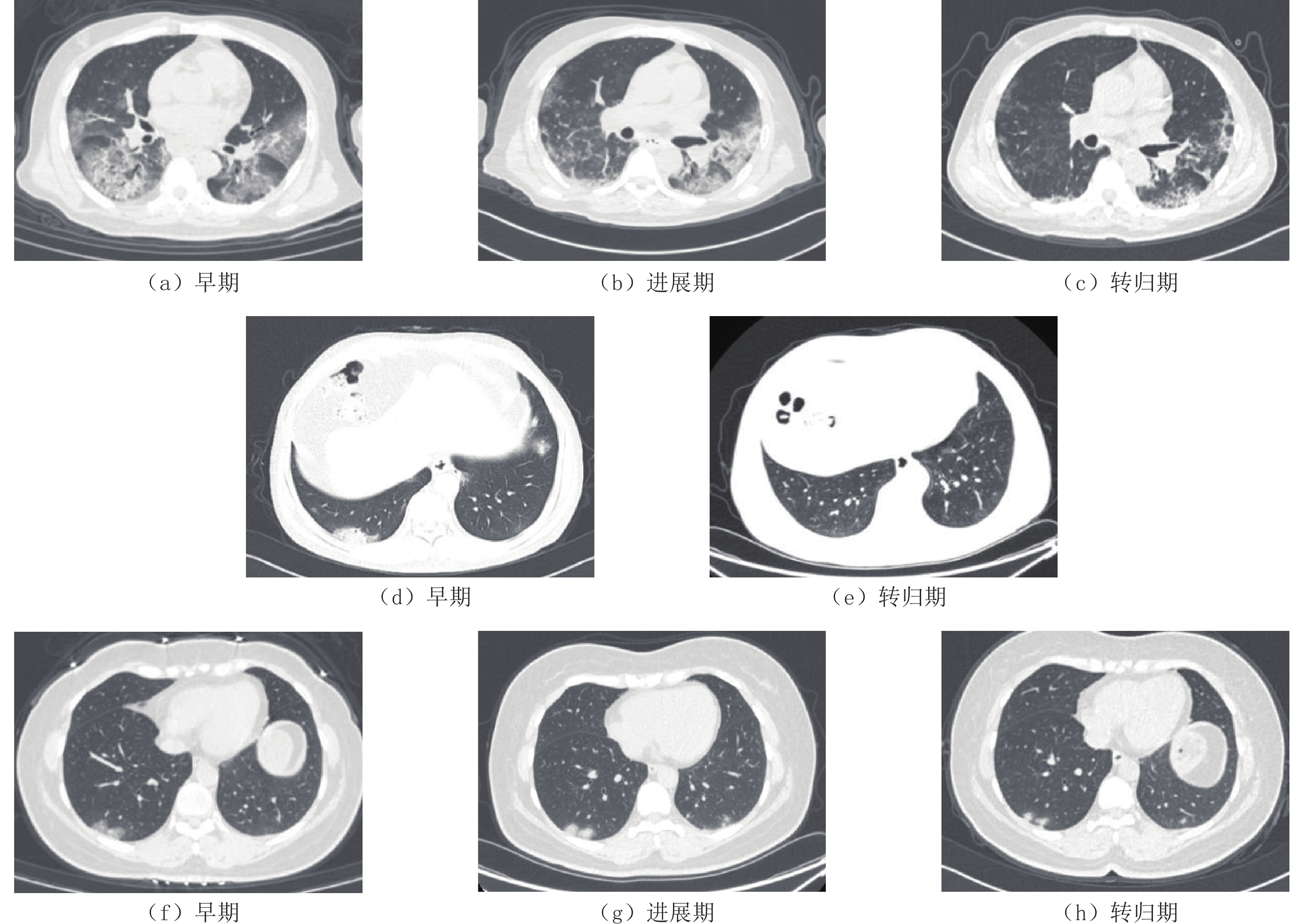
![]()
图 1 新冠肺炎不同时期CT表现
Figure 1. CT imaging features and evolution of different stages of COVID-19
表 1 不同时期CT表现
Table 1 CT findings in patients with new coronary pneumonia in different periods
CT表现 早期 进展期 转归期 磨玻璃 26(81%) 32(37%) 48(34%) 实变+磨玻璃 6(19%) 55(63%) 90(64%) 胸膜下分布 13(40%) 24(28%) 48(34%) 胸膜下+支气管血管周围分布 19(60%) 63(72%) 90(64%) 多叶 32(100%) 87(100%) 137(98%) 单叶 1(2%) 其内小叶间隔增粗 32(100%) 85(97%) 5(3%) 其内血管增粗 32(100%) 87(100%) 1(10%) 支气管充气征 1(3%) 6(7%) 2(1%) 条索 48(55%) 48(34%) 胸腔积液 6(7%) 1(1%) 肺气肿 3(3%)  下载: 导出CSV
-
[1] LI Q, GUAN X H, WU P, et al. Early transmission dynamics in wuhan, china, of novel coronavirus-infected pneumonia[J]. The New England Journal of Medicine, 2020, 382(13): 1199−1207. doi: 10.1056/NEJMoa2001316
[2] 湖南省医学会放射学专业委员会, 湖南省医学会影像技术专业委员会. 新型冠状病毒肺炎影像学检查、诊断及医院内感染预防与控制: 湖南省放射学专家共识[J]. 中南大学学报(医学版), 2020,45(3): 221−228. [3] 国家卫生健康委办公厅, 国家中医药局综合司. 《新型冠状病毒感染试行诊疗方案(试行第十版)》[J]. 全科医学临床与教育, 2023,21(1): 5−9. [4] 姚永刚, 杜静波, 廖建勇, 等. 新型冠状病毒肺炎的CT征象研究[J]. CT理论打应用研究, 2020,29(2): 169−176. DOI: 10.15953/j.1004-4140.2020.29.02.07. YAO Y G, DU J B, LIAO J Y, et al. Study of chest CT features of COVID-19[J]. CT Theory and Applications, 2020, 29(2): 169−176. DOI: 10.15953/j.1004-4140.2020.29.02.07. (in Chinese).
[5] 中华人民共和国国家卫生健康委员会医疗应急司. 《新型冠状病毒感染诊疗方案(试行第十版)》调整要点[J]. 中国医药, 2023,18(2): 167. [6] WANG W L, XU Y L, GAO R Q, et al. Detection of SARS-CoV-2 in different types of clinical specimens[J/OL]. The Journal of the American Medical Association, 2020, 323(18): 1843-1844.
[7] AI T, YANG Z L, HOU H Y, et al, et al. Correlation of chest CT and RT-PCR testing in coronavirus disease 2019 (COVID-19) in China: A report of 1014 cases[J/OL]. Radiology, 2020, 296(2): E32-E40.
[8] FANG Y, ZHANG H, XIE J, et al. et al. Sensitivity of chest CT for COVID-19: Comparison to RT-PCR[J/OL]. Radiology, 2020, 296(2): E115-E117.
[9] JEFFREY P K, BRENT P L, JONATHAN H C, et al. Essentials for radiologists on COVID-19: An update-radiology scientific expert panel[J/OL]. Radiology, 2020, 296(2): E113-E114.
[10] 杨立平, 李文贵, 高万军, 等. 3例聚集性发病新型冠状病毒肺炎CT表现[J]. 中国医学影像技术, 2020,36(2): 314−315. doi: 10.13929/j.issn.1003-3289.2020.02.037 YANG L P, LI W G, GAO W J, et al. CT manifestations of 3 cases of clustered novel coronavirus pneumonia[J]. Chinese Journal of Medical Imaging Technology, 2020, 36(2): 314−315. (in Chinese). doi: 10.13929/j.issn.1003-3289.2020.02.037
[11] 李莉, 王珂, 任美吉, 等. 新型冠状病毒肺炎早期胸部CT表现[J]. 首都医科大学学报, 2020,41(2): 174−177. doi: 10.3969/j.issn.1006-7795.2020.02.005 LI L, WANG K, REN M J, et al. Early chest CT findings of novel coronavirus pneumonia[J]. Journal of Capital Medical University, 2020, 41(2): 174−177. (in Chinese). doi: 10.3969/j.issn.1006-7795.2020.02.005
[12] 汪锴, 康嗣如, 田荣华, 等. 新型冠状病毒肺炎胸部CT影像学特征分析[J]. 中国临床医学, 2020,27(1): 27−31. WANG K, KANG S R, TIAN R H, et al. Analysis of chest CT imaging features of novel coronavirus pneumonia[J]. Chinese Journal of Clinical Medicine, 2020, 27(1): 27−31. (in Chinese).
[13] 陈月华, 张涛. 新型冠状病毒肺炎的临床特征及CT影像表现[J]. CT理论与应用研究, 2020,29(2): 155−162. DOI: 10.15953/j.1004-4140.2020.29.02.05. CHEN Y H, ZHANG T. Clinical features and CT imaging findings of patients with corona virus disease-19[J]. CT Theory and Applications, 2020, 29(2): 155−162. DOI: 10.15953/j.1004-4140.2020.29.02.05. (in Chinese).
[14] DONG D, TANG Z, WANG S, et a1. The role of imaging in the detection and management of COVID-19: A review[J]. IEEE Reviews in Biomedical Engineering, 2021, 14: 16−29. doi: 10.1109/RBME.2020.2990959
[15] 贾翠宇, 赵大伟, 冯骥良, 等. 重症COVID-19的肺部影像学表现及演变特点分析[J]. 北京医学, 2020, 42: 489-492. JIA C Y, ZHAO D W, FENG J L, et al. Analysis of lung imaging manifestations and evolution characteristics of severe COVID-19[J]. Beijing Medical Journal, 2020, 42: 489-492. (in Chinese).
[16] YING Z, LING W, SUQIN B. Meta-analysis of chest CT features of patients with COVID-19 pneumonia[J]. Journal of Medical Virology, 202l, 93(1): 241-249.
[17] 曹玉芳, 王小智, 谢晓红, 等. 新型冠状病毒、细菌和病毒性肺炎患者胸部影像学特征分析[J]. 中华危重病急救医学, 2023,35(1): 28−31. doi: 10.3760/cma.j.cn121430-20210318-00394 CAO Y F, WANG X Z, XIE X H, et al. Analysis of chest imaging features in patients with novel coronavirus, bacterial and viral pneumonia[J]. Chinese Journal of Critical Care Emergency Medicine, 2023, 35(1): 28−31. (in Chinese). doi: 10.3760/cma.j.cn121430-20210318-00394
[18] 刘祥龙, 李燕, 房凌宇, 等. CT在新型冠状病毒肺炎诊断及临床分型中的价值[J]. 中国CT和MRI杂志, 2022,20(5): 105−107. doi: 10.3969/j.issn.1672-5131.2022.05.034 LIU X L, LI Y, FANG L Y, et al. Value of CT in the diagnosis and clinical classification of novel coronavirus pneumonia[J]. Chinese Journal of CT and MRI, 2022, 20(5): 105−107. (in Chinese). doi: 10.3969/j.issn.1672-5131.2022.05.034
[19] 王苏丹, 李宏军, 张岩岩. 新型冠状病毒肺炎患者胸部CT影像动态变化及临床特征分析[J]. 北京医学, 2022,44(10): 907−913. WANG S D, LI H J, ZHANG Y Y. Dynamic changes and clinical features of chest CT images in patients with novel coronavirus pneumonia[J]. Beijing Medical Journal, 2022, 44(10): 907−913. (in Chinese).
[20] 中国研究型医院学会感染与炎症放射学专业委员会, 中国医师协会放射医师分会感染影像专业委员会, 中华医学会放射学分会传染病学组中国性病艾滋病防治协会感染(传染病)影像工作委员会, 等. 《新型冠状病毒肺炎影像诊断指南(2020 年第二版简版)》[J]. 首都医科大学学报, 2020,41(2): 168−173. doi: 10.3969/j.issn.1006-7795.2020.02.004 -
期刊类型引用(2)
1. 龙安军,陈树锋,刘窗,洪进益. 输卵管系膜囊肿的多层螺旋CT表现特征分析. 实用医学影像杂志. 2019(04): 359-361 .  百度学术
百度学术
2. 吴伟斌,彭涛,潘献伟,孟家晓,邹映文,何俊. 骨巨细胞瘤影像学特征及误诊分析. CT理论与应用研究. 2017(04): 505-510 . 本站查看
其他类型引用(0)
计量
- 文章访问数: 232
- HTML全文浏览量: 88
- PDF下载量: 22
- 被引次数: 2


