
Risk-Prediction Model Construction of Visceral Pleural Invasion in Early Lung Adenocarcinoma Based on Computed Tomography Imaging Features
-
摘要:
目的:构建基于CT影像特征早期肺腺癌脏层胸膜侵犯风险预测模型。方法:回顾性纳入2016年1月至2022年12月行手术治疗并确诊为IA期肺腺癌患者170例,根据有无脏层胸膜侵犯分为侵犯组(84例)和未侵犯组(86例);采用单因素和多因素法评价早期胸膜下肺腺癌脏层胸膜侵犯CT影像学特征相关独立危险因素,早期胸膜下肺腺癌脏层胸膜侵犯CT影像学特征预测模型效能分析。结果:单因素分析结果显示,实性成分占比、实性成分最大径、桥征、血管集束征及病灶与胸膜毗邻关系均可能与早期胸膜下肺腺癌患者脏层胸膜侵犯有关。多因素分析结果显示,桥征、血管集束征及病灶与胸膜毗邻关系Ⅱ型/Ⅲ 型均是早期胸膜下肺腺癌脏层胸膜侵犯独立危险因素。利用桥征、血管集束征、病灶与胸膜毗邻关系、Logistic模型预测概率对早期胸膜下肺腺癌脏层胸膜侵犯情况预测,最佳截断值分别为0.50、0.50、0.50和56.25%,约登指数分别为24.00%、23.95%、48.70% 和55.04%。结论:基于桥征、血管集束征及病灶与胸膜毗邻关系构建CT影像学特征模型可用于早期胸膜下肺腺癌脏层胸膜侵犯高危人群识别。
Abstract:Objective: To construct a structural risk prediction model for visceral pleural invasion in early lung adenocarcinoma based on computed tomography (CT) features. Methods: 170 patients with early lung adenocarcinoma treated surgically in our hospital were retrospectively selected between January 2016 and December 2022 and grouped according into invasion (84 cases) and non-invasion (86 cases) groups. Independent risk factors related to CT imaging features of visceral pleural invasion in early lung adenocarcinoma were evaluated using univariate and multivariate factor methods. The effectiveness of the CT imaging feature prediction model for visceral pleural invasion in early subpleural lung adenocarcinoma was analyzed. Results: Univariate analysis showed that the proportion and maximum diameter of solid components, bridging sign, vascular bundle sign, and relationship between the lesion and pleura may be related to visceral pleural invasion in patients with early subpleural lung adenocarcinoma. Multivariate analysis showed that the bridging sign, vascular bundle sign, and type II/III relationship between the lesion and pleura were independent risk factors for visceral pleural invasion in early subpleural lung adenocarcinoma. The best cutoff values for predicting visceral pleural invasion in early subpleural lung adenocarcinoma using the bridge sign, vascular bundle sign, adjacent relationship between the lesion and pleura, and logistic model prediction probability were 0.50, 0.50, 0.50, and 56.25%, respectively. The Jordan index for each of these was 24.00%, 23.95%, 48.70%, and 55.04%, respectively. Conclusion: Based on the bridge sign, vascular cluster sign, and the relationship between tumor and pleural adjacency, the CT imaging feature model can be used to identify high-risk groups for visceral pleural invasion in early lung adenocarcinoma.
-
Keywords:
- CT imaging features /
- lung adenocarcinoma /
- visceral pleural invasion /
- prediction
-
流行病学数据显示世界范围内正常人群非小细胞肺癌发病率超过10%,发病率高居恶性肿瘤第2位[1]。肺腺癌作为非小细胞肺癌最为常见病理组织学亚型,而脏层胸膜侵犯被认为是该亚型患者预后不良重要危险因素[2]。最新肺癌TNM分期标准中肺腺癌患者如出现脏层胸膜侵犯则T分期必然提高,同时脏层胸膜侵犯患者相较于无脏层胸膜侵犯者术中亦需接受更为广泛淋巴结清扫[3]。如何在术前准确识别脏层胸膜侵犯高危人群以提高早期肺癌手术治疗效果及改善远期预后已成为医学界关注的热点问题。有报道认为病灶直接接触胸膜与脏层胸膜侵犯发生关系密切[4],但对于胸膜下早期肺腺癌脏层胸膜侵犯发生与哪些因素有关尚存争议,同时与术前CT影像学特征间关系及基于上述指标构建预测模型研究亦相对较少。
因此,本研究通过构建基于CT影像学特征早期胸膜下肺腺癌脏层胸膜侵犯风险预测模型,旨在为术前识别高危人群及手术方案制定提供更多参考。
1. 资料与方法
1.1 一般资料
纳入标准:①经病理组织学确诊肺腺癌;②临床分期 cT1N0M0期;③术前 CT检查提示病灶实性成分最大径≤3 cm且位于胸膜下;④病灶与胸膜间最短距离≤1 cm;⑤完成穿刺或手术治疗;⑥年龄18~80岁。排除标准:①癌前病变或其他类型肺癌;②既往接受过抗癌治疗;②既往穿刺或手术史;③穿刺或手术禁忌症;④存在淋巴结或远处转移;⑤图像质量差无法评估;⑥CT扫描与手术间隔>2周;⑦临床资料不全。
最终纳入2016年1月至2022年12月于重庆医科大学附属大学城医院及川北医学院附属三台医院行手术治疗并确诊为IA期肺腺癌患者170例,根据病理组织学检查有无脏层胸膜侵犯分为侵犯组(84例)和未侵犯组(86例)。研究方案经医院伦理委员会批准。
1.2 研究方法
1.2.1 资料收集
登录医院电子病理系统由专人统一收集患者性别、年龄、病灶位置、CT影像学及病理组织学检查资料等。
1.2.2 CT影像学检查
采用飞利浦极速64层螺旋CT扫描仪,仰卧位下扫描整个肺野,扫描参数设置管电压、管电流、扫描层厚及重建层厚分别为120 kVp、150 mAs、5 mm、0.625 mm或1 mm,选择肺算法或标准算法完成图像重建。原始图像导入自带专用图像后处理软件(飞利浦ISP 4.6版本)完成定量参数和定性参数评估。
多平面重组肺窗图像中测量病灶最大径、病灶与胸膜间最短距离、病灶实性成分最大径及实性成分比例,其中全部影像学检查阅片均由两名副高及以上职称高年资放射科医师独立阅片,如意见不一致由科室主任综合评估后发出最终报告。
1.2.3 观察项目及分析方法
实性成分比例计算方法为肺窗病灶实性成分与病灶最大径比值,重复测量3次并计算平均值[5]。CT肺窗下评估病灶与胸膜毗邻关系类型,参考相关文献分为Ⅰ型、Ⅱ型及 Ⅲ型。Ⅰ型:病灶与胸膜有一条或多条索条影接触,无胸膜凹陷;Ⅱ型:病灶与胸膜有一条或多条索条影接触,有胸膜凹陷;Ⅲ 型:病灶与胸膜紧贴,贴合面为磨玻璃密度或实性密度[6]。
记录判断病灶与胸膜间桥征、支气管改变、血管集束征、病灶密度类型、病灶形状、边界是否模糊及内部结构情况。桥征判定标准为CT肺窗下观察到病灶与胸膜间拱形线样影,且边缘呈扁平化改变;血管集束征是指病灶周围血管向病灶方向汇集;支气管改变是指周围支气管病灶内扭曲走行,或僵硬或截断[7]。
1.2.4 病理组织学检查
穿刺活检及手术切除标本均由两位中级及以上职称专科病理医生独立阅片诊断,达成一致后由病理科主任复核。诊断及分级标准参照WHO肺部病灶组织学分类(2021版)标准,分期标准采用TNM分期(第八版)[8]。
1.3 统计学分析
选择SPSS 20.0软件处理数据。正态性评估采用Kolmogorov-Smirnov检验,其中符合正态分布计量资料比较采用独立样本t检验,以(
$ \bar x $ ±s)表示;计数资料比较采用$\chi^2 $ 检验,以频数表示;利用Logistic多因素回归分析筛选独立影响因素。利用受试者工作特征(ROC)曲线的方法对预测价值进行比较,利用Delong法比较ROC曲线下的面积。检验水准α=0.05,P<0.05为差异有统计学意义。
2. 结果
2.1 早期胸膜下肺腺癌脏层胸膜侵犯CT影像学特征相关危险因素单因素分析
单因素分析结果显示,实性成分占比、实性成分最大径、桥征、血管集束征及病灶与胸膜毗邻关系均可能与早期胸膜下肺腺癌患者脏层胸膜侵犯有关(表1)。
表 1 早期胸膜下肺腺癌脏层胸膜侵犯CT影像学特征相关危险因素单因素分析Table 1. Univariate analysis of risk factors associated with CT imaging features of visceral pleural invasion in early subpleural lung adenocarcinoma指标 组别 统计检验 未侵犯组(n=86) 侵犯组(n=84) $t/\chi^2 $ P 年龄/岁 60.54±7.30 61.31±7.86 -0.909 0.365 男/女/例 30/56 33/51 0.353 0.552 病灶位置 0.189 0.664 左肺 26(30.23) 28(33.33) 右肺 60(69.77) 56(66.67) CT影像学指标 病灶最大径/mm 24.54±4.40 25.36±4.63 -1.643 0.102 病灶与胸膜间最短距离/mm 3.01±0.54 2.95±0.50 1.113 0.267 实性成分最大径/mm 12.83±2.40 14.79±3.12 -5.826 0.000 实性成分比例(%) 53.41±4.10 57.01±6.14 -5.438 0.000 病灶密度类型/例 0.538 0.463 亚实性 76(88.37) 71(84.52) 实性 10(11.63) 13(15.48) 病灶形状/例 1.754 0.185 圆/类圆形 68(79.07) 59(70.24) 不规则 18(20.93) 25(29.76) 分叶征/例 85(98.84) 77(91.67) 3.404◆ 0.065 毛刺征/例 13(15.12) 20(23.81) 2.053 0.152 边界模糊/例 2(2.33) 4(4.76) 0.198◆ 0.656 内部结构/例 空泡 24(27.91) 27(32.14) 0.363 0.547 囊腔/空洞 4(4.65) 5(5.95) 0.001◆ 0.971 毗邻结构/例 支气管改变 54(62.79) 46(54.76) 1.131 0.288 血管集束征 7(8.14) 27(32.14) 15.302 0.000 桥征/例 5(5.81) 21(25.00) 12.074 0.001 病灶与胸膜毗邻关系类型/例 53.114 0.000 Ⅰ型 47(54.65) 5(5.95) Ⅱ型 31(36.05) 44(52.38) Ⅲ 型 8(9.30) 35(41.67) 注:◆为连续校正$\chi^2 $检验。 2.2 早期胸膜下肺腺癌脏层胸膜侵犯CT影像学特征相关危险因素多因素分析
以早期胸膜下肺腺癌脏层胸膜是否发生侵犯作为因变量(0=未侵犯,1=侵犯),以单因素分析中结果阳性的变量作为自变量,其中连续变量均采用实测值纳入计算,分类变量的赋值方法如下:桥征:0=否;1=是;病灶与胸膜毗邻关系类型:0,0=Ⅰ型;1,0=Ⅱ型;0,1=Ⅲ 型。
进行多因素分析。多因素分析结果显示,桥征、血管集束征及病灶与胸膜毗邻关系Ⅱ型/Ⅲ 型均是早期胸膜下肺腺癌脏层胸膜侵犯独立危险因素(表2)。
表 2 早期胸膜下肺腺癌脏层胸膜侵犯CT影像学特征相关危险因素多因素分析Table 2. Multivariate analysis of the risk factors related to CT imaging features of visceral pleural invasion in early subpleural lung adenocarcinoma影响因素 β SE Wald $\chi^2 $ OR值 OR 95% CI P 实性成分最大径 -0.001 0.074 0.000 0.999 0.864~1.154 0.987 实性成分比例 -0.043 0.041 1.100 0.958 0.884~1.038 0.294 血管集束征 2.074 0.653 10.087 7.954 2.212~28.601 0.001 桥征 2.162 0.668 10.469 8.687 2.345~32.184 0.001 病灶与胸膜毗邻关系类型 30.805 <0.001 Ⅱ型 2.987 0.620 23.236 19.832 5.886~66.815 <0.001 Ⅲ 型 3.742 0.696 28.930 42.170 10.786~164.873 <0.001 常量 -0.830 2.343 0.125 0.436 0.723 2.3 早期胸膜下肺腺癌脏层胸膜侵犯CT影像学特征预测模型效能分析
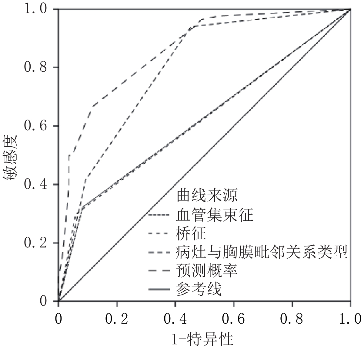
利用桥征、血管集束征、病灶与胸膜毗邻关系、Logistic模型预测概率对早期胸膜下肺腺癌脏层胸膜侵犯情况预测,最佳截断值分别为0.50、0.50、0.50、56.25%,约登指数分别为24.00%、23.95%、48.70% 和55.04%。Logistic模型预测概率对早期胸膜下肺腺癌脏层胸膜侵犯情况预测的效能最佳(表3、图1和图2)。
表 3 早期胸膜下肺腺癌脏层胸膜侵犯CT影像学特征预测价值Table 3. Predictive value of CT imaging features of visceral pleural invasion in early subpleural lung adenocarcinoma项目 最佳截断值 ROC曲线下面积 诊断灵敏度/% 诊断特异度/% 约登指数/% 血管集束征 0.50 0.620 32.14 91.86 24.00 桥征 0.50 0.620 29.76 94.19 23.95 病灶与胸膜毗邻关系类型 0.50 0.794 94.05 54.65 48.70 Logistic模型预测概率 56.25% 0.861 66.67 88.37 55.04 ![]() 图 1 早期胸膜下肺腺癌脏层胸膜侵犯CT影像学特征预测ROC曲线Figure 1. ROC curve predicted by CT imaging features of visceral pleural invasion in early subpleural lung adenocarcinoma
图 1 早期胸膜下肺腺癌脏层胸膜侵犯CT影像学特征预测ROC曲线Figure 1. ROC curve predicted by CT imaging features of visceral pleural invasion in early subpleural lung adenocarcinoma![]() 图 2 早期肺腺癌脏层胸膜侵犯典型图像Figure 2. Typical images of visceral pleural invasion in early-stage lung adenocarcinoma表 4 ROC曲线下的成对样本区域差异Table 4. Regional differences in paired samples under the ROC curve
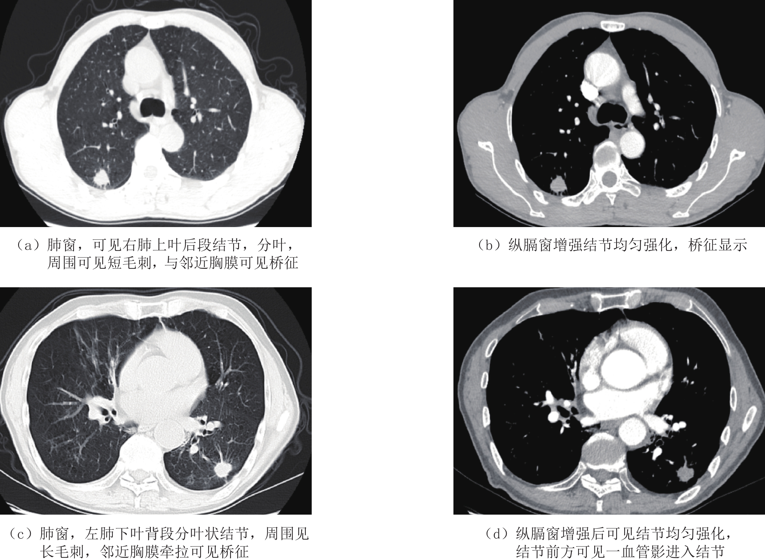
图 2 早期肺腺癌脏层胸膜侵犯典型图像Figure 2. Typical images of visceral pleural invasion in early-stage lung adenocarcinoma表 4 ROC曲线下的成对样本区域差异Table 4. Regional differences in paired samples under the ROC curve检验结果对 统计检验 Z P 血管集束征-桥征 0.007 0.995 血管集束征-病灶与胸膜毗邻关系类型 -4.189 0.000 血管集束征-Logistic模型预测概率 -7.194 0.000 桥征-病灶与胸膜毗邻关系类型 -4.155 0.000 桥征-Logistic模型预测概率 -7.694 0.000 病灶与胸膜毗邻关系类型-Logistic模型预测概率 -3.649 0.000 3. 讨论
脏层胸膜下存在丰富淋巴回流系统,并能够与气管、血管内走行中轴淋巴系统连接。故合并脏层胸膜侵犯的肺癌更易出现经肺门/纵隔淋巴结或其他跳跃性淋巴结转移,往往需通过术后辅助化疗以期控制病情进展[9]。
同时对于T1期肺癌患者如存在脏层胸膜侵犯应优先选择肺叶切除术而非常规肺段切除术,这被证实可增加患者远期临床获益[10]。故术前准确评估有无脏层胸膜侵犯发生对于早期肺癌患者治疗方案制定具有重要指导意义。
3.1 桥征及血管集束征与早期胸膜下肺腺癌脏层胸膜侵犯间的关系
以往有关CT影像学特征与肺癌脏层胸膜侵犯报道中绝大部分关注病灶本身,对于病灶形态学征象涉及较少[11]。本研究单因素和多因素分析结果显示,桥征与血管集束征均是早期胸膜下肺腺癌脏层胸膜侵犯发生独立危险因素。
有报道提示实性成分直径≤3 cm且合并脏器胸膜侵犯非小细胞肺癌患者89% 存在桥征,其形成可能与病灶与胸膜大范围接触及胸膜凹陷呈梯形改变有关[12];但本研究提示这一征象单一预测效能偏低,是否能够提高预测准确度仍需进一步探索验证。而血管集束征出现主要与病灶浸润生长至周围肺血管支气管束间,而病灶内纤维反应性增生对肺血管造成牵拉导致局部聚集造成,同时随病灶恶性程度增加牵拉效应亦更为明显,而这亦支持血管集束征与早期胸膜下肺腺癌脏层胸膜侵犯间的关系[13-14]。
3.2 胸膜毗邻关系与早期胸膜下肺腺癌脏层胸膜侵犯间的关系
已有研究显示,胸膜下肺癌但病灶与胸膜间未见影像学线样影关系,发生脏层胸膜侵犯风险极低[15]。本研究结果提示,胸膜毗邻关系分型中Ⅱ型及Ⅲ 型是早期胸膜下肺腺癌脏层胸膜侵犯独立危险因素。
有报道提示,肺癌病灶与胸膜关系中 Ⅲ型患者发生脏层胸膜侵犯风险最高[16],与本研究结果一致。我们认为胸膜毗邻关系与早期胸膜下肺腺癌脏层胸膜侵犯间的独立关系可能与以下因素有关。
早期病灶肿瘤细胞可能通过肿瘤与胸膜间线样影或条状影所建立通道向周围胸膜侵犯,但因病灶内纤维反应性增生相对轻微,并未引起邻近胸膜凹陷;而随病灶内纤维反应性增生及浸润程度加重,可导致周围胸膜牵拉凹陷且范围扩大,最终在CT扫描下呈Ⅱ型及 Ⅲ型改变[17-18]。
3.3 其他CT影像学特征与早期胸膜下肺腺癌脏层胸膜侵犯间的关系
既往研究显示实性成分比例和毛刺征均可用于预测肺癌脏层胸膜侵犯风险[19-20],但本研究并未支持这一观点。我们认为这可能与本研究纳入患者更为严格,即胸膜下肺腺癌且病灶与胸膜间最短距离应≤1 cm有关,排除既往研究多种混杂因素影响。
3.4 基于CT影像学特征构建的数学模型在早期胸膜下肺腺癌脏层胸膜侵犯预测中的价值
本文利用所构建的Logistic数学模型对各个指标的早期胸膜下肺腺癌脏层胸膜侵犯预测价值实施分析,结果显示,单项指标的预测均存在一定的错误,如果将各指标进行联合预测,则十分明显的提升预测的效能。这也提示在工作中需要综合考虑多种因素,对于提升预测的准确度具有重要的作用。
研究局限性:①属于单中心回顾性研究,无法避免选择偏倚;②单纯通过 CT定性及定量指标预测效能可能仍有改善空间。后续可进一步开展影像组学、AI深度学习等方面探索,以便更为准确识别筛选预后不良高危人群。
综上所述,基于桥征、血管集束征及病灶与胸膜毗邻关系构建CT影像学特征Logistic模型可用于早期胸膜下肺腺癌脏层胸膜侵犯高危人群识别。
-
![]()
图 1 早期胸膜下肺腺癌脏层胸膜侵犯CT影像学特征预测ROC曲线
Figure 1. ROC curve predicted by CT imaging features of visceral pleural invasion in early subpleural lung adenocarcinoma
![]()
图 2 早期肺腺癌脏层胸膜侵犯典型图像
Figure 2. Typical images of visceral pleural invasion in early-stage lung adenocarcinoma
表 1 早期胸膜下肺腺癌脏层胸膜侵犯CT影像学特征相关危险因素单因素分析
Table 1 Univariate analysis of risk factors associated with CT imaging features of visceral pleural invasion in early subpleural lung adenocarcinoma
指标 组别 统计检验 未侵犯组(n=86) 侵犯组(n=84) $t/\chi^2 $ P 年龄/岁 60.54±7.30 61.31±7.86 -0.909 0.365 男/女/例 30/56 33/51 0.353 0.552 病灶位置 0.189 0.664 左肺 26(30.23) 28(33.33) 右肺 60(69.77) 56(66.67) CT影像学指标 病灶最大径/mm 24.54±4.40 25.36±4.63 -1.643 0.102 病灶与胸膜间最短距离/mm 3.01±0.54 2.95±0.50 1.113 0.267 实性成分最大径/mm 12.83±2.40 14.79±3.12 -5.826 0.000 实性成分比例(%) 53.41±4.10 57.01±6.14 -5.438 0.000 病灶密度类型/例 0.538 0.463 亚实性 76(88.37) 71(84.52) 实性 10(11.63) 13(15.48) 病灶形状/例 1.754 0.185 圆/类圆形 68(79.07) 59(70.24) 不规则 18(20.93) 25(29.76) 分叶征/例 85(98.84) 77(91.67) 3.404◆ 0.065 毛刺征/例 13(15.12) 20(23.81) 2.053 0.152 边界模糊/例 2(2.33) 4(4.76) 0.198◆ 0.656 内部结构/例 空泡 24(27.91) 27(32.14) 0.363 0.547 囊腔/空洞 4(4.65) 5(5.95) 0.001◆ 0.971 毗邻结构/例 支气管改变 54(62.79) 46(54.76) 1.131 0.288 血管集束征 7(8.14) 27(32.14) 15.302 0.000 桥征/例 5(5.81) 21(25.00) 12.074 0.001 病灶与胸膜毗邻关系类型/例 53.114 0.000 Ⅰ型 47(54.65) 5(5.95) Ⅱ型 31(36.05) 44(52.38) Ⅲ 型 8(9.30) 35(41.67) 注:◆为连续校正$\chi^2 $检验。  下载: 导出CSV
表 2 早期胸膜下肺腺癌脏层胸膜侵犯CT影像学特征相关危险因素多因素分析
Table 2 Multivariate analysis of the risk factors related to CT imaging features of visceral pleural invasion in early subpleural lung adenocarcinoma
影响因素 β SE Wald $\chi^2 $ OR值 OR 95% CI P 实性成分最大径 -0.001 0.074 0.000 0.999 0.864~1.154 0.987 实性成分比例 -0.043 0.041 1.100 0.958 0.884~1.038 0.294 血管集束征 2.074 0.653 10.087 7.954 2.212~28.601 0.001 桥征 2.162 0.668 10.469 8.687 2.345~32.184 0.001 病灶与胸膜毗邻关系类型 30.805 <0.001 Ⅱ型 2.987 0.620 23.236 19.832 5.886~66.815 <0.001 Ⅲ 型 3.742 0.696 28.930 42.170 10.786~164.873 <0.001 常量 -0.830 2.343 0.125 0.436 0.723
下载: 导出CSV
表 3 早期胸膜下肺腺癌脏层胸膜侵犯CT影像学特征预测价值
Table 3 Predictive value of CT imaging features of visceral pleural invasion in early subpleural lung adenocarcinoma
项目 最佳截断值 ROC曲线下面积 诊断灵敏度/% 诊断特异度/% 约登指数/% 血管集束征 0.50 0.620 32.14 91.86 24.00 桥征 0.50 0.620 29.76 94.19 23.95 病灶与胸膜毗邻关系类型 0.50 0.794 94.05 54.65 48.70 Logistic模型预测概率 56.25% 0.861 66.67 88.37 55.04
下载: 导出CSV
表 4 ROC曲线下的成对样本区域差异
Table 4 Regional differences in paired samples under the ROC curve
检验结果对 统计检验 Z P 血管集束征-桥征 0.007 0.995 血管集束征-病灶与胸膜毗邻关系类型 -4.189 0.000 血管集束征-Logistic模型预测概率 -7.194 0.000 桥征-病灶与胸膜毗邻关系类型 -4.155 0.000 桥征-Logistic模型预测概率 -7.694 0.000 病灶与胸膜毗邻关系类型-Logistic模型预测概率 -3.649 0.000
下载: 导出CSV
-
[1] SUNG H, FERLAY J, SIEGEL R L, et al. Global cancer statistics 2020: Globocan estimates of incidence and mortality worldwide for 36 cancers in 185 countries[J]. CA: A Cancer Journal for Clinicians, 2021, 71(3): 209−249. DOI: 10.3322/caac.21660.
[2] CHOI H, KIM H, HONG W, et al. Prediction of visceral pleural invasion in lung cancer on CT: Deep learning model achieves a radiologist-level performance with adaptive sensitivity and specificity to clinical needs[J]. European Journal of Radiology, 2021, 31(5): 2866−2876. DOI: 10.1007/s00330-020-07431-2.
[3] ZHANG T, ZHANG J T, LI W F, et al. Visceral pleural invasion in T1 tumors (≤3 cm), particularly T1a, in the eighth tumor-node-metastasis classification system for non-small cell lung cancer: A population-based study[J]. Journal of Thoracic Disease, 2019, 11(7): 2754−2762. DOI: 10.21037/jtd.2019.06.32.
[4] WO Y, ZHAO Y, QIU T, et al. Impact of visceral pleural invasion on the association of extent of lymphadenectomy and survival in stage I non-small cell lung cance[J]. Cancer Medicine, 2019, 8(2): 669−678. DOI: 10.1002/cam4.1990.
[5] ZUO Z, LI Y, PENG K, et al. CT texture analysis-based nomogram for the preoperative prediction of visceral pleural invasion in cT1N0M0 lung adenocarcinoma: An external validation cohort study[J]. Clinical Radiology, 2022, 77(3): e215−e221. DOI: 10.1016/j.crad.2021.11.008.
[6] YUAN M, LIU J Y, ZHANG T, et al. Prognostic impact of the findings on thinsection computed tomography in stage I lung adenocarcinoma with visceral pleural invasion[J]. Scientific Reports, 2018, 8(1): 1−9.
[7] ONODA H, HIGASHI M, MURAKAMI T, et al. Correlation between pleural tags on CT and visceral pleural invasion of peripheral lung cancer that does not appear touching the pleural surface[J]. European Journal of Radiology, 2021, 31(12): 9022−9029. DOI: 10.1007/s00330-021-07869-y.
[8] WEI S H, ZHANG J M, SHI B, et al. The value of CT radiomics features to predict visceral pleural invasion in ≤3 cm peripheral type early non-small cell lung cancer[J]. Journal of X-ray Science and Technology, 2022, 30(6): 1115−1126. DOI: 10.3233/XST-221220.
[9] ZHANG Y, KWON W, LEE H Y, et al. Imaging assessment of visceral pleural surface invasion by lung cancer: Comparison of CT and contrast-enhanced radial T1-weighted gradient echo 3-Tesla MRI[J]. Korean Journal of Radiology, 2021, 22(5): 829−839. DOI: 10.3348/kjr.2020.0955.
[10] SHI J, LI F, YANG F, et al. The combination of computed tomography features and circulating tumor cells increases the surgical prediction of visceral pleural invasion in clinical T1N0M0 lung adenocarcinoma[J]. Translational Lung Cancer Research, 2021, 10(11): 4266−4280. DOI: 10.21037/tlcr-21-896.
[11] TU Z, LI C, TIAN T, et al. A risk classification system predicting the cancer-specific survival for postoperative stage IB non-small-cell lung cancer patients without lymphovascular and visceral pleural invasion[J]. Lung Cancer, 2021, 161(11): 114−121.
[12] YANG X, SUN F, CHEN L, et al. Prognostic value of visceral pleural invasion in non-small cell lung cancer: A propensity score matching study based on the SEER registry[J]. Journal Surgery Oncology, 2017, 116(3): 398−406. DOI: 10.1002/jso.24677.
[13] YU Y, HUANG R, WANG P, et al. Sublobectomy versus lobectomy for long-term survival outcomes of early-stage non-small cell lung cancer with a tumor size ≤2 cm accompanied by visceral pleural invasion: A SEER population-based study[J]. Journal of Thoracic Disease, 2020, 12(3): 592−604. DOI: 10.21037/jtd.2019.12.121.
[14] NAM J G, PARK S, PARK C M, et al. Histopathologic basis for a chest CT deep learning survival prediction model in patients with lung adenocarcinoma[J]. Radiology, 2022, 305(2): 441−451. DOI: 10.1148/radiol.213262.
[15] 汤敏, 孙丹丹, 尹柯, 等. 胸膜下肺腺癌脏层胸膜侵犯CT及临床风险因素[J]. 放射学实践, 2020, 35(10): 1243−1248. TANG M, SUN D D, YIN K, et al. CT and clinical risk factors of visceral pleural invasion in subpleural lung adenocarcinoma[J]. Radiology Practice, 2020, 35(10): 1243−1248. (in Chinese).
[16] YANG S, YANG L, TENG L, et al. Visceral pleural invasion by pulmonary adenocarcinoma ≤3 cm: The pathological correlation with pleural signs on computed tomography[J]. Journal of Thoracic Disease, 2018, 10(7): 3992−3999. DOI: 10.21037/jtd.2018.06.125.
[17] WANG F, LI P, LI F. Nomogram for predicting the relationship between the extent of visceral pleural invasion and survival in non-small-cell lung cancer[J]. Canadian Respiratory Journal, 2021, 20(7): 8816860.
[18] WANG Y, LYU D, ZHOU T, et al. Multivariate analysis based on the maximum standard unit value of 18F-fluorodeoxyglucose positron emission tomography/computed tomography and computed tomography features for preoperative predicting of visceral pleural invasion in patients with subpleural clinical stage IA peripheral lung adenocarcinoma[J]. Diagnostic and Interventional Radiology, 2023, 29(2): 379−389. DOI: 10.4274/dir.2023.222006.
[19] KIM H J, CHO J Y, LEE Y J, et al. Clinical significance of pleural attachment and indentation of subsolid nodule lung cancer[J]. Cancer Research and Treatment, 2019, 51(4): 1540−1548. DOI: 10.4143/crt.2019.057.
[20] HEIDINGER B H, SCHWARZ-NEMEC U, ANDERSON K R, et al. Visceral pleural invasion in pulmonary adenocarcinoma: Differences in CT patterns between solid and subsolid cancers[J]. Radiology-Cardiothoracic Imaging, 2019, 1(3): e190071. DOI: 10.1148/ryct.2019190071.


计量
- 文章访问数: 120
- HTML全文浏览量: 26
- PDF下载量: 24


