
Application Study of Contrast-enhanced CT in Renal Cystic Lesions with the Bosniak Classification Version 2019
-
摘要: 目的:本研究拟采用多期增强CT对肾囊性病变的2019版Bosniak分类细节进行评估。方法:共入组88例肾囊性病变,两名不同的观察者对Bosniak v.2019分类评判标准中的细节进行评测,并将最终分类结果与病理进行对照。结果:两名观察者对肾囊性病变的大小、病灶内均质程度、各期CT值、分隔数量及结节大小的评测一致性很好(ICC=0.97,ICC=0.96,ICC=0.98,ICC=0.97,ICC=0.96,ICC=0.97,ICC=0.90,ICC=0.96),对囊壁厚度的评测一致性良好(ICC=0.73),而对病变分隔厚度及不规则突起厚度的评测一致性中等(ICC=0.41,ICC=0.50)。与病理结果进行对照,我们发现对于肾囊性病变良恶性,其病灶大小、均质度、各期CT值、囊壁厚度、分隔厚度及分隔数量存在统计学意义,钙化大小则无明显统计学意义。结论:对Bosniak v.2019版CT分类细节的评估有利于判断肾囊性病变的良恶性,但不同观察者对于细节的评测存在一定程度的差异。Abstract: This study aimed to use multiphase-enhanced CT to evaluate the classification details of the Bosniak classification version 2019 of renal cystic lesions. A total of 88 cases of renal cystic lesions were enrolled. Two reviewers evaluated the details of the Bosniak v. 2019 classification and then compared the final classifications with pathological results. We found that the two reviewers had excellent consistency in evaluating the size, degree of homogeneity within the lesion, CT values of each scanning phase, number of septations, and nodule size of renal cystic lesions, while the consistency in evaluating the thickness of the cyst wall was good. The consistency in evaluating lesion septal thickness and irregular protrusion thickness was moderate. Compared to pathological results, we found that for benign and malignant renal cystic lesions, there were statistical significance in lesion size, homogeneity, CT values of each scanning phase, cyst wall thickness, septal thickness, and number of septations, while there was no significant statistical significance in calcification size. Therefore, the evaluation of the classification details of Bosniak classification v. 2019 CT is beneficial in determining the benign and malignant nature of renal cystic lesions, but there are differences in observer consistency.
-
Keywords:
- X-ray computed tomography /
- renal cystic disease /
- classification
-
冠状动脉CT血管成像(coronary computed tomography angiography,CCTA)凭借快速、准确、无创的特点,已成为筛查和诊断心血管疾病的一线影像学手段[1-4]。然而,CCTA临床应用存在辐射暴露的生物效应问题[5],引发公众广泛关注。研究表明,辐射剂量与管电压平方呈正相关[6],合理调节管电压可在保证图像质量的前提下降低辐射剂量[7]。CARE kV技术能够基于患者体型、组织衰减特性及预设图像质量要求,自动生成个体化管电压-管电流组合参数[8-11],为辐射剂量的精准调控奠定技术基础。
当前CARE kV技术相关研究聚焦其在正常体型患者中的应用效果,且剂量评价体系局限于容积CT剂量指数(volume CT dose index,CTDIvol)、剂量长度乘积(dose-length product,DLP)以及基于DLP的有效剂量(effective dose,ED)[8,12-13],以上指标无法反映体型和性别等因素对实际辐射剂量的影响[14]。针对上述局限性,本研究在传统剂量评价指标基础上,纳入体型特异性剂量估计值(size-specific dose estimates,SSDE)和基于蒙特卡罗模拟的ED,分析不同辐射剂量评价间差异及其与管电压设置的相关性,通过分析不同管电压设置下辐射剂量评价指标及剂量比值间差异来源,探讨CARE kV技术对CCTA辐射剂量的影响,并明确不同剂量指标的临床应用价值,为精准化辐射剂量管理提供理论依据。
1. 资料与方法
1.1 资料收集与分组
本研究为回顾性研究,经重庆医科大学附属第一医院伦理委员会批准(伦理号:K2024-222-01),免除患者知情同意。收集2023年1月至2024年12月于重庆医科大学附属第一医院行CCTA检查患者的影像学资料。
纳入标准:①年龄≥18岁;②存在胸痛、心前区不适等临床症状或心血管疾病高危因素;③图像符合临床诊断要求。排除标准:①多部位联合扫描;②扫描部位存在金属植入物或金属外固定物;③临床资料不完整。最终纳入
1529 例患者,其中男性727例(47.55%),年龄中位数59(49,68)岁;女性802例(52.45%),年龄中位数62(55,70)岁。根据CARE kV技术应用后设备自动选择的管电压(70、80、90和100 kV)对患者进行分组。1.2 检查方法
使用西门子双源Somatom Force CT扫描仪进行CCTA检查。患者取仰卧位,头先进,身体置于检查床正中,双手上举过头并伸直,连接心电门控。扫描范围为气管隆突下方1 cm至心脏膈面水平,使用对比剂智能追踪技术,监测左冠状动脉发出层面的降主动脉,吸气后屏气扫描。CCTA扫描技术参数:使用自动管电流调控技术(Care Dose 4D,Quality ref,mAs为80 mAs)及CARE kV技术(Ref.kV为120 kV),探测器准直为192×0.6 mm,螺距为0.15,转速为0.25 s/周,采用回顾性心电门控扫描,并自动选择最佳期相进行重建。重建层厚/层间距为0.75 mm/0.5 mm,视野(field of view,FOV)为200 mm,重建算法为Br36重建核和ADMIRE算法(强度3)。
1.3 数据收集
将数据从影像归档与通信系统(Picture Archiving and Communication System,PACS)传输至Radimetrics软件进行辐射剂量计算。基于定位像(包括完整胸廓)计算水当量直径(Water Equivalent Diameter,WED),并依据AAPM Task Group 220[15]提供的转换因子获取平均SSDE。通过蒙特卡罗模拟(基于年龄、性别、体型匹配内置体模库)计算器官吸收剂量,加权求和生成ED,记作ED_Radimetrics。最终软件输出SSDE、ED_Radimetrics等剂量学指标。从设备剂量报告中提取CTDIvol、DLP等常规剂量指标,并采用k因子法基于DLP计算ED,记作ED_DLP,计算公式:ED_DLP=DLP×k(胸部k=0.014 mSv/(mGy∙cm)[16]);为量化不同剂量评价指标间差异,进一步计算SSDE/CTDIvol、ED_DLP/ED_Radimetrics比值。
1.4 统计学分析
使用R软件(V4.3.2)进行统计分析。通过Shapiro-Wilk正态性检验评估连续变量的分布特性,结果显示所有指标均不符合正态分布(P < 0.05),故采用中位数(四分位数间距)描述数据。采用Kruskal-Wallis H检验分析不同组间辐射剂量指标及剂量比值的差异(显著性水平α=0.05),对存在统计学差异的指标进行Bonferroni校正的多重比较(校正后显著性水平α=0.008)。使用Spearman相关分析评估管电压与各剂量学指标及比值间的相关性强度。
2. 结果
2.1 kV分布及辐射剂量特征
根据实际使用的管电压将患者分为四组:70 kV组(n=775,占比53.78%)、80 kV组(n=508,占比32.25%)、90 kV组(n=60,占比4.16%)、100 kV组(n=98,占比6.80%)。各组辐射剂量水平见表1。所有组别ED_DLP均低于《心脏冠状动脉CT血管成像技术规范化应用中国指南》[17]推荐值5.00 mSv。
表 1 CCTA患者辐射剂量数据(根据CARE kV技术自动选择的管电压分组)Table 1. Radiation dose data for patients undergoing CCTA (grouped by tube voltage)项目 组别 70 kV(N=815) 80 kV(N=541) 90 kV(N=67) 100 kV(N=106) CTDlvol/mGy 11.40[4.07] 15.80[7.92] 16.70[10.40] 22.60[10.70] DLP/(mGy·cm) 139.00[56.40] 190.00[103.00] 193.00[138.00] 289.00[141.00] SSDE/mGy 17.10[5.42] 22.90[9.89] 24.00[14.50] 33.50[13.30] ED_DLP/mSv 1.95[0.79] 2.66[1.44] 2.70[1.93] 4.05[1.98] ED_Radimetrics/mSv 3.70[2.11] 5.20[2.84] 5.41[3.41] 8.22[4.33] 2.2 辐射剂量指标与剂量比值的组间比较
Kruskal-Wallis H检验结合Bonferroni校正的事后检验结果(表2)显示,各组剂量学指标(CTDIvol、DLP、SSDE、ED_DLP、ED_Radimetrics)及剂量比值(SSDE/CTDIvol、ED_Radimetrics/ED_DLP)差异均具有统计学意义(Kruskal-Wallis H检验p < 0.05,Bonferroni校正后P < 0.008)。各剂量学指标(CTDIvol、DLP、SSDE、ED_DLP和ED_Radimetrics )由70 kV组至100 kV组呈递增趋势;剂量比值未观察到随kV值的变化趋势。
表 2 组间辐射剂量指标与比值差异的评估Table 2. Differences in radiation dose index and ratio between groups组别 例数 辐射剂量指标与比值 CTDIvol DLP SSDE ED_DLP ED_Radimetrics SSDE/CTDIvol ED_Radimetrics
/ED_DLP70 kV 815 11.40[4.07] 139.00[56.40] 17.10[5.42] 1.95[0.79] 3.70[2.11] 1.52[0.12] 2.25[1.19] 80 kV 541 15.80[7.92] 190.00[103.00] 22.90[9.89] 2.66[1.44] 5.20[2.84] 1.46[0.16] 2.19[1.20] 90 kV 67 16.70[10.40] 193.00[138.00] 24.00[14.50] 2.70[1.93] 5.41[3.41] 1.44[0.19] 2.31[1.28] 100 kV 106 22.60[10.70] 289.00[141.00] 33.50[13.30] 4.05[1.98] 8.22[4.33] 1.45[0.16] 1.70[1.18] Kruskal-Wallis H
检验chi-squared 805.54 701.79 463.17 701.79 569.02 152.22 131.91 p-value < 0.05 < 0.05 < 0.05 < 0.05 < 0.05 < 0.05 < 0.05 Bonferroni校正 p-value < 0.008 < 0.008 < 0.008 < 0.008 < 0.008 < 0.008 < 0.008 2.3 kV与辐射剂量指标及剂量比值的相关性分析
Spearman相关性分析结果显示(表3),管电压与CTDIvol(rho=0.53,P < 0.05)、DLP(rho=0.49,P < 0.05)、SSDE(rho=0.54,P < 0.05)、ED_DLP(rho=0.49,P < 0.05)和ED_Radimetrics(rho=0.46,P < 0.05)呈中等强度正相关,且具有统计学意义;管电压与SSDE/CTDIvol呈弱负相关(rho=−0.30,P < 0.05),与ED_Radimetrics/ED_DLP呈微弱正相关(rho=0.07,P < 0.05)。
表 3 管电压与剂量指标及剂量比值的相关性Table 3. Correlation between tube voltage, dose index, and dose ratio辐射剂量指标与比值 Spearman相关性分析结果 CTDIvol rho 0.53 p-value < 0.05 DLP rho 0.49 p-value < 0.05 SSDE rho 0.54 p-value < 0.05 ED_DLP rho 0.49 p-value < 0.05 ED_Radimetrics rho 0.46 p-value < 0.05 SSDE/CTDIvol rho −0.30 p-value < 0.05 ED_Radimetrics/ED_DLP rho 0.07 p-value < 0.05 3. 讨论
本研究系统评估了CARE kV技术在CCTA检查中对辐射剂量的影响,并在该技术背景下,深入比较了传统剂量指标(CTDIvol、DLP、ED_DLP)与基于患者个体特征的剂量指标(SSDE、ED_Radimetrics)之间的差异及其与管电压的相关性。主要发现包括:①CARE kV技术倾向于选择较低的管电压(70 kV和80 kV占比达86.03%),且ED_DLP(1.95~4.03 mSv)低于指南推荐值5.00 mSv;②所有评估的辐射剂量指标均随管电压升高而显著增加;③基于患者体型和器官敏感性校正的SSDE和ED_Radimetrics显著高于对应的传统指标CTDIvol和ED_DLP,且差异与CARE kV自动选择的管电压相关性较弱。这些发现对于理解自动化kV技术下的辐射剂量特征及优化临床剂量管理策略具有重要意义。
个体化扫描方案设置是CT检查优化的核心理念。多项研究强调应根据患者体型等个性化指标调整扫描参数[18-21]。本研究应用CARE kV技术,实现基于体型和组织衰减的扫描参数个体化优化,在辐射剂量控制与图像质量保障间达成平衡。本研究中,CARE kV技术优先选择70 kV、80 kV的低管电压设置。这一表现与Fan等[18]给出的建议相吻合,后者指出在正常体型患者中使用低管电压方案,可以在保证图像质量的前提下降低辐射剂量。提示临床应积极采用CARE kV等自动化工具优化CCTA 扫描参数,在适用患者中优先考虑低管电压方案。此外,各组ED_DLP均低于指南推荐值5.00 mSv,体现了CARE kV技术在剂量控制方面的优势。
本研究发现,各组间的剂量学指标及剂量比值差异显著,CTDIvol、DLP、SSDE、ED_DLP、ED_Radimetrics与管电压呈中等强度正相关(rho=0.47~0.54),主要归因于管电压对辐射剂量的直接影响以及CARE kV技术的参数优化机制,体型较小的患者由CARE kV技术自动分配低管电压,从而实现更低的辐射剂量,大体型患者需要采用较高管电压以保证X射线穿透力和图像质量,导致辐射剂量升高。以上发现进一步说明管电压设置在扫描参数优化中的重要性,未来有必要纳入体型因素,构建“体型-kV-剂量”预测模型,辅助技师快速制定个性化扫描方案。
本研究还观察到,尽管CARE kV技术能根据患者衰减特性自动选择最优管电压以平衡图像质量和辐射剂量,但其并未消除传统剂量指标(CTDIvol、ED_DLP)相对个体化剂量指标(SSDE、ED_Radimetrics)的系统性低估。SSDE通过基于WED的体型转换系数校正患者体型及组织衰减系数差异对辐射剂量的影响,能更准确的反映实际剂量水平[22];由于大多数患者胸部WED小于CTDIvol标准测试模体(320 mm圆柱形水模),当WED < 350 mm时,转换系数 > 1,导致CTDIvol小于SSDE[23];ED_Radimetrics不仅考虑了体型因素,还纳入了不同器官辐射敏感性差异对有效剂量的影响[24],CCTA扫描范围包括乳腺这一高辐射敏感器官,其剂量贡献不能忽略(ICRP103号报告[25]将乳腺组织的加权因子调整为0.12),因此ED_Radimetrics大于ED_DLP。传统剂量指标CTDIvol、ED_DLP基于标准模体设计而忽略个体差异,SSDE和ED_Radimetrics通过体型和器官敏感性进行校正,修正比例由患者特异性参数决定,与管电压调整的关联性弱,因此剂量比值接近于常数。这表明,无论CARE kV技术选择何种管电压,依赖CTDIvol和DLP/ED_DLP进行剂量评估都可能显著低估患者承受的实际剂量。基于上述分析,本研究提出剂量评价指标的差异化应用策略:建议将CTDIvol、DLP、ED_DLP作为检查过程中粗略评估患者辐射剂量是否符合剂量管理要求的评价指标;将SSDE和ED_Radimetrics作为扫描参数优化的重要参考指标并纳入剂量评价体系。这一策略与Kopp等[14]、Brindhaban等[14,26]提出的个体化剂量管理框架类似,同样强调应结合设备输出指标与患者特异性指标以实现精准剂量控制。
本研究存在以下局限性:①单中心研究且样本量有限,结论外推性受限,未来需开展多中心、大样本量的研究;②本研究仅纳入了一种CT设备和一种CCTA扫描方案,不同设备、不同扫描模式(如前瞻性触发)下的结果可能不同;③基于定位像的SSDE未纳入不同组织衰减系数差异,准确性低于基于断层图像的SSDE[26],有必要开发基于CCTA平扫断层图像WED的SSDE计算方法,提高辐射剂量评估精度;④未系统分析不同体型患者在CARE kV技术应用下的剂量学特征差异,后续应量化体型因素对CARE kV技术下辐射剂量的影响;⑤本研究未能评估不同kV设置下的图像质量。辐射剂量的降低必须以保证诊断信息为前提,未来的研究应将客观图像质量指标和主观评分纳入分析,探讨CARE kV在剂量与图像质量之间的最佳平衡点。
综上所述,CARE kV技术通过基于患者体型和组织衰减特性的参数自适应优化,能有效降低CCTA检查的辐射剂量。在该技术应用下,各剂量学指标与管电压之间存在中等正相关,提示在扫描参数设定时应重视管电压选择对辐射剂量的影响;常规剂量指标CTDIvol和ED_DLP会系统性地低估患者实际剂量,且低估程度与CARE kV选择的管电压的相关性较弱。推荐将SSDE和ED_Radimetrics等更精确的指标纳入剂量管理体系,以实现更精准的辐射防护和风险评估。
-
![]()
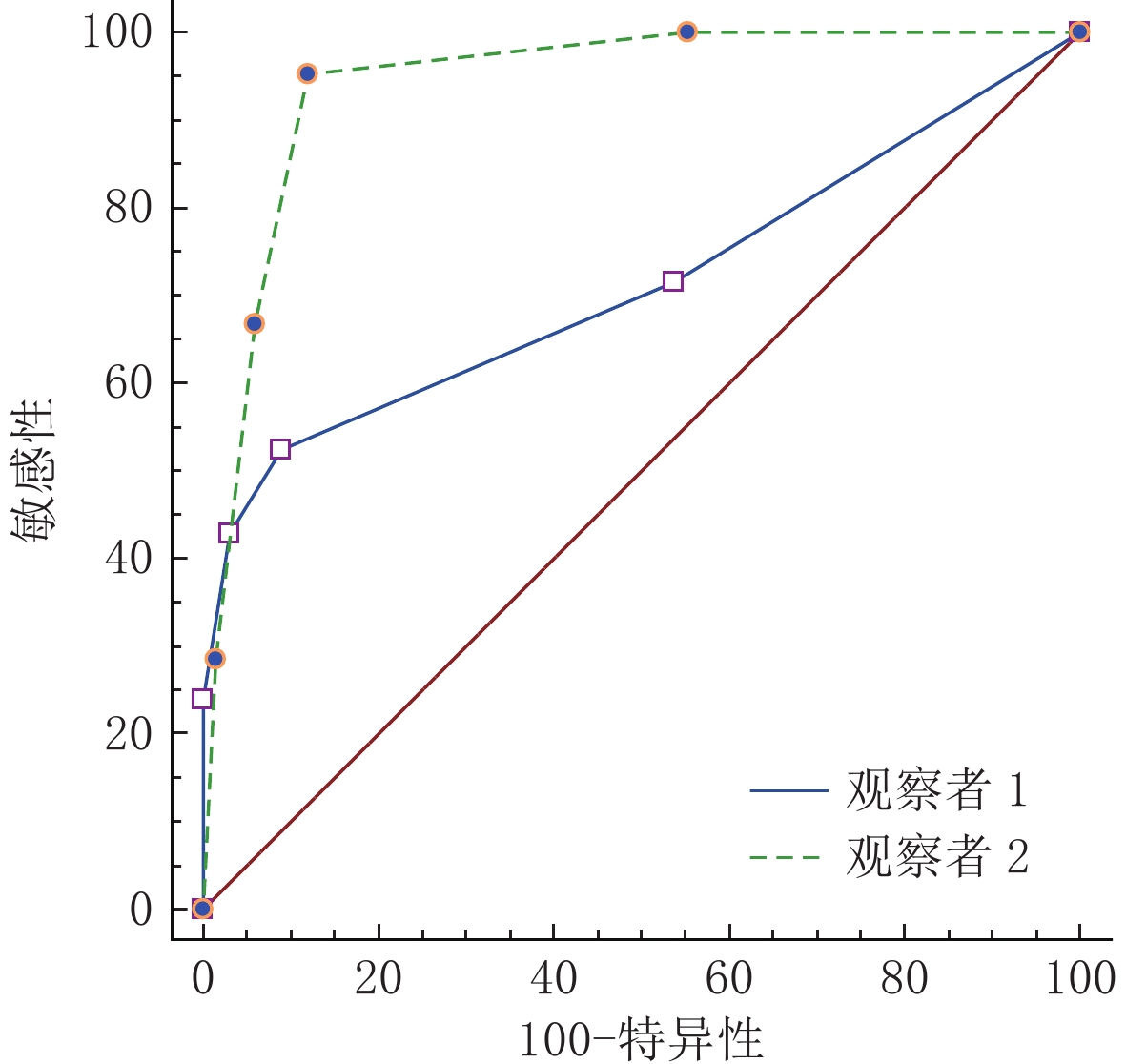
图 1 两名观察者应用Bosniak v.2019诊断肾囊性病变良恶性的ROC曲线图
Figure 1. ROCs of Bosniak v. 2019 in the diagnosis of benign and malignant renal cystic lesions by two reviewers
![]()
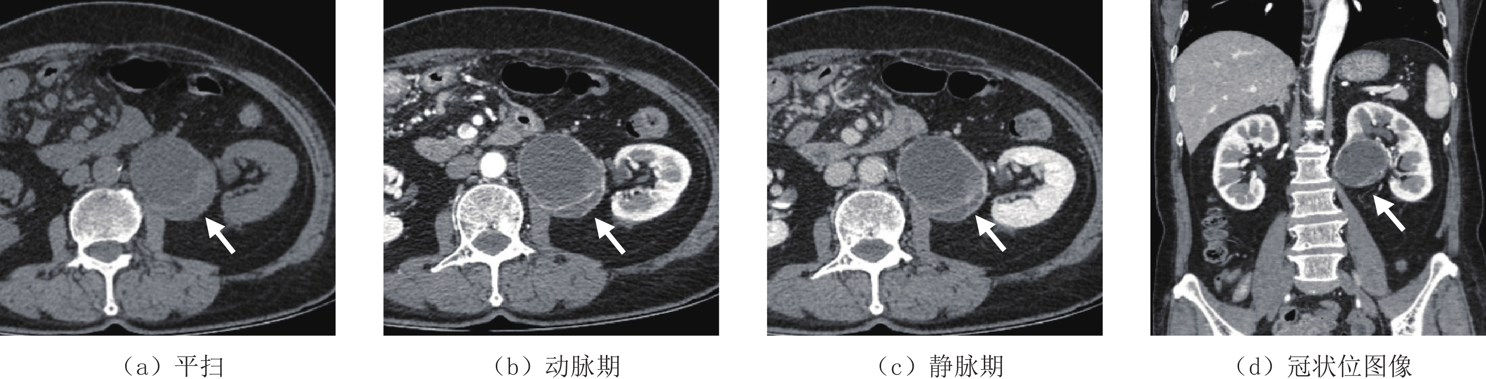
图 2 62岁女性,两名观察者对该囊性病变(白箭头)的分类分别为ⅡF和 Ⅲ 类,最终病理类型为透明细胞性肾细胞癌(WHO/ISUP 分级:2级)
Figure 2. Female, 62 years old, the two observers classified the cystic lesion (white arrow) as ⅡF and Ⅲ, respectively, with the final pathological type being clear cell renal cell carcinoma (WHO/ISUP grade: 2)
表 1 肾囊性病变CT细节特征的评测结果
Table 1 Evaluation results of CT details of renal cystic lesions
CT细节 观察者1 观察者2 ICC值 大小/mm 52.30±18.20 51.90±18.50 0.97 均质程度 1.56±0.69 1.60±0.72 0.96 CT值/HU 15.01±11.90 15.24±11.54 0.98 动脉期CT值/HU 16.16±13.61 17.92±13.71 0.97 静脉期CT值/HU 16.66±13.94 18.85±13.78 0.96 钙化大小/mm 2.60±2.51 2.48±2.14 0.97 囊壁厚度/mm 1.19±0.45 1.31±0.53 0.73 分隔数量/mm 2.20±2.33 2.70±2.35 0.90 分隔厚度/mm 1.47±0.87 1.60±0.86 0.41 结节/mm 4.23±4.30 3.9±2.84 0.96 不规则突起厚度/mm 1.67±0.58 1.25±0.50 0.50  下载: 导出CSV
表 2 两名观察者对肾囊性病变的Bosniak分类结果
Table 2 Bosniak classification results for renal cystic lesions by the two reviewers
Bosniak v.2019 肾囊性病变分类(总计(恶性病例)) Kappa值 观察者1 观察者2 Ⅰ 37(0) 32(0) 0.834 Ⅱ 34(5) 31(1) 0.781 ⅡF 6(5) 10(6) 0.727 Ⅲ 6(6) 9(8) 0.782 Ⅳ 5(5) 6(6) 0.903
下载: 导出CSV
表 3 肾囊性病变良、恶性CT细节差异
Table 3 Differences in CT details between benign and malignant renal cystic diseases
CT细节 病变情况 P 良性 恶性 大小/mm 55.70±17.80 40.00±15.30 0.000 均质程度 1.46±0.61 2.05±0.86 0.001 CT值/HU 12.60±8.33 23.67±15.89 0.000 动脉期CT值/HU 14.00±8.90 30.43±18.44 0.005 静脉期CT值/HU 14.76±8.90 31.91±18.14 0.001 钙化大小/mm 2.68±2.40 1.83±0.75 0.408 囊壁厚度/mm 1.15±0.40 1.81±0.60 0.000 分隔数量/mm 1.74±1.71 4.78±2.24 0.000 分隔厚度/mm 1.28±0.69 2.28±0.83 0.000
下载: 导出CSV
-
[1] MENSEL B, KUHN J P, KRACHT F, et al. Prevalence of renal cysts and association with risk factors in a general population: An MRI-based study[J]. Abdominal radiology (New York), 2018, 43(11): 3068−3074. doi: 10.1007/s00261-018-1565-5
[2] YAN J H, CHAN J, OSMAN H, et al. Bosniak Classification version 2019: Validation and comparison to original classification in pathologically confirmed cystic masses[J]. European Radiology, 2021, 31(12): 9579−9587.
[3] BOSNIAK M A. The current radiological approach to renal cysts[J]. Radiology, 1986, 158(1): 1−10. doi: 10.1148/radiology.158.1.3510019
[4] GRAUMANN O, OSTHER S S, KARSTOFT J, et al. Bosniak classification system: Inter-observer and intra-observer agreement among experienced uroradiologists[J]. Acta Radiologica, 2015, 56(3): 374−383. doi: 10.1177/0284185114529562
[5] 刘柳, 周印, 李庆姝, 等. 肾脏囊性病变2019版Bosniak分级的应用及一致性分析[J]. 中国医学影像学杂志, 2022,30(11): 1161−1165. LIU L, ZHOU Y, LI Q S, et al. Application and consistency analysis of the 2019 edition of bosniak classification of renal cystic lesions[J]. Chinese Journal of Medical Imaging, 2022, 30(11): 1161−1165. (in Chinese).
[6] 周航, 胡杉, 李树荣, 等. 基于2019版Bosniak分级系统对肾囊性病变的MSCT与病理对照研究[J]. 放射学实践, 2022,37(5): 556−559. ZHOU H, HU S, LI S R, et al. Correlative study between MSCT and histopathology based on bosniak classification version 2019[J]. Radiol Practice, 2022, 37(5): 556−559. (in Chinese).
[7] SILVERMAN S G, PEDROSA I, ELLIS J H, et al. Bosniak classification of cystic renal masses, version 2019: An update proposal and needs assessment[J]. Radiology, 2019, 292(2): 475−488. doi: 10.1148/radiol.2019182646
[8] ARITA Y, YOSHIDA S, KWEE T C, et al. Clinical utility of the Bosniak classification version 2019: Diagnostic value of adding magnetic resonance imaging to computed tomography examination[J]. European Journal of Radiology, 2022, 148: 110163. doi: 10.1016/j.ejrad.2022.110163
[9] PACHECO E O, TORRES U S, ALVES A M A, et al. Bosniak classification of cystic renal masses version 2019 does not increase the interobserver agreement or the proportion of masses categorized into lower Bosniak classes for non-subspecialized readers on CT or MR[J]. European Journal of Radiology, 2020, 131: 109270.
[10] SMITH A D, ABOU E A. Approach to renal cystic masses and the role of radiology[J]. Radiologic Clinics of North America, 2020, 58(5): 897−907.
[11] SHAMPAIN K L, SHANKAR P R, TROOST J P, et al. Interrater agreement of bosniak classification version 2019 and version 2005 for cystic renal masses at CT and MRI[J]. Radiology, 2022, 302(2): 357−366.
[12] EDNEY E, DAVENPORT M S, CURCI N, et al. Bosniak classification of cystic renal masses, version 2019: Interpretation pitfalls and recommendations to avoid misclassification[J]. Abdominal Radiology, 2021, 46(6): 2699−2711. doi: 10.1007/s00261-020-02906-8
-
期刊类型引用(4)
1. 尹振琪,吴俊峰,朱石柱. 人工智能辅助诊断系统结合低剂量CT在肺小结节筛查中的应用进展. 影像研究与医学应用. 2024(24): 7-9 .  百度学术
百度学术
2. 王璟琛,柴军. 人工智能体积密度法判断肺亚实性结节的浸润性研究. CT理论与应用研究. 2023(02): 241-248 . 本站查看
3. 王和良,苏良宝,蔡少辉,杜丽珠. 人工智能辅助诊断系统在肺结节CT检测中的应用分析. 中国医疗器械信息. 2023(05): 91-93+161 . 百度学术
4. 苏寅晨,张晓琴. 人工智能在肺结节检测及诊断中的应用进展. 内蒙古医学杂志. 2023(04): 506-508 . 百度学术
其他类型引用(1)
计量
- 文章访问数: 256
- HTML全文浏览量: 81
- PDF下载量: 15
- 被引次数: 5


