
Computed Tomography and Magnetic Resonance Imaging Diagnosis of Intracranial Solitary Fibrous Tumor: A Clinical Case Analysis
-
摘要:
颅内孤立性纤维瘤(ISFT)是一种间叶组织来源的梭形细胞肿瘤,临床表现依据病程和肿瘤发生部位不同表现各异,发病罕见,影像及临床医生对其缺乏全面认识,术前误诊率高。本文报告1例颅内孤立性纤维瘤病例,52岁男性,临床表现为头痛及双下肢无力;CT及MRI影像学检查诊断为左侧听神经瘤。后经术后病理组织活检确诊为ISFT。患者术后1年半复查,左侧面神经麻痹,左侧面部憋胀,余无明显不适。本文回顾性分析该患者的影像学表现及临床资料,旨在总结此种罕见病的CT及MRI影像表现,以提高医生的术前诊断准确率,为临床精准治疗提供重要的帮助。
Abstract:Intracranial solitary fibroma tumor (ISFT) is a kind of mesenchymal tissue-derived spindle cell tumor. Patients’ disease progression vary according to the course and location of the tumor. Due to its rarity, radiologists and clinicians lack a comprehensive understanding of ISFT. Hence, the preoperative misdiagnosis rate is high. This case report describes a 52-year-old male patient with intracranial solitary fibroma who presented with headache and weakness of both lower extremities. He underwent radiological examination, including computed tomography (CT) and magnetic resonance imaging (MRI), and was diagnosed with acoustic neuroma. After postoperative pathological tissue biopsy, he was diagnosed with ISFT. He was reviewed one and a half years after surgery; there was no significant discomfort in addition to the paralysis of the left facial nerve and swelling of the left side of the face. This case report retrospectively analyzes the radiological scans and the clinical data of the patient to summarize the key CT and MRI features of ISFT, improving the accuracy of the preoperative diagnosis of this rare disease, and contributing to current knowledge of the precise treatment of ISFT.
-
Keywords:
- tomography /
- X-ray computed /
- magnetic resonance imaging /
- solitary fibrous tumors
-
颅内孤立性纤维瘤(intracranial solitary fibrous tumor,ISFT)是一种间叶组织来源的梭形细胞肿瘤,发病罕见,尽管近年来国内外相关报道的病例逐渐增多,但是影像及临床医生对其仍缺乏全面认识,术前误诊率高。
本文介绍1例经术后病理证实的颅内孤立性纤维瘤,结合其病例资料进行相关分析,以提高医生对此种罕见病的术前诊断准确率,对手术方案制定及预后有重要价值,具有报道意义。
1. 病例资料
1.1 临床资料
患者,男,52岁,因“双下肢无力,步态不稳20天”入院。入院前10年无明显诱因出现头痛,自行口服药物治疗,未系统治疗。近20天感下肢行走无力,伴踩棉花感,发病以来精神食欲及睡眠较差,大小便正常。
1.2 实验室检查
白蛋白37.3↓(40~55 g/L),天门冬氨酸转移酶9.8↓(15~40 U/L),胱抑素C 1.49↑(0~1.4 mg/L),补体C1 q138.9↓(159~233 mg/L);余(-)。
1.3 影像学表现
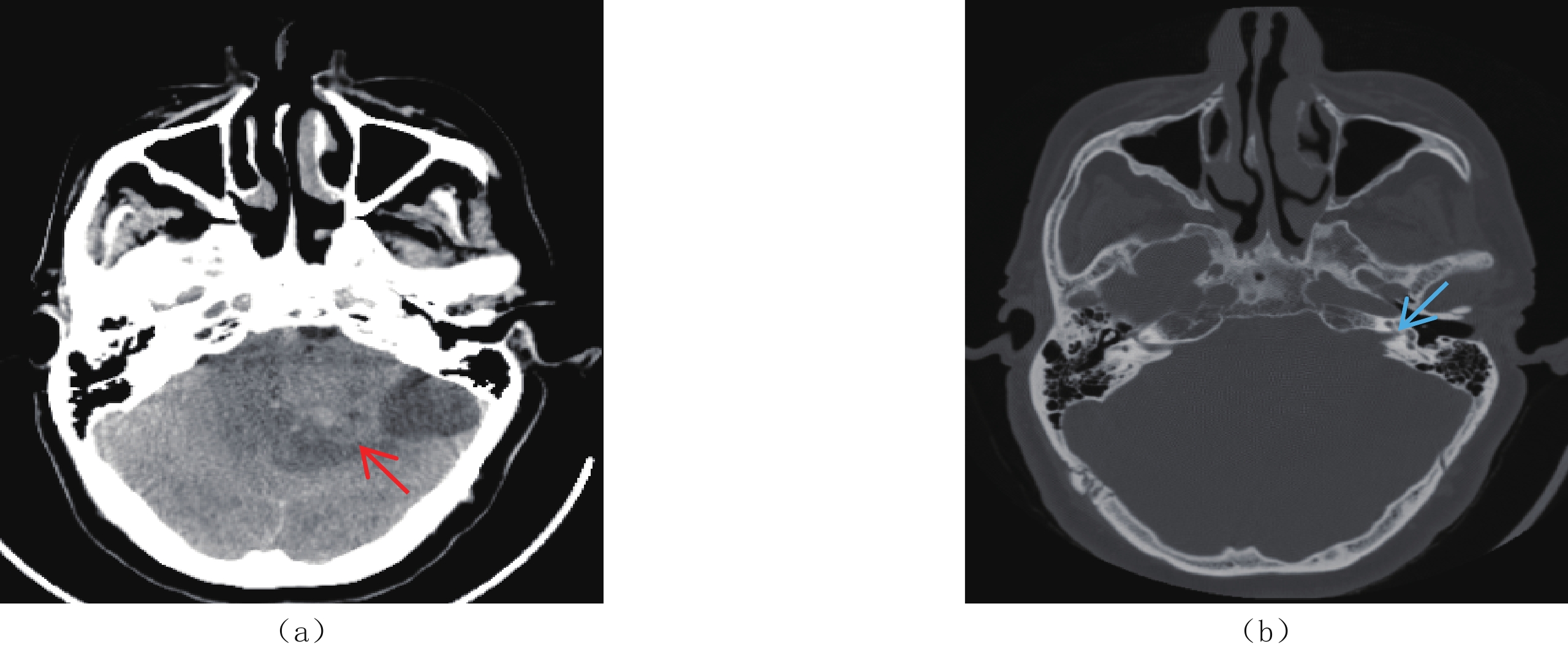
CT:平扫左侧桥小脑角区片状低密度影(图1)。MRI:左侧桥小脑角区听神经根部可见大小约4.3 cm×5.9 cm×4.8 cm,囊实性混杂信号影,实性部分T1WI、T2WI呈混杂信号,边界清晰,边缘欠规整,DWI呈混杂信号。增强扫描实性成分呈不均匀明显持续性强化(图2),听神经增粗,左侧内听道扩大。影像诊断为左侧听神经瘤。
![]() 图 1 ISFT患者CT平扫图像(a)和(b)轴位CT平扫示左侧桥小脑角区片状低密度影(红箭),周围水肿明显,左侧内听道扩大(蓝箭)。Figure 1. CT plain scan of the patient with ISFT
图 1 ISFT患者CT平扫图像(a)和(b)轴位CT平扫示左侧桥小脑角区片状低密度影(红箭),周围水肿明显,左侧内听道扩大(蓝箭)。Figure 1. CT plain scan of the patient with ISFT![]() 图 2 ISFT患者MRI平扫及增强图像(a)~(c)MRI平扫示左侧桥小脑角区听神经根部大小约4.3 ㎝×5.9 ㎝×4.8 ㎝囊实性混杂信号影(红箭),实性部分T1WI(a)、T2WI(b)呈混杂信号,边界清晰,边缘欠规整,DWI(c)呈混杂信号,听神经增粗,左侧内听道扩大,邻近脑实质及第四脑室受压推移,瘤周水肿明显,并可见流空血管影;(d)~(g)增强扫描图像,(d)和(e)为轴位,(f)和(g)分别为矢状位和冠状位,肿瘤实性成分呈不均匀明显持续性强化(红箭),窄基底与硬脑膜相连,T2WI低信号区强化明显。Figure 2. MRI plain and contrast-enhanced images of the patient with ISFT
图 2 ISFT患者MRI平扫及增强图像(a)~(c)MRI平扫示左侧桥小脑角区听神经根部大小约4.3 ㎝×5.9 ㎝×4.8 ㎝囊实性混杂信号影(红箭),实性部分T1WI(a)、T2WI(b)呈混杂信号,边界清晰,边缘欠规整,DWI(c)呈混杂信号,听神经增粗,左侧内听道扩大,邻近脑实质及第四脑室受压推移,瘤周水肿明显,并可见流空血管影;(d)~(g)增强扫描图像,(d)和(e)为轴位,(f)和(g)分别为矢状位和冠状位,肿瘤实性成分呈不均匀明显持续性强化(红箭),窄基底与硬脑膜相连,T2WI低信号区强化明显。Figure 2. MRI plain and contrast-enhanced images of the patient with ISFT1.4 诊疗过程
手术记录:术中见肿瘤色淡红,包膜完整,血供丰富,肿瘤向上方生长到小脑幕孔,压迫脑干,与脑干粘连紧密,并包绕面神经。
术后病理:脑部梭形细胞肿瘤(图3),免疫组化结果支持孤立性纤维性肿瘤。免疫组化结果:Vim(+),GFAP(-),CD34(+),EMA(-),CK(-),S-100(-),SMA(-),Calponin(-),CD31(-),Bcl-2(+),CD99(少部分+),CD68(散+),Lysozyme(散+),P53(-),Ki-67(+3~5%)。
![]() 图 3 ISFT患者术后病理图像(a)和(b)镜下肿瘤细胞呈梭形,稀疏区与密集区交替分布(HE,×200)。Figure 3. Postoperative pathological images of the patient with ISFT
图 3 ISFT患者术后病理图像(a)和(b)镜下肿瘤细胞呈梭形,稀疏区与密集区交替分布(HE,×200)。Figure 3. Postoperative pathological images of the patient with ISFT1.5 随访
患者术后1年半门诊复查,左侧面神经麻痹,左侧面部憋胀,余无明显不适。
2. 讨论
孤立性纤维瘤(solitary fibrous tumor,SFT)是一种间叶组织来源的梭形细胞肿瘤,起源于结缔组织中的CD34阳性的树突状细胞,既可发生于胸膜腔,也可发生于全身各部位,如肝脏、腹膜、头颈部等。颅内孤立性纤维瘤由Carneiro等[1]于1996年首先报道,发病率极低,目前国内外文献报道相关病例较少。
2021年WHO中枢神经系统肿瘤将其划分为间叶性非脑膜上皮肿瘤,分为Ⅰ~Ⅲ 级;核分裂象<5/10 HPF时,若镜下有致密胶原纤维伴相对较低密度的梭形细胞则为Ⅰ级,若梭形肿瘤细胞多而胶原较少,且有“鹿角”状脉管系统则为Ⅱ级,核异型性明显,核分裂象≥5/10个高倍镜视野(high power field,HPF)时,为 Ⅲ 级。该分类取消了2016版“孤立性纤维瘤/血管外皮细胞瘤(hemangiopericytoma,HPC)”的混合术语,并将所谓HPC合并于“孤立性纤维瘤”[2-3]。
2.1 病因及发病机制
孤立性纤维瘤的病因及发病机制尚不明确,Miettinen等[4]认为SFT的发生可能与环境因素或基因变异相关,其可能的机制是包含NAB2-STAT6融合基因的染色体12q13臂内倒置。
2.2 临床特征
ISFT在人群中的发病率并无显著性别差异,可发生于任何年龄段,以51~60岁年龄组占比最大,本例ISFT为中老年男性,发病年龄与以往文献报道一致。ISFT大多位于小脑幕(16%),其次是大脑额凸、桥小脑角区(CPA)、脑室、大脑镰和颅后窝[5]。
临床表现依据病程和发生的部位不同表现各异,初期可无明显症状,肿瘤体积增大时发生在幕上常有头痛、头晕、恶心、呕吐、癫痫等症状;幕下可有听力下降、脑干受压或行走不稳等症状。本例发生于左侧桥小脑角区,双下肢无力,步态不稳,符合幕下ISFT临床特征。
2.3 CT与MRI影像学表现
CT平扫检查ISFT表现为孤立性、实质性肿块,多呈圆形、类圆形,一般体积较大,边界多光整,境界清楚,无分叶或浅分叶,呈软组织密度,囊变坏死区呈低密度,钙化少见。增强扫描病灶实性部分明显强化,体积较大时可见无强化的囊变坏死区[6]。
MRI检查具有良好的软组织分辨能力,在ISFT诊断中具有明显优势。T1WI多呈等低或等信号,T2WI可表现为高信号、稍高信号或低信号。T2WI上出现高低信号混杂的现象称为“阴阳征”或“黑白征”,是影像诊断ISFT一个相当重要的典型征象,即高信号区反映黏液坏死变性及血管间质细胞堆积,稍高信号区反映肿瘤细胞密集区域,低信号区反映致密胶原纤维[7-8]。动态增强扫描肿瘤多为不均匀持续性强化或进行性延迟强化,T2WI低信号区明显强化[9-10]。
此外,ISFT由于瘤体及瘤周血运丰富,常出现流空血管影,也是典型征象之一。但是部分脑膜瘤也可见血管流空,区别在于ISFT多表现为“蛇形流空效应”,而脑膜瘤以“光芒征”多见,其原因是ISFT主要来源于颈内动脉或椎动脉分支供血,而脑膜瘤主要来源于颈外动脉的脑膜中动脉供血[11-12],因此可以为二者鉴别诊断提供思路。部分ISFT可见“脑膜尾征”,但该征象少见,不具有特异性,只能说明肿瘤与硬脑膜关系密切,并不能对肿瘤来源进行鉴别。
本例患者出现“阴阳征”与流空血管影,并且肿瘤以窄基底与硬膜相连,呈“脑膜尾征”,符合文献报道。
2.4 诊断与鉴别诊断
尽管颅内孤立性纤维瘤患者的MRI表现具有相对特异性,但是未出现“阴阳征”等典型表现时较难做出明确诊断,确诊依赖于病理及免疫组织化学检查。ISFT病理主要表现为梭形细胞呈束状或不规则排列在透明样病变血管周围,伴有宽大的胶原纤维[13]。免疫组化检查常表现为CD34(+)、Bcl-2(+)和Vimentin(+);EMA(-)和S-100(-)[14],其中CD34可作为特异性免疫标志物之一,本例患者免疫组化结果与文献一致,符合颅内孤立性纤维瘤诊断。
随着对SFT的深入研究,有学者发现STAT6诊断SFT有高度敏感性和特异性[15],STAT6与CD34等传统标记物联合诊断SFT可能成为新趋势。
ISFT在影像学上主要与脑膜瘤、神经鞘瘤和转移瘤等鉴别。①脑膜瘤:起源于蛛网膜帽状细胞,多呈圆形或类圆形,CT平扫肿瘤多为均匀的略高密度,钙化多见,相邻颅骨可见增生,增强扫描肿瘤呈均匀一致的显著强化,边缘锐利;在T1WI上多数为等信号,少数低信号,T2WI可为高、等或低信号,增强扫描明显均匀强化并有“脑膜尾征”。而ISFT信号多不均匀,稍高信号内可见小片状低信号,不均匀持续性强化或进行性延迟强化;相邻颅骨可有侵蚀,增生少见。②神经鞘瘤:ISFT发生于桥小脑角区时需与听神经鞘瘤鉴别,后者可见特征性桥小脑角区“冰激凌征”,与周围组织分界清楚,囊变坏死多见,多呈不均匀T1WI低、T2WI高信号,增强扫描实性部分明显强化,较早出现颅神经功能缺损症状。本例患者肿瘤发生于桥小脑角区,影像可见听神经增粗、内听道扩大等表现,故术前误诊为听神经瘤。
综上所述,颅内孤立性纤维瘤是一种临床罕见的中枢神经系统肿瘤,当瘤内出现“阴阳征”或“黑白征”、T2WI低信号区明显强化及流空血管影等时应考虑诊断ISFT,最终确诊需病理及免疫组织化学检查。
-
![]()
图 1 ISFT患者CT平扫图像
(a)和(b)轴位CT平扫示左侧桥小脑角区片状低密度影(红箭),周围水肿明显,左侧内听道扩大(蓝箭)。
Figure 1. CT plain scan of the patient with ISFT
![]()
图 2 ISFT患者MRI平扫及增强图像
(a)~(c)MRI平扫示左侧桥小脑角区听神经根部大小约4.3 ㎝×5.9 ㎝×4.8 ㎝囊实性混杂信号影(红箭),实性部分T1WI(a)、T2WI(b)呈混杂信号,边界清晰,边缘欠规整,DWI(c)呈混杂信号,听神经增粗,左侧内听道扩大,邻近脑实质及第四脑室受压推移,瘤周水肿明显,并可见流空血管影;(d)~(g)增强扫描图像,(d)和(e)为轴位,(f)和(g)分别为矢状位和冠状位,肿瘤实性成分呈不均匀明显持续性强化(红箭),窄基底与硬脑膜相连,T2WI低信号区强化明显。
Figure 2. MRI plain and contrast-enhanced images of the patient with ISFT
-
[1] CARNEIRO S S, SCHEITHAUER B W, NASCIMENTO A G, et al. Solitary fibrous tumor of the meninges: A lesion distinct from fibrous meningioma. A clinicopathologic and immunohistochemical study[J]. American Journal of Clinical Pathology, 1996, 106(2): 217−224. DOI: 10.1093/ajcp/106.2.217.
[2] LOUIS D N, PERRY A, WESSELING P, et al. The 2021 WHO classification of tumors of the central nervous system: A summary[J]. Neuro-Oncology, 2021, 23(8): 1231−1251. DOI: 10.1093/neuonc/noab106.
[3] 吴虹林, 苏伟杰, 李西西, 等. 中枢神经系统孤立性纤维瘤临床特征及术后复发因素[J]. 中国神经精神疾病杂志, 2023,49(2): 85−91. DOI: 10.3969/j.issn.1002-0152.2023.02.003. WU H J, SU W J, LI X X, et al. Clinical features and recurrent factors of solitary fibrous tumor in central nervous system[J]. Chinese Journal of Nervous and Mental Diseases, 2023, 49(2): 85−91. DOI: 10.3969/j.issn.1002-0152.2023.02.003. (in Chinese).
[4] MIETTINEN M M, EL-RIFAI W, SARLOMO-RIKALA M, et al. Tumor size-related DNA copy number changes occur in solitary fibrous tumors but not in hemangiopericytomas[J]. Modern Pathology, 1997, 10(12): 1194−1200.
[5] FARGEN K M, OPALACH K J, WAKEFIELD D, et al. The central nervous system solitary fibrous tumor: A review of clinical, imaging and pathologic findings among all reported cases from 1996 to 2010[J]. Clinical Neurology and Neurosurgy, 2011, 113(9): 703−710. DOI: 10.1016/j.clineuro.2011.07.024.
[6] 高文华, 黄玉芳, 朱小贵, 等. 颅内孤立性纤维瘤影像表现[J]. 现代医用影像学, 2019,28(7): 1472−1474. doi: 10.3969/j.issn.1006-7035.2019.07.005 GAO W H, HUANG Y F, ZHU X G, et al. Imaging characters of intracranial solitary fibrous tumor[J]. Modern Medical Imageology, 2019, 28(7): 1472−1474. (in Chinese). doi: 10.3969/j.issn.1006-7035.2019.07.005
[7] 梅磊磊, 聂蕾, 唐文英, 等. 孤立性纤维瘤的影像表现及临床病理特征[J]. 放射学实践, 2022,37(5): 566−570. DOI: 10.13609/j.cnki.1000-0313.2022.05.006. MEI L L, NIE L, TANG W Y, et al. Imaging and clinicopathologic features of solitary fibrous tumor[J]. Radiologic Practice, 2022, 37(5): 566−570. DOI: 10.13609/j.cnki.1000-0313.2022.05.006. (in Chinese).
[8] 曾禹莉, 易熙, 赵洁, 等. 颅内孤立性纤维瘤的CT及MRI表现[J]. 影像研究与医学应用, 2022,6(4): 163−165. doi: 10.3969/j.issn.2096-3807.2022.04.055 [9] CLARENCON F, BONNEVILLE F, ROUSSEAU A, et al. Intracranial solitary fibrous tumor: Imaging findings[J]. European Journal of Radiology, 2011, 80(2): 387−394. DOI: 10.1016/j.ejrad.2010.02.016.
[10] 何文杰, 雷益, 焦娟, 等. 颅内孤立性纤维瘤/血管外皮瘤的影像表现与病理分析[J]. 放射学实践, 2019,34(12): 1299−1303. DOI: 10.13609/j.cnki.1000-0313.2019.12.004. HE W J, LEI Y, JIAO J, et al. Imaging features and pathological analysis of intracranial solitary fibrous tumor/hemangiopericytoma[J]. Radiologic Practice, 2019, 34(12): 1299−1303. DOI: 10.13609/j.cnki.1000-0313.2019.12.004. (in Chinese).
[11] 冯加和, 杨云竣, 杨粤龙, 等. 颅内孤立性纤维瘤/血管外皮瘤瘤内血管流空特征研究[J]. 医学影像学杂志, 2022,32(8): 1273−1276. doi: 10.3969/j.issn.1006-9011.2022.8.yxyxxzz202208002 FENG J H, YANG Y J, YANG Y L, et al. Study on the characteristics of intratumoral vascular flow void in solitary fibrous tumor/hemangiopericytoma[J]. Journal of Medical Imaging, 2022, 32(8): 1273−1276. (in Chinese). doi: 10.3969/j.issn.1006-9011.2022.8.yxyxxzz202208002
[12] 张宇泽, 杨云竣, 陈静勿, 等. 血管瘤型脑膜瘤与颅内孤立性纤维瘤的MRI征象分析[J]. 临床放射学杂志, 2022,41(11): 2010−2014. DOI: 10.13437/j.cnki.jcr.2022.11.007. ZHANG Y Z, YANG Y J, CHEN J W, et al. A comparative study on MRI signs between angiomatous meningioma and solitary fibrous tumor[J]. Journal of Clinical Radiology, 2022, 41(11): 2010−2014. DOI: 10.13437/j.cnki.jcr.2022.11.007. (in Chinese).
[13] 陶静雄, 梁奕, 王佳, 等. 颅内孤立性纤维瘤的影像学和病理学特征[J]. 医学影像学杂志, 2019,29(11): 1976−1978. TAO J X, LIANG Y, WANG J, et al. Imaging and pathological features of intracranial solitary fibrous tumor[J]. Journal of Medical Imaging, 2019, 29(11): 1976−1978. (in Chinese).
[14] 柯代波, 刘文科, 张思, 等. 颅内孤立性纤维瘤的MRI表现[J]. 华西医学, 2017,32(1): 46−50. DOI: 10.7507/1002-0179.201511166. KE D B, LIU W K, ZHANG S, et al. MRI manifestations of intracranial solitary fibrous tumor[J]. West China Medical Journal, 2017, 32(1): 46−50. DOI: 10.7507/1002-0179.201511166. (in Chinese).
[15] DOYLE L A, VIVERO M, FLETCHER C D, et al. Nuclear expression of STAT6 distinguishes solitary fibrous tumor from histologic mimics[J]. Modern Pathology, 2014, 27(3): 390−395. DOI: 10.1038/modpathol.2013.164.
-
期刊类型引用(1)
1. 杨庆东,彭泱泱,李绿洲. 腹壁侵袭性纤维瘤误诊为子宫肌瘤1例及文献复习. 中国当代医药. 2025(09): 138-142 .  百度学术
百度学术
其他类型引用(0)
 下载:
下载:


计量
- 文章访问数: 165
- HTML全文浏览量: 79
- PDF下载量: 45
- 被引次数: 1


