
Morphological Characteristics of Intracranial Aneurysms and Applicability of CT Angiography in Risk Assessment of Rupture
-
摘要:
目的:探讨CT血管造影(CTA)在颅内动脉瘤破裂风险评估中的应用价值及形态学特征。方法:选取2018年10月至2021年10月蚌埠医学院第三附属医院颅内动脉瘤患者82例,所有研究对象均通过西门子64排128层螺旋CT及西门子数字减影血管造影(DSA)机实施CTA检查及DSA检查。统计分析本组患者瘤体破裂情况、不同瘤体破裂情况者形态学特征,并分析颅内动脉瘤瘤体破裂影响因素。结果:本组82例颈动脉患者瘤体破裂率为74.39%(61/82)、瘤体未破裂率为25.61%(21/82);瘤体破裂者子囊数目(60.66%)、面积比、SR、AR、瘤体长度大于瘤体未破裂者,瘤体破裂者动脉瘤单发情况(91.80%)、动脉瘤分布位置与瘤体未破裂者比较无显著差异。Logistic回归分析证实,面积比、SR、AR、瘤体长度均是导致颅内动脉瘤瘤体破裂的重要危险因素。结论:通过CTA检查可明确颅内动脉瘤患者瘤体形态学特征,评估瘤体破裂风险。
Abstract:Objective: To explore the application value and morphological characteristics of CT angiography (CTA) in the risk assessment of intracranial aneurysm rupture. Method: We included 82 patients with intracranial aneurysms from the Third Affiliated Hospital of Bengbu Medical College from October 2018 to October 2021. All study participants underwent CTA and digital subtraction angiography (DSA) examinations using Siemens 64 row 128 slice spiral CT and Siemens DSA machines. Statistical analyses were conducted on the condition of tumor rupture. The morphological characteristics of different tumor rupture cases, and the factors influencing intracranial aneurysm rupture were analyzed. Result: The rupture rate of 82 carotid artery patients in this group was 74.39% (61/82), and the non-rupture rate of the tumor was 25.61% (21/82). The number of sub-capsules (60.66%), area ratio, Size ration (SR), Aspect ratio (AR) and length of the ruptured aneurysm were greater than that of the unruptured aneurysm. The ruptured and unruptured aneurysms did not differ significantly in the incidence of single aneurysm (91.80%) and the distribution of aneurysms. Logistic regression analysis confirmed that area ratio, SR, AR, and tumor length were important risk factors for intracranial aneurysm rupture. Conclusion: CTA examination can clarify the morphological characteristics of intracranial aneurysms and evaluate the risk of tumor rupture.
-
-
![]()
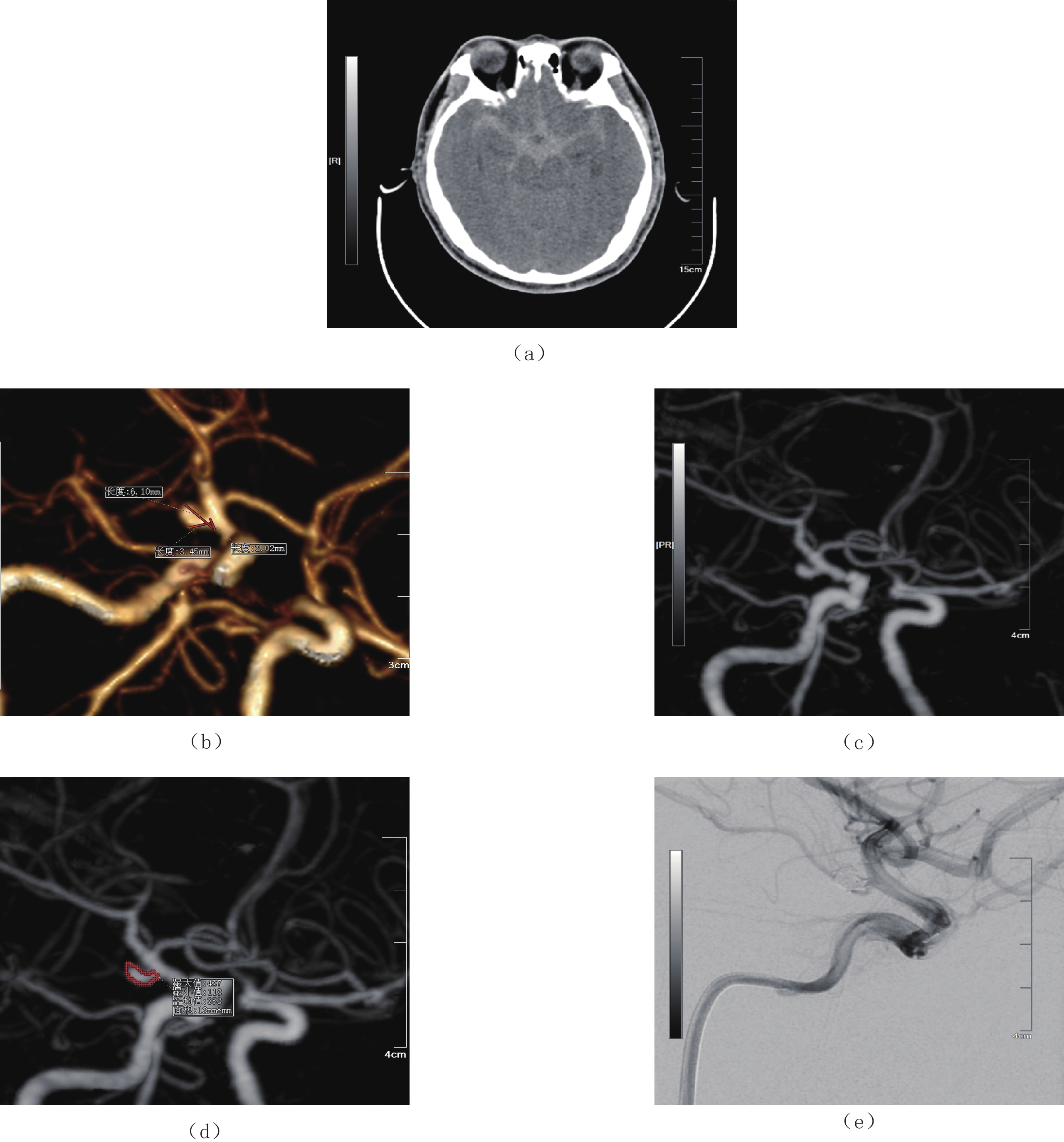
图 2 男性,46岁,突发头晕、头痛伴恶心呕吐
注:(a)CT横轴位平扫提示蛛网膜下腔出血;(b)CTA重建VRT提示右侧颈内动脉C7段动脉瘤,进行瘤高、瘤颈、瘤深测量;(c)和(d)MIP图提示右侧颈内动脉C7段动脉瘤以及瘤颈处动脉瘤面积测量;(e)DSA证实右侧颈内动脉C7段动脉瘤并进行栓塞处理。
Figure 2. Male, 46 years old, Sudden dizziness, headache、nausea and vomiting
![]()
图 3 男性,51岁,突发意识不清
注:(a)CT横轴位平扫提示蛛网膜下腔出血;(b)CTA重建VRT提示右侧颈内动脉C7段动脉瘤,进行瘤高、瘤颈、瘤深测量;(c)和(d)MIP图提示右侧颈内动脉C7段动脉瘤以及瘤颈处动脉瘤面积测量;(e)DSA证实右侧颈内动脉C7段动脉瘤。
Figure 3. Male, 51 years old, Sudden unconsciousness
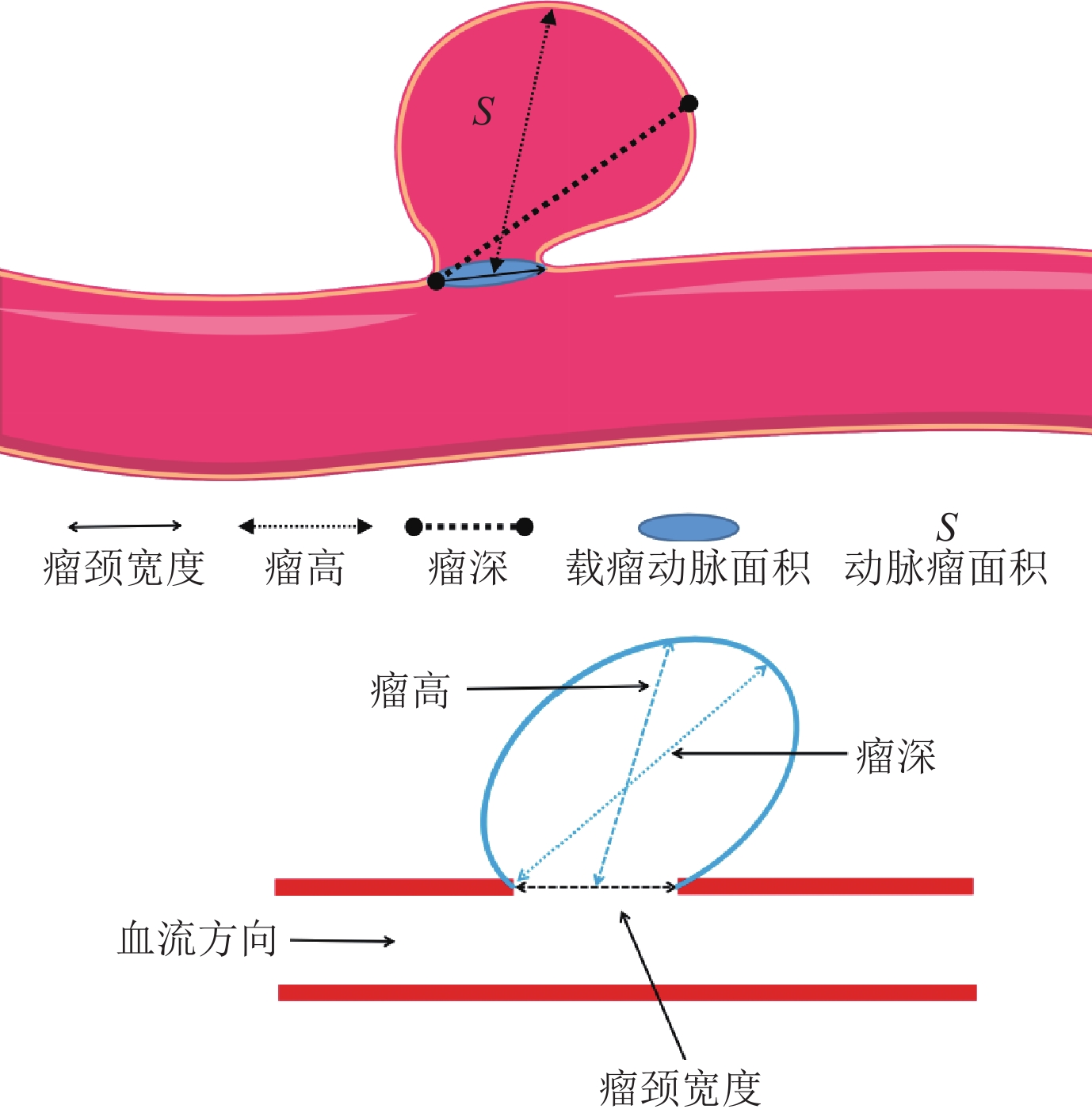
表 1 不同瘤体破裂情况者形态学特征分析
Table 1 Morphological characteristics analysis of patients with different tumor rupture situations
形态学特征 组别 统计检验 瘤体破裂 瘤体未破裂 $\chi^2/t $ P 例数 61 21 - - 子囊数目n/% 37(60.66) 5(23.81) 8.489 0.004 面积比 2.98±0.84 1.89±0.56 5.527 <0.001 SR 2.31±0.86 1.59±0.64 3.511 0.001 AR 1.71±0.72 1.30±0.59 2.349 0.021 瘤体长度/mm 6.11±2.13 4.49±1.64 3.172 0.002 动脉瘤单发n/% 56(91.80) 20(95.24) 0.272 0.602 动脉瘤分布位置n/% 大脑前动脉 7(11.48) 5(23.81) 1.902 0.168 大脑中动脉 12(19.67) 4(19.05) 0.004 0.950 前交通动脉 15(24.59) 3(14.29) 0.968 0.325 后交通动脉 20(32.79) 3(14.29) 2.650 0.104 后循环 7(11.48) 6(28.57) 3.423 0.064  下载: 导出CSV
表 2 颅内动脉瘤瘤体破裂影响因素分析
Table 2 Analysis of factors influencing intracranial aneurysm rupture
变量 β S.E. Wald $\chi^2 $ P OR 95% CI 面积比 1.280 0.414 9.556 <0.001 3.596 1.892~6.834 SR 1.215 0.406 8.954 <0.001 3.370 1.781~6.376 AR 1.597 0.613 6.790 <0.001 4.940 3.067~7.957 瘤体长度 1.534 0.571 7.216 <0.001 4.636 2.564~8.382
下载: 导出CSV
-
[1] 樊志红, 邢瑞欣. 双源CT血管造影应用于颅内动脉瘤诊断中的价值研究[J]. 中国实用医药, 2022, 17(1): 77−80. FAN Z H, XING R X. The value study of dual-source CT angiography in the diagnosis of intracranial aneurysms[J]. China Practical Medicine, 2022, 17(1): 77−80. (in Chinese).
[2] SCARABELLO M, ALEXANDRE A, LOZUPONE E, et al. Intra- and inter-observer variability in intracranial aneurysm segmentation: Comparison between CT angiography (semi-automated segmentation software stroke VCAR) and digital subtraction angiography (3D rotational angiography)[J]. La Radiologia Medica, 2021, 126(3): 484−493. DOI: 10.1007/s11547-020-01275-y.
[3] 唐运军, 韦典君, 卢忠武. CT血管造影与MR血管造影对颅内动脉瘤诊断价值的对比探讨[J]. 影像研究与医学应用, 2021, 5(18): 94−95. DOI: 10.3969/j.issn.2096-3807.2021.18.045. TANG Y J, WEI D J, LU Z W. Investigation of the comparative value of CT angiography and MR angiography for the diagnosis of intracranial aneurysms[J]. Imaging Research and Medical Application, 2021, 5(18): 94−95. DOI: 10.3969/j.issn.2096-3807.2021.18.045. (in Chinese).
[4] 刘莹, 曾辉, 姜磊, 等. CT血管造影、血清SICAM-1检测在颅内动脉瘤及术后复查中的意义[J]. 中国CT和MRI杂志, 2021, 19(7): 9−11. LIU Y, ZENG H, JIANG L, et al. Significance of CT angiography and serum SICAM-1 detection in intracranial aneurysms and postoperative review[J]. China CT and MRI Magazine, 2021, 19(7): 9−11. (in Chinese).
[5] GU Y, ZHANG Y, LUO M, et al. Risk factors for asymptomatic intracranial small aneurysm rupture determined by electrocardiographic-gated 4D computed tomographic (CT) angiography[J]. Medical Science Monitor: International Medical Journal of Experimental and Clinical Research, 2020, 26(1): e921835.
[6] 时玉春. 术前三维CT血管造影对颅内动脉瘤破裂出血的诊断及预后评估效能观察[J]. 中国医学工程, 2021, 29(1): 58−60. SHI Y C. Evaluation of the efficacy of diagnosis and prognosis of intracranial aneurysm[J]. The Medical Engineering of China, 2021, 29(1): 58−60. (in Chinese).
[7] 杨明, 肖刚. 256层三维螺旋CT血管造影对颅内动脉瘤的临床诊断价值分析[J]. 心血管病防治知识, 2020, 10(30): 21−22. YANG M, XIAO G. Analysis of the clinical diagnostic value of the 256-layer three-dimensional spiral CT angiography for intracranial aneurysms[J]. Knowledge of Cardiovascular Disease Prevention and Control, 2020, 10(30): 21−22. (in Chinese).
[8] 马英剑. 3D-CTA与3D-DSA对颅内动脉瘤破裂出血早期诊断价值分析[J]. 中国实用神经疾病杂志, 2020, 23(9): 808−811. MA Y J. Analysis of the value of 3D-CTA and 3D-DSA in the early diagnosis of ruptured and bleeding from intracranial aneurysms[J]. Chinese Journal of Practical Neurological Diseases, 2020, 23(9): 808−811. (in Chinese).
[9] 中国医师协会神经介入专业委员会, 中国颅内动脉瘤计划研究组. 中国颅内破裂动脉瘤诊疗指南2021[J]. 中国脑血管病杂志, 2021, 18(8): 546−574. Neurological Intervention Professional Committee of the Chinese Medical Doctor Association, China Intracranial Aneurysm Program Research Group. Guidelines for the diagnosis and treatment of ruptured intracranial aneurysms in China 2021[J]. Chinese Journal of Cerebrovascular Diseases, 2021, 18(8): 546−574. (in Chinese).
[10] 朱光斌, 杜国新, 刘燕, 等. 多层螺旋CT血管造影术与数字减影血管造影术对颅内动脉瘤诊断价值的对照分析[J]. 华北理工大学学报(医学版), 2019, 21(3): 197−201. ZHU G B, DU G X, LIU Y, et al. A controlled analysis of the diagnostic value of multilayer spiral CT angiography versus digital subtraction angiography for intracranial aneurysms[J]. Journal of North China University of Science and Technology (Medical Edition), 2019, 21(3): 197−201. (in Chinese).
[11] CALISKAN E, ONCEL D. CT angiography evaluation of intracranial aneurysms: Distribution, characteristics, and association with subarachnoid hemorrhage[J]. Nigerian Journal of Clinical Practice, 2021, 24(6): 833−840. DOI: 10.4103/njcp.njcp_97_20.
[12] 王三锋, 王筱璇. 三维CT血管造影诊断微小颅内动脉瘤的价值观察[J]. 中国肿瘤临床与康复, 2018, 25(11): 1357−1359. WANG S F, WANG X X. The value observation of 3D CT angiography for the diagnosis of tiny intracranial aneurysms[J]. Clinical and Rehabilitation of Oncology in China, 2018, 25(11): 1357−1359. (in Chinese).
[13] 刘要先, 宋太民, 王海增, 等. 三维CT血管造影与平板脑血管数字减影血管造影对颅内动脉瘤诊断价值的对比分析[J]. 中国医学工程, 2018, 26(10): 12−14. LIU Y X, SONG T M, WANG H Z, et al. Comparative analysis of the diagnostic value of 3D CT angiography with the digital subtraction angiography of flat brain vessels for intracranial aneurysms[J]. The Medical Engineering of China, 2018, 26(10): 12−14. (in Chinese).
[14] 冯锐元. 64排螺旋CT血管造影在颅内动脉瘤中的临床应用价值[J]. 临床医学研究与实践, 2018, 3(17): 148−149. FENG R Y. Clinical utility of 64-row spiral CT angiography in intracranial aneurysms[J]. Clinical Medicine Research and Practice, 2018, 3(17): 148−149. (in Chinese).
[15] CUI Y, XING H, ZHOU J, et al. Aneurysm morphological prediction of intracranial aneurysm rupture in elderly patients using four-dimensional CT angiography[J]. Clinical Neurology and Neurosurgery, 2021, 208(1): 106877.
[16] 王坤, 杨尚文, 顾康康, 等. 头颅电子计算机断层扫描血管造影和头颅磁共振血管成像在颅内动脉瘤中诊断价值比较的回顾性研究[J]. 现代生物医学进展, 2021, 21(20): 3912−3916. WANG K, YANG S W, GU K K, et al. A retrospective study comparing the diagnostic value of cranial electronic computed tomography angiography and cranial magnetic resonance vascular imaging in intracranial aneurysms[J]. Advances in Modern Biomedicine, 2021, 21(20): 3912−3916. (in Chinese).
[17] ZHANG J, LI X, ZHAO B, et al. Irregular pulsation of intracranial unruptured aneurysm detected by four-dimensional CT angiography is associated with increased estimated rupture risk and conventional risk factors[J]. Journal of Neurointerventional Surgery, 2021, 13(9): 854−859. DOI:10.1136/neurintsurg- 2020-016811.
[18] 李波. 256排螺旋CT血管造影检查在颅内动脉瘤中的应用价值[J]. 中国药物与临床, 2019, 19(2): 237−238. LI B. Value of 256-spiral CT angiography in intracranial aneurysms[J]. Chinese Drug and Clinical, 2019, 19(2): 237−238. (in Chinese).
[19] ZWANZGER C, LOPEZ-RUEDA A, CAMPODONICO D, et al. Usefulness of CT angiography for characterizing cerebral arteriovenous malformations presenting as hemorrhage: Comparison with digital subtraction angiography[J]. Radiologia, 2020, 62(5): 392−399. DOI: 10.1016/j.rx.2020.01.006.
[20] 梁满球, 陈妙玲, 徐文, 等. 3D-CTA在急性破裂颅内动脉瘤诊断和治疗中的应用价值分析[J]. 中国数字医学, 2019, 14(12): 76−79. LIANG M Q, CHEN M L, XU W, et al. Analysis of the application value of 3D-CTA in the diagnosis and treatment of acute ruptured intracranial aneurysms[J]. Digital Medicine in China, 2019, 14(12): 76−79. (in Chinese).
[21] MOCCO J, BROWN R D J R, TORNER J C, et al. Aneurysm morphology and prediction of rupture: An international study of unruptured intracranial aneurysms analysis[J]. Neurosurgery, 2018, 82(4): 491−496. DOI: 10.1093/neuros/nyx226.

计量
- 文章访问数: 152
- HTML全文浏览量: 49
- PDF下载量: 24


