
Differences in Volume Rendering Imaging Based on Different Algorithms in Assisting Detection of Linear Fracture of Nasal Bone Area
-
摘要:
目的:探索最佳的容积再现成像(VR)重建算法,以提高对鼻区线性骨折的诊断效能。方法:回顾性纳入2022年8月至2023年8月的成人鼻骨CT影像资料,随机选取100例鼻区线性骨折和35例无骨折患者,分别行平滑算法(Smooth)、标准算法(Standard)、锐利算法(Sharp)、骨算法(Bone)的VR后处理。两名放射医师以双盲法对VR有无骨折、鼻骨孔显示及图像质量进行评分。采用相同协议对CT质控模体进行扫描,并测量不同重建算法模体图像的噪声功率谱(NPS),任务传递函数(TTF)和可检测性指数(
$ d'$ )。结果:医师对鼻区线性骨折的诊断效能在VR_Standard、VR_Sharp和VR_Bone之间存在差异,VR_Sharp的鼻骨孔显示评分高于VR_Standard,并且VR_Sharp的图像质量评分高于VR_Standard和VR_Bone;随着重建算法锐利程度的增加,噪声量和空间分辨力逐渐增加;Standard组、Sharp组和Bone组的NPS峰值和TTF50%分别为(225.85 HU2·mm2,0.42),(416.67 HU2·mm2,0.53)和(1 888.20 HU2·mm2,0.8)。当待测目标直径为1 mm时,Sharp组的$d' $ 值最高。结论:VR_Sharp对鼻区线性骨折的诊断效能最佳,能更好的发挥VR在辅助诊断中的价值。Abstract:Objective: To explore the optimal reconstruction algorithm for volume rendering imaging (VR), improving the diagnostic efficacy of linear fractures of nasal bone area. Methods: Adult CT images of the nasal bone from August 2022 to August 2023 were retrospectively included, and 100 patients with linear fracture and 35 patients without fracture in the nasal region were randomly selected and underwent post-processing of VR with Smooth, Standard, Sharp, and Bone algorithms, respectively. Two radiologists scored the VR with and without fracture, the display of the nasal foramen, and the image quality in a double-blind method. The CT phantom was used for measuring the noise power spectrum (NPS), task transfer function (TTF) and detectability index
$(d') $ of the CT images of different reconstruction algorithms using the same scanning protocol. Results: The diagnostic efficacy for linear nasal fractures varied between VR_Standard, VR_Sharp, and VR_Bone, with higher scores for the display of the nasal foramen in VR_Sharp than in VR_Standard and higher image quality scores in VR_Sharp than in VR_Standard and VR_Bone. As the sharpness of the reconstruction algorithm increased, the amount of noise and spatial resolution gradually increased. The NPSpeak and TTF50% for the Standard, Sharp, and Bone groups were (225.85 HU2·mm2, 0.42), (416.67 HU2·mm2, 0.53), and (1888.20 HU2·mm2, 0.8), respectively. The Sharp group had the highest$d' $ value when the diameter of the target to be measured was 1 mm. Conclusion: VR_Sharp has the best diagnostic efficacy for linear fractures in the nasal region, which better utilizes the value of VR in aiding diagnosis.-
Keywords:
- tomography /
- nasal bone /
- linear fracture /
- volume rendering (VR)
-
-
![]()
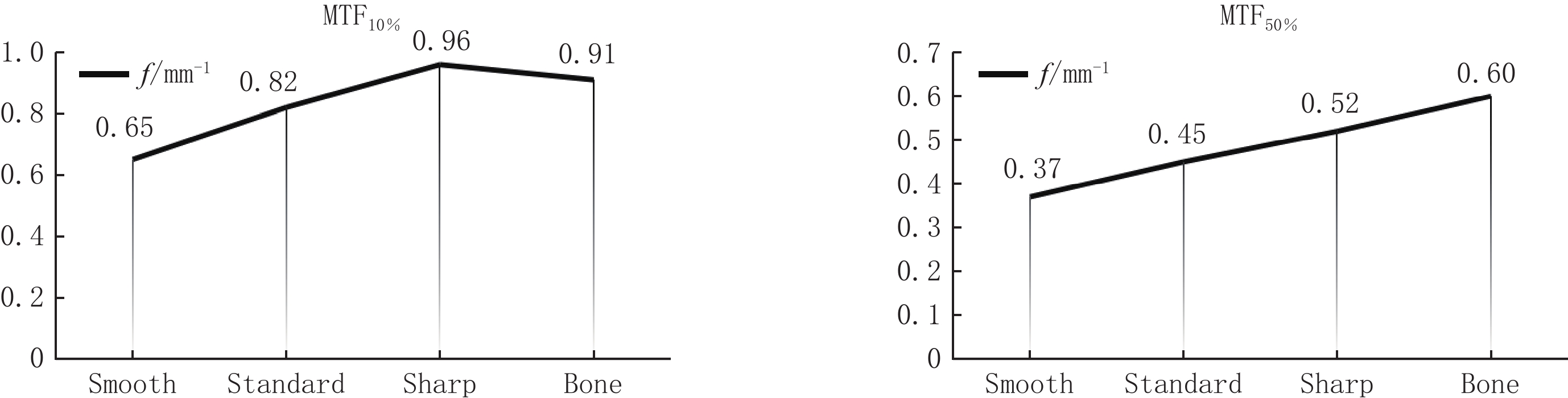
图 2 不同重建算法的模体CT图像的MTF值
Figure 2. The MTF of CT phantom images with different reconstruction algorithms
![]()
图 3 不同重建算法的模体CT图像的NPS,TTF和
$d' $ 值Figure 3. The NPS, TTF and
$d' $ values of CT phantom images with different reconstruction algorithms![]()
图 4 基于不同重建算法的VR对鼻区骨折的显示情况
注:女,49岁,右侧鼻骨前端骨折。从左至右依次为VR_Standard(a),VR_Smooth(b),VR_Sharp(c),VR_Bone(d)对骨折的显示(黑箭)。
Figure 4. Display of nasal region fractures by VR based on different reconstruction algorithms
![]()
图 5 基于Standard、Sharp和Bone重建算法的VR对横行骨折的显示情况
注:男,47岁,双侧鼻骨横行骨折。VR_Standard(a),VR_Sharp(b),VR_Bone(c)对鼻骨孔及线性骨折的显示,VR_Bone(c)对鼻骨孔及骨折线显示最好(黑箭),但受噪声影响,冠状面(d)可见横行骨折线(白箭)。
Figure 5. Display of transverse fractures by VR based on standard, sharp and bone reconstruction algorithms
![]()
图 6 基于Bone重建算法的VR在诊断鼻区线性骨折中容易出现假阳性
注:女,57岁,无骨折。VR_Standard(a),VR_Sharp(b)诊断无骨折,VR_Bone(c)将左侧上颌骨额突处(黑箭)误认为线性骨折,假阳性,横断面(d)显示无骨折发生(白箭)。
Figure 6. VR based on Bone reconstruction algorithm is prone to false positives in the diagnosis of linear fractures in the nasal region
![]()
图 7 基于Standard重建算法的VR在诊断鼻区线性骨折中容易出现假阴性
注:女,41岁,右侧上颌骨额突骨折。VR_Standard(a),VR_Sharp(b),VR_Bone(c)对鼻区线性骨折的显示,VR_Standard(a)易将右侧上颌骨额突处(黑箭)线性骨折漏掉,假阴性,横断面(d)可见骨折断端(白箭)。
Figure 7. VR based on the Standard reconstruction algorithm is prone to false negatives in the diagnosis of linear fractures in the nasal region
表 1 不同重建算法模体CT图像的高对比分辨力
Table 1 The high contrast resolution of CT phantom images with different reconstruction algorithms
Smooth Standard Sharp Bone 线对数LP/cm 8 9 10 10  下载: 导出CSV
表 2 两名医师对不同重建算法VR的主观评价得分和一致性分析
Table 2 Subjective evaluation scores and consistency analysis of VR with different reconstruction algorithms by two radiologists
项目 组别 医师A/例 医师B/例 评分/% 一致性分析 3分 2分 1分 3分 2分 1分 3分 2分 1分 Kappa P 图像质量评分 VR_Standard组 95 33 7 83 41 11 65.93 27.41 6.67 0.652 <0.001 VR_Sharp组 121 14 0 119 14 2 88.89 10.37 0.74 0.628 <0.001 VR_Bone组 96 36 3 94 28 13 70.37 23.70 5.93 0.637 <0.001 鼻骨孔显示评分 VR_Standard组 61 62 12 60 56 19 44.81 43.70 11.48 0.751 <0.001 VR_Sharp组 83 51 1 91 39 5 64.44 33.33 2.22 0.767 <0.001 VR_Bone组 95 33 7 99 28 8 71.85 22.59 5.56 0.645 <0.001
下载: 导出CSV
表 3 医师在VR_Standard、VR_Sharp、VR_Bone中对鼻区骨折的诊断效能比较
Table 3 Comparison of diagnostic efficacy for linear fractures in the nasal region between two radiologists in VR_Standard, VR_Sharp, and VR_Bone
医师 效能指标 VR_Standard VR_Sharp VR_Bone 三者总体
差异PVR_Standard
vs VR_Sharp
差异校正PVR_Standard
vs VR_Bone
差异校正PVR_Sharp
vs VR_Bone
差异校正P医师A 敏感度 0.84(84/100) 0.92(92/100) 0.92(92/100) 0.039 0.043 1.000 0.199 特异度 0.83(29/35) 0.86(30/35) 0.69(24/35) 准确率 0.84(113/135) 0.90(122/135) 0.85(116/135) 医师B 敏感度 0.85(85/100) 0.95(95/100) 0.95(95/100) 0.034 0.028 0.447 0.745 特异度 0.80(28/35) 0.77(27/35) 0.66(23/35) 准确率 0.84(113/135) 0.90(122/135) 0.87(118/135)
下载: 导出CSV
-
[1] DAVARI R, PIRZADEH A, SATTARI F. Etiology and epidemiology of nasal bone fractures in patients referred to the otorhinolaryngology section, 2019[J]. International Archives of Otorhinolaryngology, 2023, 27(2): e234−e239. DOI: 10.1055/s-0043-1768208.
[2] ZHANG P, ZHAO J, ZANG M, et al. Etiology of nasal bone fracture: A retrospective analysis of 1441 patients in China[J]. The Journal of Craniofacial Surgery, 2022, 33(4): 1185−1189. DOI: 10.1097/SCS.0000000000008479.
[3] CHUKWULEBE S, HOGREFE C. The diagnosis and management of facial bone fractures[J]. Emergency Medicine Clinics of North America, 2019, 37(1): 137−151. DOI: 10.1016/j.emc.2018.09.012.
[4] LANDEEN KC, KIMURA K, STEPHAN S J. Nasal fractures[J]. Facial Plastic Surgery Clinics of North America, 2022, 30(1): 23−30. DOI: 10.1016/j.fsc.2021.08.002.
[5] LI L F, ZANG H R, HAN D M, et al. Nasal bone fractures: Analysis of 1193 cases with an emphasis on coincident adjacent fractures[J]. Facial Plastic Surgery & Aesthetic Medicine, 2020, 22(4): 249−254. DOI: 10.1089/fpsam.2020.0026.
[6] 陶建华, 曲晓霞, 康天良, 等. 容积再现成像在鼻区线性骨折中的诊断效能[J]. 实用放射学杂志, 2022, 38(8): 1233−1237. DOI: 10.3969/j.issn.1002-1671.2022.08.004. TAO J H, QU X X, KANG T L, et al. Diagnostic efficacy of volume rendering imaging in assisting detection of linear fracture of nasal bone area[J]. Journal of Practical Radiology, 2022, 38(8): 1233−1237. DOI: 10.3969/j.issn.1002-1671.2022.08.004. (in Chinese).
[7] 汪茂文, 檀思蕾, 刘霞, 等. 鼻区骨折MSCT图像后处理显示与诊断探讨[J]. 中国司法鉴定, 2017, (6): 56−60. DOI: 10.3969/j.issn.1671-2072.2017.06.009. WANG M W, TAN S L, LIU X, et al. Post processing of MSCT Images in forensic examination of nasal and paranasal bone fracture[J]. Chinese Journal of Forensic Sciences, 2017, (6): 56−60. DOI: 10.3969/j.issn.1671-2072.2017.06.009. (in Chinese).
[8] SANDEEP R B, NAIK D, KENKERE D. Role of multidetector computed tomography in the evaluation of maxillofacial trauma[J]. Cureus, 2023, 15(2): e35008. DOI: 10.7759/cureus.35008.
[9] SAMEI E, BAKALYAR D, BOEDEKER K L, et al. Performance evaluation of computed tomography systems: Summary of AAPM task group 233[J]. Medical Physics. 2019, 46(11): e735-e756. DOI: 10.1002/mp.13763.
[10] 曾令明, 邓涵, 吕琴, 等. 偏离等中心点对CT图像质量影响的体模研究[J]. 中华放射学杂志, 2022, 56(11): 1237−1241. DOI: 10.3760/cma.j.cn112149-20220710-00596. ZENG L M, DENG H, LV Q, et al. A phantom study of the effect of deviation from isocentric points on CT image quality[J]. Chinese Journal of Radiology, 2022, 56(11): 1237−1241. DOI:10.3760/cma.j.cn112149- 20220710-00596. (in Chinese).
[11] GREFFIER J, FRANDON J, LARBI A, et al. CT iterative reconstruction algorithms: A task-based image quality assessment[J]. European Radiology, 2020, 30(1): 487−500. DOI: 10.1007/s00330-019-06359-6.
[12] 杨政君, 张昂, 陈勇, 等. 辐射剂量和管电压对CT图像质量的影响: 基于任务的图像质量评价[J]. CT理论与应用研究, 2022, 31(2): 211−217. DOI: 10.15953/j.ctta.2021.060. YANG Z J, ZHANG A, CHEN Y, et al. The effect of radiation dose and tube potential on image quality of CT: A task-based image quality assessment[J]. CT Theory and Applications, 2022, 31(2): 211−217. DOI: 10.15953/j.ctta.2021.060. (in Chinese).
[13] 陶建华, 曲晓霞, 张怀宇, 等. 成人鼻骨末端、鼻骨孔、鼻骨其他孔及咬合缝间骨型鼻颌缝的多层螺旋CT影像特征: 附1600例分析[J]. 中华解剖与临床杂志, 2022, 27(1): 1−7. DOI: 10.3760/cma.j.cn101202-20210428-00118. TAO J H, QU X X, ZHANG H Y, et al. Multi-slice spiral computed tomography of the morphology of the nasal bone end, foramen of nasal bone, accessory foramen of nasal bone, and nasomaxillary suture in 1600 cases[J]. Chinese Journal of Anatomy and Clinics, 2022, 27(1): 1−7. DOI: 10.3760/cma.j.cn101202-20210428-00118. (in Chinese).
[14] 原媛, 卢东生, 钟朝辉. 基于噪声功率谱的不同重建类型CT图像噪声分析[J]. 中国医学装备, 2017, 14(4): 32−35. DOI: 10.3969/J.ISSN.1672-8270.2017.04.007. YUAN Y, LU D S, ZHONG Z H. The noise analysis of CT imaging based on noise power spectrum of different reconstruction type[J]. China Medical Equipment, 2017, 14(4): 32−35. DOI: 10.3969/J.ISSN.1672-8270.2017.04.007. (in Chinese).
[15] 余晓锷, 高海英, 蔡凡伟, 等. 基于噪声功率谱的CT图像噪声评价[J]. 中国医学影像技术, 2014, 30(8): 1243−1246. DOI: 10.13929/j.1003-3289.2014.08.035. YU X E, GAO H Y, CAI F W, et al. Noise power spectrum-based evaluation of CT image noise[J]. Chinese Journal of Medical Imaging Technology, 2014, 30(8): 1243−1246. DOI: 10.13929/j.1003-3289.2014.08.035. (in Chinese).
-
期刊类型引用(5)
1. 李月莉,张海三. 多层螺旋CT与1.5T磁共振成像技术诊断肝脏血管瘤的价值观察. 大医生. 2024(04): 118-120 .  百度学术
百度学术
2. 葛欣. 多层螺旋CT在肝脏血管肉瘤和血管瘤鉴别诊断检测中的影像学特点. 中国民康医学. 2020(03): 113-115 . 百度学术
3. 李宜亮,张春霞,李勇. CT平扫及增强扫描鉴别肝脏血管瘤与肝脏血管肉瘤的价值. 中国医药科学. 2020(07): 211-214 . 百度学术
4. 朱锡勋,李志伟,郑宇. 原发性肝血管肉瘤脾转移破裂出血1例报告. 临床肝胆病杂志. 2019(03): 608-610 . 百度学术
5. 张强,霍建婷,王春艳. 原发性肝血管肉瘤自发破裂出血1例. 中南大学学报(医学版). 2018(11): 1276-1280 . 百度学术
其他类型引用(1)


计量
- 文章访问数: 273
- HTML全文浏览量: 57
- PDF下载量: 31
- 被引次数: 6


