
The Value of Constructing a Nomogram Model Based on Arterial-phase Imaging Radiomics Features Combined with Clinical-CT Features in Differentiating Lung Squamous Carcinoma and Adenocarcinoma
-
摘要:
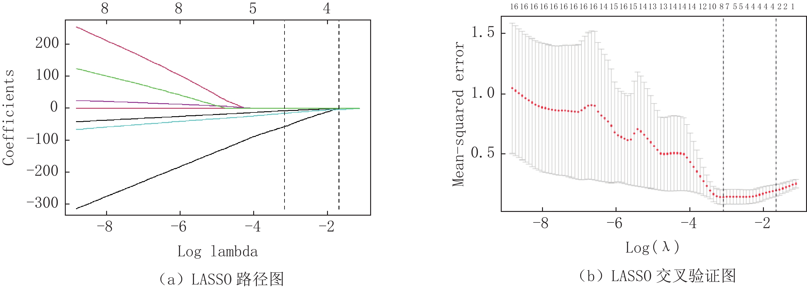
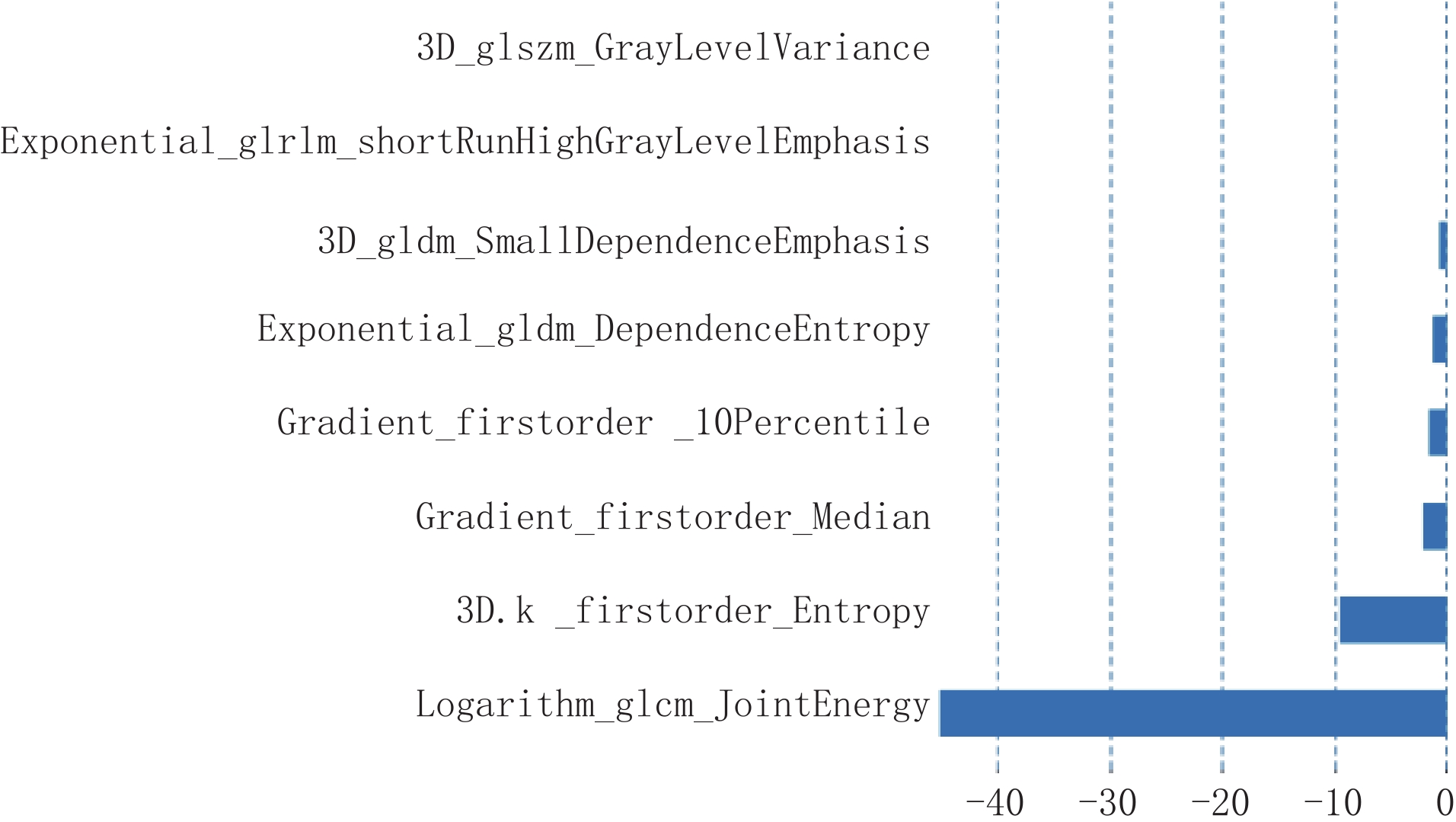
目的:探讨基于动脉期影像组学特征联合临床CT特征构建列线图模型在鉴别肺鳞癌(SCC)与肺腺癌(ADC)中的应用价值。方法:回顾性收集2021年8月至2023年9月在我院进行穿刺病理活检或手术的肺癌患者85例作为训练集,同时收集2023年5月至2024年6月经病理证实的肺癌患者40例作为验证集,所有患者均行胸部CT增强检查。根据病理结果将训练集分为SCC组(n=29)和ADC组(n=56)。比较两组患者一般临床资料和CT图像特征的差异,采用单因素和多因素Logistic回归分析筛选出独立预测因素,构建临床-CT模型。应用ITK Snap软件提取训练集动脉期图像影像组学特征,依次采用组内相关系数(ICC)、Roruta特征筛选和最小绝对收缩和选择算子(LASSO)对提取的影像组学特征进行降维处理,筛选出有意义的特征。采用Logistic回归构建动脉期影像组学模型,计算该模型的影像组学评分(Rad-score)。以多因素Logistic回归分析筛选出临床-CT特征有意义的自变量与Rad-score构建联合模型,绘制列线图。应用ROC曲线、校正曲线、H-L检验、Delong检验及临床决策曲线(DCA)对临床-CT模型、影像组学模型及列线图模型进行评价。结果:单因素分析结果显示,分叶征、坏死空洞征均多于ADC组,癌胚抗原(CEA)、血管集束征、胸膜牵拉及毛刺征均少于ADC组。将上述自变量纳入多因素Logistic进一步筛选,结果显示,CEA、分叶征、胸膜牵拉及毛刺征为独立危险因素,基于此构建临床-CT模型的训练集和验证集AUC值分别为0.623和0.786。影像组学特征经降维后共筛选出的有意义特征有8个,分别为一阶特征3个、二阶特征5个。经ROC曲线分析显示,影像组学模型训练集和验证集AUC值分别为0.830和0.846;列线图模型训练集和验证集AUC值分别为0.913和0.922。经Delong检验显示,列线图模型AUC值均明显高于临床-CT模型和影像组学模型;Hosmer-Lemeshow检验结果显示,临床-CT模型、影像组学模型及列线图模型的拟合度均良好;校准曲线分析显示,列线图模型的预测概率曲线与理想曲线最接近,预测精准度更好;DCA分析结果显示,列线图模型的曲线下面积最大,临床净收益最高。结论:基于动脉期影像组学特征联合临床-CT特征构建列线图模型在鉴别SCC与ADC中具有一定的诊断价值,为无创鉴别SCC与ADC提供一种新的诊断方式。
Abstract:Objective: To investigate the value of constructing a nomogram model based on arterial-phase imaging radiomics features combined with clinical-CT features for the differentiation between squamous lung cancer (SCC) and adenocarcinoma of the lung (ADC). Methods: Retrospectively, 85 patients with lung cancer who underwent puncture pathology biopsy or surgery in our hospital, from August 2021 to September 2023, were collected as a training set. Concurrently, 40 patients with pathologically confirmed lung cancer in our hospital, from May 2023 to June 2024, were collected as a validation set. All patients underwent chest CT enhancement. The training set was divided into the SCC group (n=29) and the ADC group (n=56) based on the pathology. General clinical data and CT image characteristics of the two groups of patients were compared and differences were identified. Independent predictors were screened using unifactorial and multifactorial logistic regression analyses, and a clinical-CT model was constructed. ITK Snap software was applied to extract the radiomics features of the arterial-phase images, and the intragroup correlation coefficient (ICC), Roruta feature screening, and least absolute shrinkage and selection operator (LASSO) were sequentially used to downsize the extracted radiomics features, screen out the meaningful features, construct the arterial-phase image radiomics model using Logistic regression, and compute the model's image radiomics score (Rad-score). A multifactor logistic regression analysis was used to screen the independent variables with meaningful clinical-CT characteristics and Rad-score to construct a joint model, and a nomogram graph was plotted. ROC curves, calibration curves, H-L test, Delong test, and clinical decision curves (DCA) were applied to evaluate the clinical-CT, radiomics, and nomogram models. Results: The results of univariate analysis showed that there were more lobular signs and necrotic cavity signs, and fewer carcinoembryonic antigen (CEA), vascular cluster signs, pleural pulling, and burr signs in the SCC than in the ADC group. The above independent variables were included in the multifactorial Logistic analysis for further screening, and the results showed that CEA, lobular sign, pleural pull, and burr sign were independent risk factors. The area under the curve (AUC) values for the training and validation sets of the clinical-CT model constructed based on this were 0.623 and 0.786, respectively. A total of eight meaningful features were screened after dimensionality reduction of the radiomics features, which were three first-order features and five second-order features. The ROC curve analysis showed that the AUC values for the training and validation sets of the radiomics model were 0.830 and 0.846, respectively; and the AUC values for the training and validation sets of the nomogram model were 0.913 and 0.922, respectively. The Delong test showed that the AUC values of the nomogram model were all significantly higher than those of the clinical-CT model and the radiomics model; the Hosmer-Lemeshow test showed that the clinical-CT model, the radiomics model, and the nomogram model were all well fitted; calibration curve analysis showed that the predictive probability curve of the nomogram model was closest to the ideal curve, with better predictive accuracy; and DCA analysis showed that the AUC of the nomogram model was the largest, with the highest net clinical benefit. Conclusion: Constructing a nomogram model based on arterial-phase imaging radiomics features combined with clinical-CT features has some diagnostic value in differentiating SCC from ADC, providing a new diagnostic modality for noninvasive differentiation.
-
Keywords:
- arterial phase /
- radiomics /
- clinical CT features /
- squamous cell carcinoma /
- adenocarcinoma /
- nomogram model
-
-
![]()
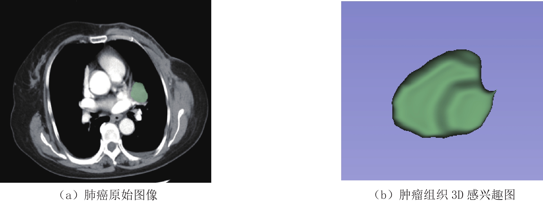
图 1 ITK-Snap软件提取肿瘤组织3D感兴趣区图
Figure 1. ITK-Snap software extracts 3D region of interest map of tumor tissue
![]()
图 2 使用 LASSO 回归降维分析筛选影像组学特征
Figure 2. Filtering radiomics features using LASSO regression dimensionality reduction analysis
![]()
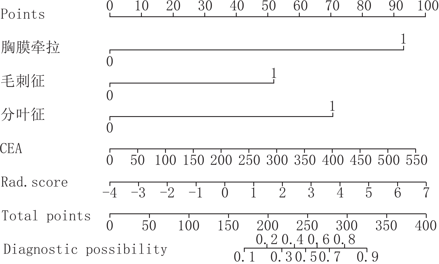
图 4 临床-CT模型联合Rad-Score构建列线图
Figure 4. Clinical-CT modeling combined with Rad-Score to construct nomogram
![]()
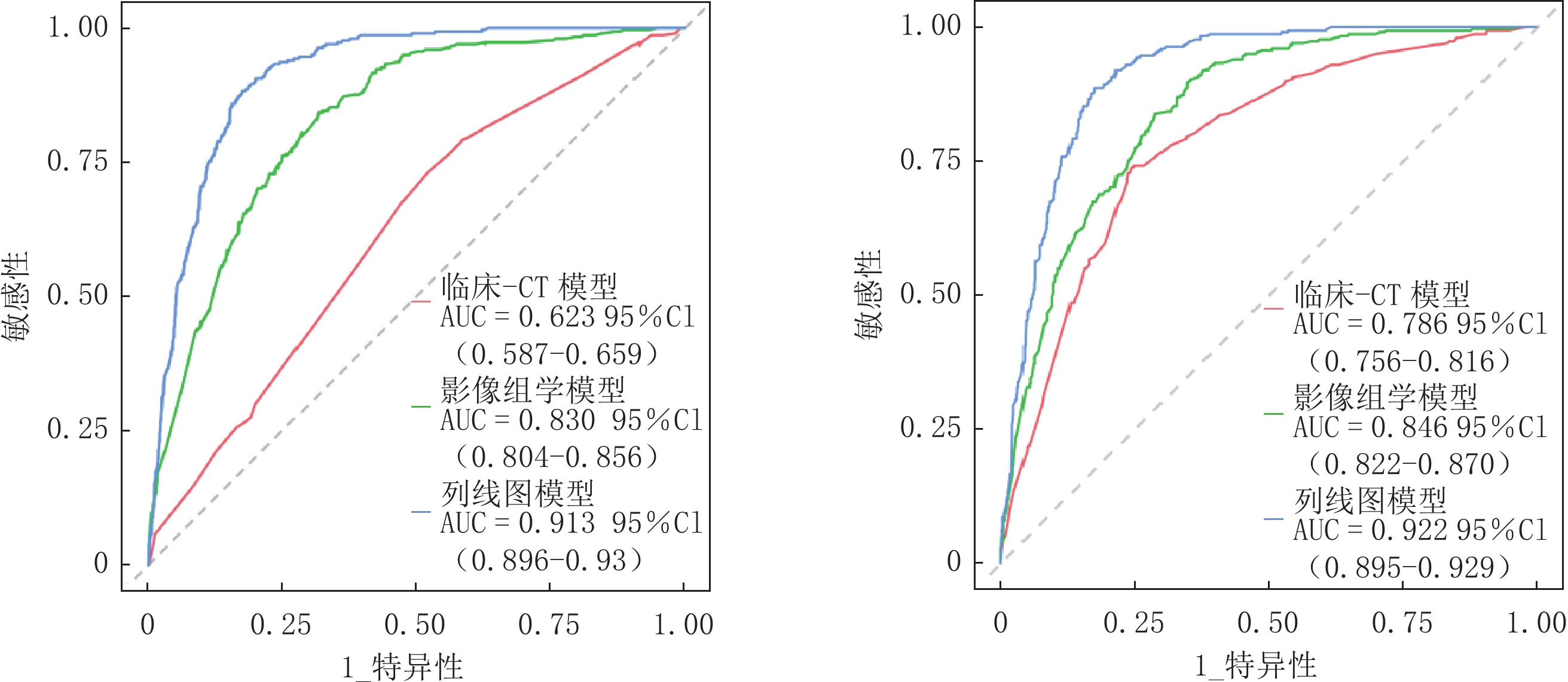
图 5 列线图模型分别在训练集和验证集ROC曲线分析结果
Figure 5. Results of ROC curve analysis of the nomogram model in the training and validation sets
![]()
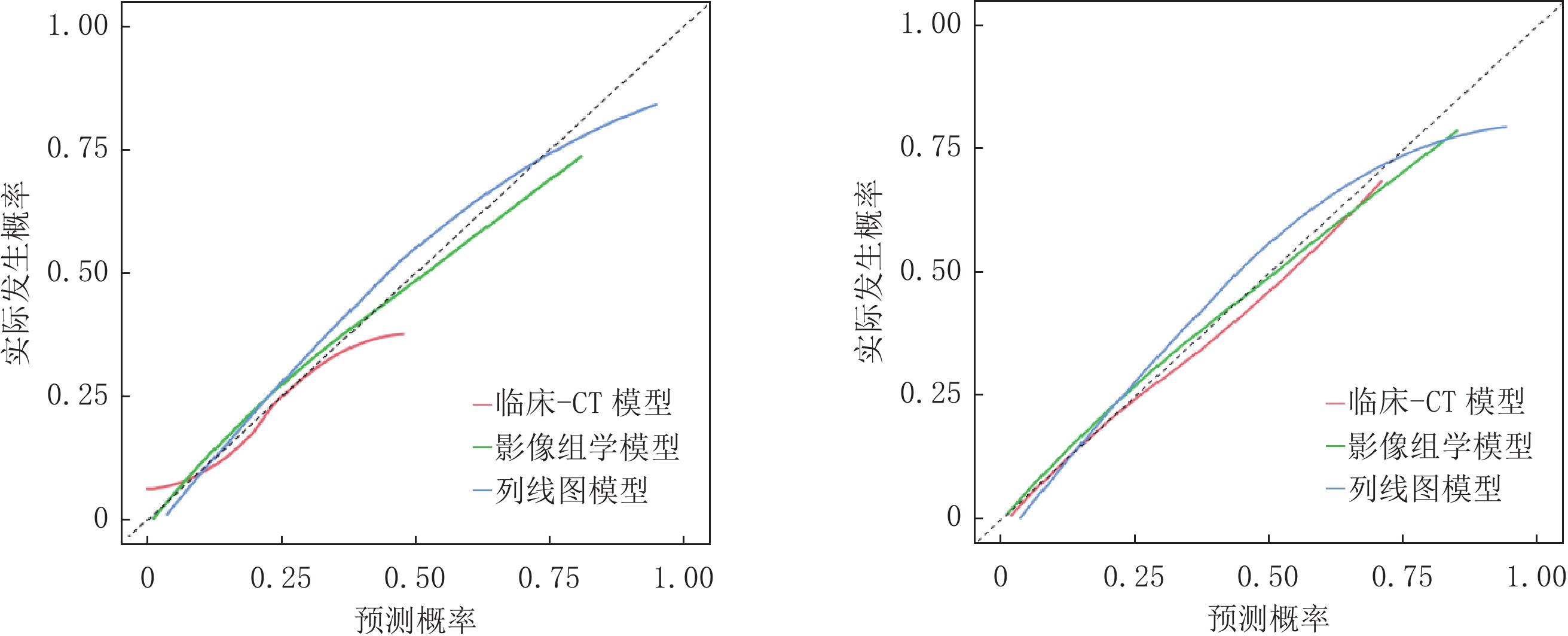
图 6 列线图模型分别在训练集和验证集校准曲线分析结果
Figure 6. Results of calibration curve analysis of the nomogram model in the training and validation sets
![]()
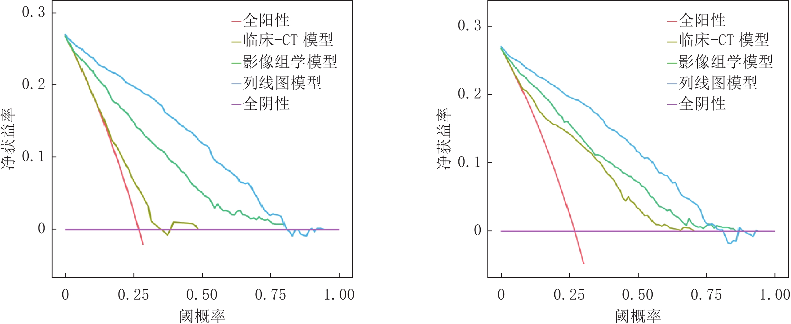
图 7 列线图模型分别在训练集和验证集DCA曲线分析结果
Figure 7. Results of DCA curve analysis of the nomogram model in the training and validation sets
表 1 训练集两组患者临床资料与CT影像特征结果比较(
$\bar x\pm s $ )Table 1 Comparison of clinical data and results of CT imaging characteristics between the two groups of patients in the training set
指标 组别 统计检验 SCC组(n=29) ADC组(n=56) t/${\chi}^2 $/Z P 年龄 72.21±9.27 67.84±9.64 1.206 0.144 性别 男 24(82.76) 28(50.00) 1.231 0.121 女 5(17.24) 28(50.00) 吸烟史 是 19(65.52) 34(60.71) 0.188 0.665 否 10(34.48) 22(39.29) 家族肿瘤史 是 12(41.38) 26(46.43) 0.197 0.657 否 17(58.62) 30(53.57) NSE(ng/mL) 4.56(3.04, 7.38) 6.24 (4.12, 8.33) 0.413 0.115 CEA(ng/mL) 96.53±8.15 198.22±21.51 6.495 <0.001 CA242(IU/mL) 28.42±4.37 37.49±5.84 1.037 0.189 CA199(U/mL) 51.17±9.53 65.27±10.51 0.202 0.565 坏死空洞 有 25(86.21) 13(23.21) 35.325 <0.001 无 2(6.89) 43(76.79) 毛刺征 有 10(34.48) 45(80.36) 17.606 <0.001 无 19(65.52) 11(19.64) 分叶征 有 26(89.66) 10(17.86) 40.340 <0.001 无 3(10.34) 46(82.14) 空泡征 有 22(75.86) 32(57.14) 2.890 0.089 无 7(24.14) 24(42.86) 胸膜牵拉 有 16(55.17) 44(78.57) 4.574 0.032 无 13(44.83) 12(21.43) 血管集束征 有 13(44.83) 41(73.21) 6.645 0.010 无 16(55.17) 15(26.79) 肿瘤位置 上叶 18(62.07) 30(53.57) 2.011 0.093 中叶 3(10.34) 9(16.07) 下叶 8(27.59) 17(30.36)  下载: 导出CSV
表 2 ADC及SCC多因素Logistic回归分析结果
Table 2 Results of Multifactor Logistic Regression Analysis of ADC and SCC
变量 β SE Wald $\chi^2 $ OR 95% CI Z P CEA 0.005 0.003 1.880 1.005 (1.000~1.010) 1.987 0.047 分叶征 1.992 0.719 4.712 7.328 (1.931~33.680) 2.768 0.006 胸膜牵拉 1.413 0.599 2.883 4.109 (1.340~14.58) 2.359 0.018 毛刺征 1.466 0.736 2.346 4.333 (1.103~20.300) 1.993 0.046 常数 −5.977 1.355 0.587 0.003 (0.000~0.025) −4.410 <0.001
下载: 导出CSV
表 3 临床-CT模型、影像组学模型及列线图模型诊断效能比较(%)
Table 3 Comparison of diagnostic efficacy of clinical-CT model, radiomics model, and nomogram model(%)
数据集 模型 AUC (95% CI) 敏感性 特异性 阳性预测值 阴性预测值 训练集 临床-CT模型 0.623(0.587~0.659) 76.79 71.82 76.58 75.45 影像组学模型 0.830(0.804~0.856) 83.78 80.63 81.55 79.92 列线图模型 0.913(0.896~0.930) 92.59 88.74 90.77 86.01 验证集 临床-CT模型 0.786(0.756~0.816) 73.83 75.58 73.53 88.34 影像组学模型 0.846(0.822~0.870) 83.89 81.10 82.52 92.05 列线图模型 0.922(0.895~0.929) 93.01 82.48 89.84 90.99
下载: 导出CSV
-
[1] 傅睿, 吴一龙, 钟文昭. 2020版肺癌多学科团队诊疗中国专家共识解读[J]. 中国肿瘤临床, 2022, 49(4): 163−167. DOI: 10.12354/j.issn.1000-8179.2022.20211201. FU R, WU Y L, ZHONG W Z. Interpretation of Chinese expert consensus on multidisciplinary team diagnosis and treatment of lung cancer (2020 version)[J]. Chinese Tumor clinical, 2022, 49(4): 163−167. DOI: 10.12354/j.issn.1000-8179.2022.20211201. (in Chinese).
[2] 王馨慧, 张笑雪, 聂奔, 等. 联合免疫模式在晚期非小细胞肺癌治疗中的研究进展[J]. 临床肿瘤学杂志, 2023, 28(3): 263−269. DOI: 10.3969/j.issn.1009-0460.2023.03.011. WANG X H, ZHANG X X, NIE B, et al. Research progress of combined immunotherapy mode in the treatment of advanced non-small cell lung cancer[J]. Chinese Clinical Oncology, 2023, 28(3): 263−269. DOI: 10.3969/j.issn.1009-0460.2023.03.011. (in Chinese).
[3] 吕祥瑞, 皇甫娟, 王孟丽, 等. 血清肿瘤标志物与肺癌病理类型的相关性研究[J]. 癌症进展, 2021, 19(14): 1451−1455. DOI: 10.11877/j.issn.1672-1535.2021.19.14.13. LV X R, HUANG P J, WANG M L, et al. Study on the correlation between serum tumor markers and pathological types of lung cancer[J]. Oncology Progress, 2021, 19(14): 1451−1455. DOI: 10.11877/j.issn.1672-1535.2021.19.14.13. (in Chinese).
[4] 张盼, 黄庆, 汪扬, 等. 非小细胞肺癌动态增强CT扫描下临床表现特征与其病理类型的关系[J]. 西部医学, 2023, 35(4): 584-587. DOI: 10.3969/j.issn.1672-3511.2023.04.023. ZHANG P, HUANG Q, WANG Y, et al. Clinical characteristics of dynamic contrast-enhanced CT scan and their relationship with pathological types in non-small cell lung cancer[J]. Western Medicine, 2023, 35(4): 584-587. DOI:10.3969/j.issn.1672-3511.2023.04.023. (in Chinese).
[5] 陈杰, 洪悦, 王艳, 等. 基于CT平扫影像组学特征在预测胸腺上皮性肿瘤WHO简化病理分型中的价值[J]. 中国CT和MRI杂志, 2024, 22(1): 71−73. DOI: 10.3969/j.issn.1672-5131.2024.01.023. CHEN J, HONG Y, WANG Y, et al. Value of simplified pathological classification of thymus epithelial tumors based on plain CT imaging features[J]. Chinese Journal of CT and MRI, 2024, 22(1): 71−73. DOI: 10.3969/j.issn.1672-5131.2024.01.023. (in Chinese).
[6] 党俊明, 朱超华, 黄慧娴, 等. 基于MRI影像组学特征预测宫颈腺癌与鳞癌病理分型的研究[J]. 医疗卫生装备, 2022, 43(5): 54−59. DOI: 10.19745/j.1003-8868.2022099. DANG J M, ZHU C H, HUANG X H, et al. Prediction of pathological classification of cervical adenocarcinoma and squamous cell carcinoma based on MRI radiomics characteristics[J]. Medical and Health Equipment, 2022, 43(5): 54−59. DOI: 10.19745/j.1003-8868.2022099. (in Chinese).
[7] 高琳, 于鑫鑫, 康冰, 等. CT 影像组学对肺纯磨玻璃结节浸润性的预测价值[J]. 山东大学学报(医学版), 2022, 60(5): 87−97. DOI: 10.6040/j.issn.1671-7554.0.2022.0500. GAO L, YU X X, KANG B, et al. Predictive value of CT-based radiomics nomogram for the invasiveness of lung pure ground-glass nodules[J]. Journal of Shandong University (Health Sciences), 2022, 60(5): 87−97. DOI: 10.6040/j.issn.1671-7554.0.2022.0500. (in Chinese).
[8] 中华医学会, 中华医学会肿瘤学分会, 中华医学会杂志社. 中华医学会肺癌临床诊疗指南(2019版)[J]. 中华肿瘤杂志, 2020, 42(4): 257−287. DOI: 10.3760/cma.j.cn112152-20200120-00049. Chinese Medical Association, Chinese Medical Association Oncology Branch, Journal of Chinese Medical Association. Chinese Medical Association guidelines for clinical diagnosis and treatment of lung cancer (2019 edition)[J]. Chinese Journal of Tumors, 2020, 42(4): 257−287. DOI: 10.3760/cma.j.cn112152-20200120-00049. (in Chinese).
[9] 朱玲玲, 伍娟, 王艇, 等. 2023年第2版NCCN肺癌筛查临床实践指南更新解读[J]. 实用肿瘤杂志, 2023, 38(5): 399−407. DOI: 10.13267/j.cnki.syzlzz.2023.063. ZHU L L, WU J, WANG T, et al. Updated interpretation of NCCN clinical practice guidelines for lung cancer screening, version 2.2023[J]. Journal of Practical Oncology, 2023, 38(5): 399−407. DOI: 10.13267/j.cnki.syzlzz.2023.063. (in Chinese).
[10] 蒋会东, 谢强, 李军, 等. 64排CT联合NSE、ProGRP在肺癌鉴别诊断及TNM分期中的应用[J]. 中国现代医学杂志, 2022, 32(14): 95−100. DOI: 10.3969/j.issn.1005-8982.2022.14.017. JIANG H D, XIE Q, LI J, et al. Application of 64-detector-row CT combined with NSE and ProGRP in differential diagnosis and TNM staging of lung cancer[J]. China Journal of Modern Medicine, 2022, 32(14): 95−100. DOI: 10.3969/j.issn.1005-8982.2022.14.017. (in Chinese).
[11] 孔令芹, 陈苏媛, 李西川, 等. 早中期小细胞肺癌与非小细胞肺癌CT影像及肿瘤标记物对比[J]. 中国医学创新, 2023, 20(17): 42−46. DOI: 10.3969/j.issn.1674-4985.2023.17.010. KONG L Q, CHEN S Y, LI X C, et al. Comparison of CT images and tumor markers between early and middle stage small cell lung cancer and non-small cell lung cancer[J]. MedicaI Innovation of China, 2023, 20(17): 42−46. DOI: 10.3969/j.issn.1674-4985.2023.17.010. (in Chinese).
[12] 唐新, 梁江涛, 向柏林, 等. PET/MRI影像组学特征预测肺腺癌与肺鳞癌病理分型价值[J]. 浙江医学, 2022, 44(6): 580−584. DOI: 10.12056/j.issn.1006-2785.2022.44.6.2021-2731. TANG X, LIANG J T, XIANG B L, et al. Application of PET/MRI radiomics in differentiating the pathological types of lung adenocarcinoma and squamous cell carcinoma[J]. Zhejiang Medicine, 2022, 44(6): 580−584. DOI: 10.12056/j.issn.1006-2785.2022.44.6.2021-2731. (in Chinese).
[13] 孙士鹤, 侯艳娟, 刘芬, 等. CT诊断老年肺癌患者病理亚型与病理学诊断的一致性及其影像学特征[J]. 中国老年学杂志, 2023, 43(24): 5915−5918. DOI: 10.3969/j.issn.1005-9202.2023.24.007. SUN S H, HOU Y J, LIU F, et al. Concordance between pathologic subtypes and pathologic diagnoses and their imaging features in CT-diagnosed elderly patients with lung cancer[J]. Chinese Journal of Gerontology, 2023, 43(24): 5915−5918. DOI: 10.3969/j.issn.1005-9202.2023.24.007. (in Chinese).
[14] 王守玉, 屈开新, 王庆龙, 等. 不典型肺癌CT诊断与鉴别诊断[J]. 中国医药科学, 2020, 10(15): 187−190. DOI: 10.3969/j.issn.2095-0616.2020.15.051. WANG S Y, QU K X, WANG Q L, et al. CT diagnosis and differential diagnosis of atypical lung cancer[J]. China Medicine and Pharmacy, 2020, 10(15): 187−190. DOI: 10.3969/j.issn.2095-0616.2020.15.051. (in Chinese).
[15] 彭弘, 李圣博, 赵旭. 周围型肺癌CT征象与病理的对照相关性研究[J]. 罕少疾病杂志, 2022, 29(12): 42−43. DOI: 10.3969/j.issn.1009-3257.2022.12.019. PENG H, LI S B, ZHAO X. A comparative study of CT signs and pathology of peripheral lung cancer[J]. Journal of Rare and Uncommon Diseases, 2022, 29(12): 42−43. DOI: 10.3969/j.issn.1009-3257.2022.12.019. (in Chinese).
[16] 张英惠. 96例早期周围型肺癌的CT影像特征分析[J]. 中国现代药物应用, 2022, 16(4): 87-90. DOI: 10.14164/j.cnki.cn11-5581/r.2022.04.033. ZHANG Y H. Analysis of CT imaging features of 96 cases of early-stage peripheral lung cancer[J]. Chinese Modern Drug Application, 2022, 16(4): 87-90. DOI:10.14164/j.cnki.cn11-5581/r.2022.04.033.(in Chinese).
[17] 谭淦纹, 曹欢, 王玲. 肺癌MSCT增强扫描影像学征象与CA199、CA242、NSE表达的相关性及临床应用价值研究[J]. 中国CT和MRI杂志, 2022, 20(3): 49−51. DOI: 10.3969/j.issn.1672-5131.2022.03.016. TAN J W, CAO H, WANG L. Correlation between imaging signs of MSCT enhanced scanning of lung cancer and the expression of CA199, CA242, NSE, and its clinical application value[J]. Chinese Journal of CT and MRI, 2022, 20(3): 49−51. DOI: 10.3969/j.issn.1672-5131.2022.03.016. (in Chinese).
[18] 陈宝华, 段强军. 血清 CEA、NSE 联合影像学特征在肺癌分期及预后中的价值[J]. 分子诊断与治疗杂志, 2022, 14(9): 1615−1619. DOI: 10.3969/j.issn.1674-6929.2022.09.040. CHEN B H, DUANG Q J. The value of serum CEA and NSE combined with imaging features in lung cancer staging and prognosis[J]. Journal of Molecular Diagnosis and Treatment, 2022, 14(9): 1615−1619. DOI: 10.3969/j.issn.1674-6929.2022.09.040. (in Chinese).
[19] 肖蓉, 潘频华. 老年肺癌CT影像学特征与特异性标记物的相关性研究及联合诊断[J]. 国际老年医学杂志, 2020, 41(6): 391−394. DOI: 10.3969/j.issn.1674-7593.2020.06.013. XIAO R, PAN P H. Correlation between CT imaging features and specific markers and their combined diagnostic value in lung cancer in older patients[J]. International Journal of Geriatrics, 2020, 41(6): 391−394. DOI: 10.3969/j.issn.1674-7593.2020.06.013. (in Chinese).
[20] ZHU X, DONG D, CHEN Z, et al. Radiomic signature as a diagnostic factor for histologic subtype classification of non-small cell lung cancer[J]. Eur Radiol, 28(7): 2772-2778. DOI: 10.1007/s00330-017-5221-1.
[21] 唐聪聪, 陈艾琪, 曹胜男, 等. CT 影像组学在非小细胞肺癌病理分级中的应用[J]. 蚌埠医学院学报, 2023, 48(6): 783-786. DOI: 10.13898/j.cnki.issn.1000-2200.2023.06.017. TANG C C, CHEN A Q, CAO S N, et al. Application of CT radiomics in the pathological grading of non-small cell lung cancer[J]. Journal of Bengbu Medical College, [21]2023, 48(6): 783-786. DOI:10.13898/j.cnki.issn.1000-2200.2023.06.017. (in Chinese).
[22] 方家杨, 梁长宇, 陶俊利, 等. 基于临床、影像组学和计算机视觉特征鉴别肺鳞癌和腺癌[J]. 中国医学影像学杂志, 2022, 30(7): 691−696,702. DOI: 10.3969/j.issn.1005-5185.2022.07.010. FANG J Y, LIANG C Y, TAO J L, et al. Differentiation of lung squamous cell carcinoma and adenocarcinoma based on clinical, radiomic and computer vision features[J]. Chinese Journal of Medical Imaging, 2022, 30(7): 691−696,702. DOI: 10.3969/j.issn.1005-5185.2022.07.010. (in Chinese).
[23] 陆亮, 徐圆. 影像组学联合CT特征对周围型肺腺癌及鳞癌的鉴别价值研究[J]. 医疗卫生装备, 2021, 42(10): 48−52, 63. DOI: 10.19745/j.1003-8868.2021211. LU L, XU Y. Diagnostic value of radiomics combined with CT features for differentia-ting peripheral lung adenocarcinoma from squamous cell carcinoma[J]. Chinese Medical Equipment Journal, 2021, 42(10): 48−52, 63. DOI: 10.19745/j.1003-8868.2021211. (in Chinese).
[24] 汪靖婷, 钟飞扬, 甘甜, 等. 基于临床及 CT 影像组学特征构建周围型小细胞肺癌与肺腺癌诊断模型的研究[J]. 临床放射学杂志, 2023, 42(3): 406-410. DOI: 10.13437/j.cnki.jcr.2023.03.035. WANG T T, ZHONG F Y, GAN T, et al. Study on the differential diagnosis model between periph eral SCLC and ADC based on clinical and CT radiomics features[J]. Journal of Clinical Radiology, 2023, 42(3): 406-410. DOI:10.13437/j.cnki.jcr.2023.03.035. (in Chinese).
[25] WEN C L, MENG P L, WEN Y H, et al. A practical dynamic nomogram model for predicting bone metastasis in patients with thyroid cancer[J]. Front Endocrinol (Lausanne), 2023, 14: 1142796. DOI: 10.3389/fendo.2023.1142796.
[26] FENG H, YU Q S, WANG J X, et al. Establishment and validation of nomogram prediction model for complicated acute appendicitis[J]. Zhonghua Wai Ke Za Zhi, 2023, 61(12): 1074−1079. DOI: 10.3760/cma.j.cn112139-20230104-00005.
-
期刊类型引用(4)
1. 张元飞. 动态增强磁共振成像联合弥散加权成像对前列腺癌的诊断效能. 实用临床医药杂志. 2024(24): 26-30 .  百度学术
百度学术
2. 殷帅涛,王嘉南,蒋召强,王向阳,姬彤宇,单磊. PI-RADS v2.1评分联合前列腺特异性抗原衍生指标对灰区前列腺癌的诊断价值. 中华实用诊断与治疗杂志. 2023(04): 372-376 . 百度学术
3. 李媛媛,张显利. f-PSA/t-PSA联合基于PI-RADS v2评分的多参数MRI在前列腺癌中的诊断价值. 临床医学研究与实践. 2022(28): 119-122+136 . 百度学术
4. 孙东方,姚伟根,刘洁,鲁鹏聪,康聪. 基于PI-RADS联合PSA及衍生物构建前列腺癌定量评分体系的研究. 现代实用医学. 2022(12): 1564-1566 . 百度学术
其他类型引用(2)

计量
- 文章访问数: 106
- HTML全文浏览量: 38
- PDF下载量: 11
- 被引次数: 6


