
The Effect of Radiation Dose and Tube Potential on Image Quality of CT: A Task-based Image Quality Assessment
-
摘要: 目的:通过基于任务的图像质量评价参数,研究对比不同辐射剂量和管电压对CT图像的影响。方法:使用GE Revolution Apex扫描美国放射学会(ACR)质量控制体模Gammex 464。采用3种剂量(5、10和20 mGy)和3种管电压(80、100和120 kVp)的扫描方案并重建9组CT图像。选取体模module 1中骨和丙烯酸测量各组图像的任务传递函数(task-based transfer function,TTF,代表空间分辨率)并记录其TTF50%。选取体模module 3测量噪声功率谱(noise power spectrum,NPS,代表噪声)并记录噪声值、空间频率(f-peak)和 NPS peak值。在图像TTF和NPS的基础上进一步计算图像的可检测能力指数(
${d}'$ ,代表对病灶的可检出能力)。剂量和管电压对图像的影响采用单因素方差分析,P值的多重比较采用 FDR校正。结果:管电压较剂量对TTF50%的影响较为明显,但两者在骨和丙烯酸物质中的差异均无统计学意义。噪声和NPS peak随着剂量上升而显著减小;随着管电压的增加而减小,但差异不具有统计学意义。剂量较管电压对f-peak的影响较大,但两者差异均无统计学意义。图像的检出能力随着剂量的增加而显著升高;各管电压下图像的检出能力差异无统计学意义。结论:剂量相比管电压更能影响CT图像质量;随着剂量的增加,图像噪声显著改善,对病灶的检出能力显著提升。基于任务为基础的评价指标可以较为全面地反映CT图像质量。Abstract: Purpose: To compare the effect of radiation dose and tube potential on image quality of CT through the task-based image quality assessment parameters. Methods: We scanned Gammex 464 (the ACR quality assurance phantom) with GE Revolution Apex CT. Three radiation doses (5, 10, 20 mGy) and three tube potentials (80, 100, 120 kVp) were used to reconstruct nine sets of image. Bone and acrylic inserts from module 1 of the phantom was selected for the measurement of task-based transfer function (TTF, representing spatial resolution) and TTF50% was recorded for each set of images. Module 3 was selected for the measurement of noise power spectrum (NPS, representing image noise) and noise value, spatial frequency (f-peak) and NPS peak value were recorded for each set of images. Detestability index (${d}'$ representing lesion detestability) was furtherly calculated based on TTF and NPS of images. The effect of radiation dose and tube potential on image quality was evaluated by One-way Anova analysis. Multiple comparisons for P value were corrected by FDR. Results: Compared with radiation dose, the effect of tube potential on TTF50% was more obvious, but there was no significant difference between them in bone and acrylic substances. Noise and NPS peak significantly decreased with the increase of both radiation dose and tube potential but no statistical difference was found. Compared with tube potential, radiation dose showed greater impact on f-peak, but no statistical difference was found. d’ was significantly improved as radiation dose increased; while no statistical difference was found under different tube potentials. Conclusion: Image quality is predominantly influenced by radiation dose rather than tube potential. Image noise and lesion detestability is signifcantly improved as radiation dose elevates. Image quality could be comprehensively inflected by the task-based image quality assessment.-
Keywords:
- CT image quality /
- radiation dose /
- image quality assessment
-
-
![]()
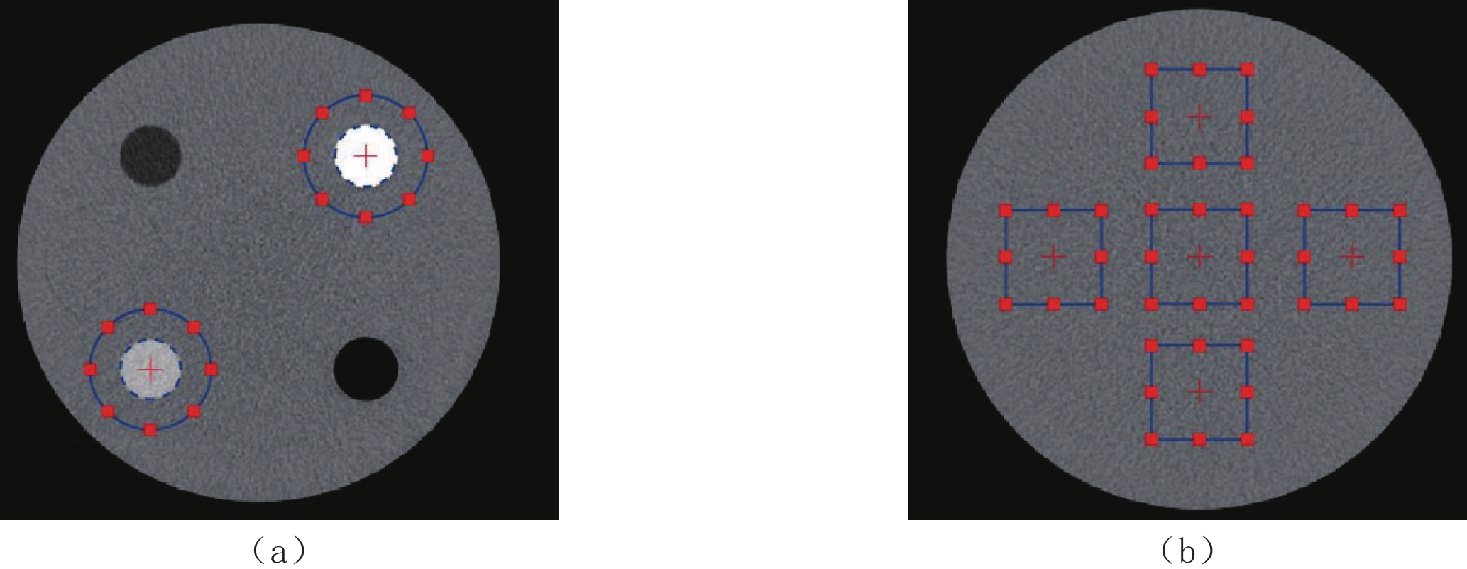
图 1 任务传递函数TTF和噪声功率谱NPS的测量图示
Figure 1. Diagram for task-based transfer function and noise power spectrum
表 1 本研究CT扫描参数
Table 1 CT Scanning parameters in this study
分组 管电压/kVp 管电流/mA 剂量/mGy 旋转时间/s 螺距 A 80 285 5 0.8 0.984 B 80 570 10 0.8 0.984 C 80 795 20 0.6 0.516 D 100 145 5 0.8 0.984 E 100 290 10 0.8 0.984 F 100 405 20 0.6 0.516 G 120 90 5 0.8 0.984 H 120 180 10 0.8 0.984 I 120 215 20 0.7 0.516  下载: 导出CSV
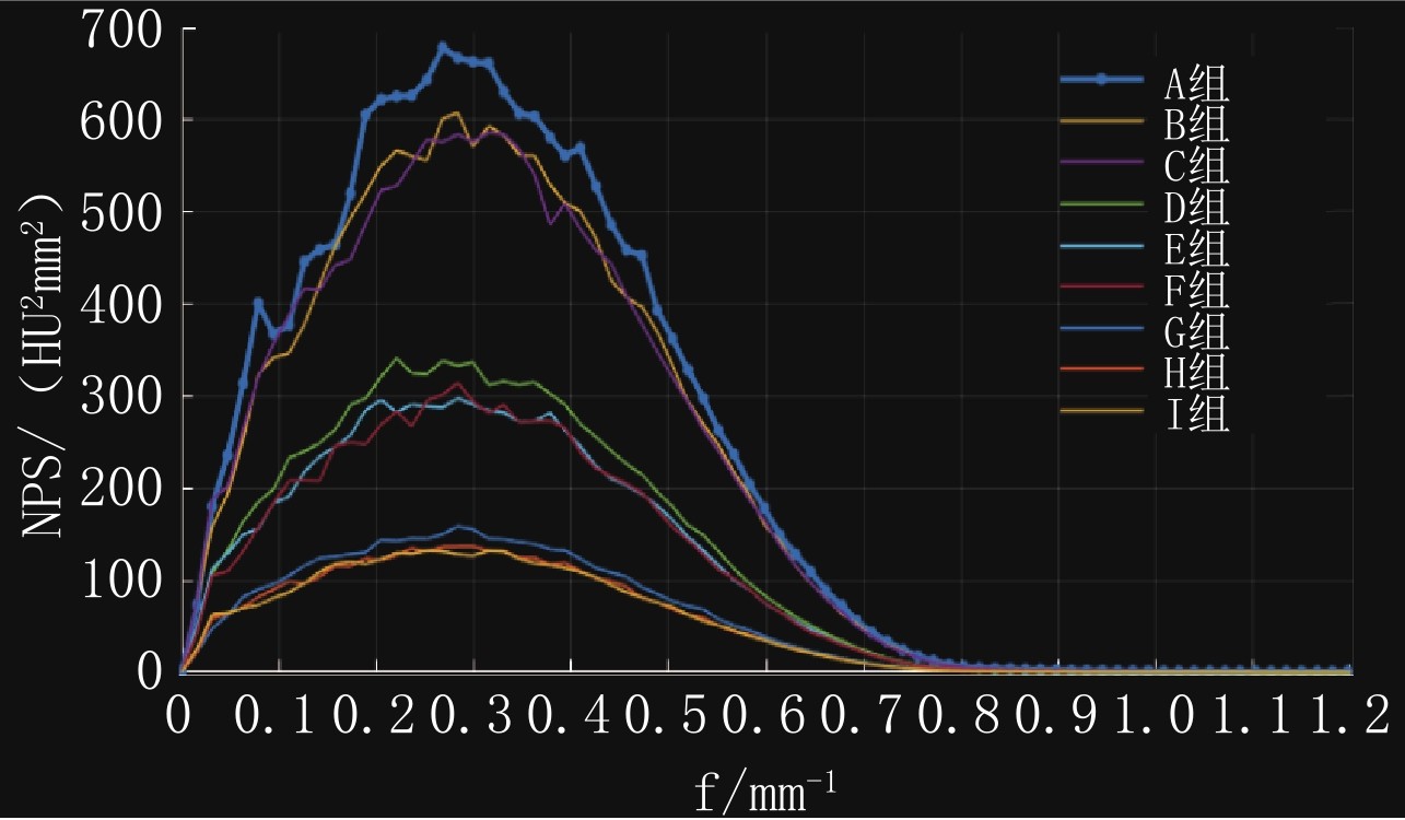
表 2 各组图像基于任务的图像质量评价参数结果
Table 2 Assessment results for images based on task-based image image quality assessment
分组 骨TTF50%/mm-1 丙烯酸TTF50%/mm-1 噪声/HU f-peak/mm-1 NPS peak /HU2mm2 ${d}'$ A 0.43 0.46 24.2 0.28 678.5 13.07 B 0.43 0.40 23.0 0.30 607.8 15.45 C 0.43 0.40 22.8 0.30 586.3 16.85 D 0.42 0.42 17.2 0.25 316.2 16.73 E 0.44 0.42 16.2 0.27 297.6 19.57 F 0.43 0.40 16.1 0.28 314.7 21.63 G 0.42 0.42 11.6 0.27 158.9 24.01 H 0.44 0.43 10.9 0.27 137.3 27.95 I 0.43 0.40 10.8 0.28 132.5 32.09
下载: 导出CSV
-
[1] LI K, GOMEZ-CARDONA D, HSIEH J, et al. Statistical model based iterative reconstruction in clinical CT systems. Part III: Task-based kV/mAs optimization for radiation dose reduction[J]. Medical Physics, 2015, 42(9): 5209−5221. DOI: 10.1118/1.4927722.
[2] KHOBRAGADE P, RUPCICH F, FAN J, et al. CT automated exposure control using a generalized detectability index[J]. Medical Physics, 2019, 46(1): 140−151. DOI: 10.1002/mp.13286.
[3] GREFFIER J, FRANDON J, LARBI A, et al. CT iterative reconstruction algorithms: A task-based image quality assessment[J]. European Radiology, 2020, 30(1): 487−500. DOI: 10.1007/s00330-019-06359-6.
[4] LARBI A, ORLIAC C, FRANDON J, et al. Detection and characterization of focal liver lesions with ultra-low dose computed tomography in neoplastic patients[J]. Diagnostic and Interventional Imaging, 2018, 99(5): 311−320. DOI: 10.1016/j.diii.2017.11.003.
[5] 张俊, 刘佳怿, 梁志鹏. 低管电压结合迭代重建在下肢动脉CT血管成像中的临床应用[J]. CT理论与应用研究, 2020,29(1): 55−60. DOI: 10.15953/j.1004-4140.2020.29.01.07. ZHANG J, LIU J Y, LIANG Z P. Clinical application of low tube voltage combined with iterative reconstruction in lower extremity arterial CTA[J]. CT Theory and Applications, 2020, 29(1): 55−60. DOI: 10.15953/j.1004-4140.2020.29.01.07. (in Chinese).
[6] 胡启云, 董越, 张瑞, 等. 结合迭代重建算法的低剂量CT在颈部的应用研究[J]. CT理论与应用研究, 2018,27(4): 447−453. DOI: 10.15953/j.1004-4140.2018.27.04.04. HU Q Y, DONG Y, ZHANG R, et al. The study of low-dose computed tomography with iterative reconstruction in neck[J]. CT Theory and Applications, 2018, 27(4): 447−453. DOI: 10.15953/j.1004-4140.2018.27.04.04. (in Chinese).
[7] SAMEI E, BAKALYAR D, BOEDEKER K L, et al. Performance evaluation of computed tomography systems: Summary of AAPM task group 233[J]. Medical Physics, 2019, 46(11): e735−e56. DOI: 10.1002/mp.13763.
[8] VAISHNAV J Y, JUNG W C, POPESCU L M, et al. Objective assessment of image quality and dose reduction in CT iterative reconstruction[J]. Medical Physics, 2014, 41(7): 071904. DOI: 10.1118/1.4881148.
[9] YU L, VRIEZE T J, LENG S, et al. Technical note: Measuring contrast- and noise-dependent spatial resolution of an iterative reconstruction method in CT using ensemble averaging[J]. Medical Physics, 2015, 42(5): 2261−2267. DOI: 10.1118/1.4916802.
[10] SAMEI E, RICHARD S. Assessment of the dose reduction potential of a model-based iterative reconstruction algorithm using a task-based performance metrology[J]. Medical Physics, 2015, 42(1): 314−323. DOI: 10.1118/1.4903899.
[11] SOLOMON J, MARIN D, Roy CHOUDHURY K, et al. Effect of radiation dose reduction and reconstruction algorithm on image noise, contrast, resolution, and detectability of subtle hypoattenuating liver lesions at multidetector CT: Filtered back projection versus a commercial model-based iterative reconstruction algorithm[J]. Radiology, 2017, 284(3): 777−787. DOI: 10.1148/radiol.2017161736.
[12] McCOLLOUGH C H, YU L, KOFLER J M, et al. Degradation of CT low-contrast spatial resolution due to the use of iterative reconstruction and reduced dose levels[J]. Radiology, 2015, 276(2): 499−506. DOI: 10.1148/radiol.15142047.
[13] MILETO A, GUIMARAES L S, McCOLLOUGH C H, et al. State of the art in abdominal CT: The limits of iterative reconstruction algorithms[J]. Radiology, 2019, 293(3): 491−503. DOI: 10.1148/radiol.2019191422.
[14] GREFFIER J, HAMARD A, PEREIRA F, et al. Image quality and dose reduction opportunity of deep learning image reconstruction algorithm for CT: A phantom study[J]. European Radiology, 2020, 30(7): 3951−3959. DOI: 10.1007/s00330-020-06724-w.
[15] GREFFIER J, SI-MOHAMED S, DABLI D, et al. Performance of four dual-energy CT platforms for abdominal imaging: A task-based image quality assessment based on phantom data[J]. European Radiology, 2021, 31(7): 5324−5334. DOI: 10.1007/s00330-020-07671-2.
-
期刊类型引用(2)
1. 袁婷婷,万胜洪. MRI磁敏感加权成像技术在颅内海绵状血管瘤中的诊断效能及影像学征象分析. 医学信息. 2024(10): 105-108 .  百度学术
百度学术
2. 李丽,阮玖根,高阳,肖琼. 磁敏感加权成像在颅内海绵状血管瘤诊断中的应用价值分析. 现代诊断与治疗. 2022(17): 2535-2537 . 百度学术
其他类型引用(0)


计量
- 文章访问数: 779
- HTML全文浏览量: 104
- PDF下载量: 78
- 被引次数: 2


