ISSN 1004-4140
CN 11-3017/P
| Citation: |
HAN Z F, SHANGGUAN H, ZHANG X, et al. Advances in research on low-dose CT imaging algorithm based on deep learning[J]. CT Theory and Applications, 2022, 31(1): 117-134. DOI: 10.15953/j.1004-4140.2022.31.01.14. (in Chinese).

|
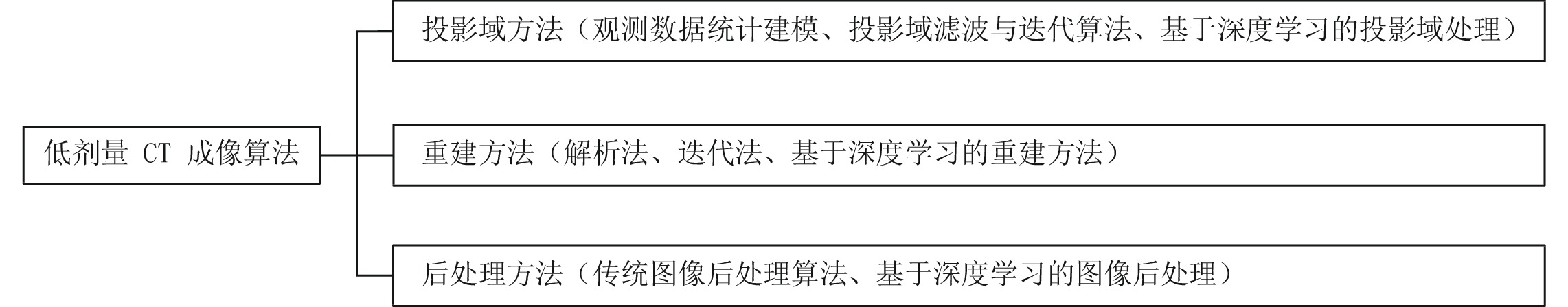
Computed tomography (CT) is widely used in clinical diagnosis because of its fast imaging speed and high resolution. However, higher doses of radiation will cause damages to human tissues and organs, while lower doses will lead to serious deterioration of imaging quality. In order to solve the above contradiction, researchers have focused on the low-dose CT imaging technology to study how to reduce the harm caused by radiation to the human body to the greatest extent under the condition of ensuring the imaging quality to meet the needs of clinical diagnosis. In recent years, deep learning has developed rapidly in the field of artificial intelligence, and has been widely used in image processing, pattern recognition, signal processing fields. Driven by big data, LDCT imaging algorithms based on deep learning have made great progress. This paper studies the development of low-dose CT imaging algorithms in recent years in terms of three aspects: the process of CT imaging, the noise modeling of low-dose CT, and the design of imaging algorithms. In particular, the imaging algorithms in the field of deep learning are systematically elaborated and analyzed. Finally, future developments in the field of LDCT image artifact suppression are also prospected.
| [1] |
BRENNER D J, HALL E J. Computed tomography: An increasing source of radiation exposure[J]. New England Journal of Medicine, 2007, 357(22): 2277−2284.
|
| [2] |
SMITH-BINDMAN R, LIPSON J, MARCUS R, et al. Radiation dose associated with common computed tomography examinations and the associated lifetime attributable risk of cancer[J]. Archives of Internal Medicine, 2009, 169(22): 2078−2086. doi: 10.1001/archinternmed.2009.427
|
| [3] |
HART D, WALL B F. UK population dose from medical X-ray examinations[J]. European Journal of Radiology, 2004, 50(3): 285−291. doi: 10.1016/S0720-048X(03)00178-5
|
| [4] |
HSIEH J. Computed tomography: Principles, design, artifacts, and recent advances[M]. SPIE Press, 2003.
|
| [5] |
SHRIMPTON P C, HILLIER M C, LEWIS M A, et al. Doses from computed tomography (CT) examinations in the UK-2003 review[M]. National Radiological Protection Board, 2005.
|
| [6] |
SIGAL-CINQUALBRE A B, HENNEQUIN R, ABADA H T, et al. Low-kilovoltage multi-detector row chest CT in adults: Feasibility and effect on image quality and iodine dose[J]. Radiology, 2004, 231(1): 169−174. doi: 10.1148/radiol.2311030191
|
| [7] |
YEDDER H B, CARDOEN B, HAMARNEH G. Deep learning for biomedical image reconstruction: A survey[J]. Artificial Intelligence Review, 2021, 54(1): 215−251.
|
| [8] |
柳澄, 秦维昌. 多层螺旋CT(一)[J]. 医学影像学杂志, 2000,10(3): 194−195.
|
| [9] |
SHANGGUAN H. Study on statistical iterative reconstruction methods for low-dose X-ray CT[D]. Taiyuan: North University of China, 2016.
|
| [10] |
MA J, LIANG Z, FAN Y, et al. Variance analysis of X-ray CT sinograms in the presence of electronic noise background[J]. Medical Physics, 2012, 39(7): 4051−4065.
|
| [11] |
ZHANG H, OUYANG L, MA J, et al. Noise correlation in CBCT projection data and its appli-cation for noise reduction in low-dose CBCT[J]. Medical Physics, 2014, 41(3): 031906. doi: 10.1118/1.4865782
|
| [12] |
XIE S P, LUO L M, YANG L F, et al. Scatter correction method for cone beam CT using beam attenuation grid[C]//Key Engineering Materials. Trans Tech Publications Ltd, 2011, 480: 341-346.
|
| [13] |
HSIEH J. Adaptive streak artifact reduction in computed tomography resulting from excessive X-ray photon noise[J]. Medical Physics, 1998, 25(11): 2139−2147.
|
| [14] |
KACHELRIEβ M, WATZKE O, KALENDER W A. Generalized multi-dimensional adaptive filtering for conventional and spiral single-slice, multi-slice, and cone-beam CT[J]. Medical Physics, 2001, 28(4): 475−490.
|
| [15] |
SAHINER B, YAGLE A E. Image reconstruction from projections under wavelet constraints[J]. IEEE Transactions on Signal Processing, 1993, 41(12): 3579−3584. doi: 10.1109/78.258101
|
| [16] |
YAZDI M, BEAULIEU L. Artifacts in spiral X-ray CT scanners: Problems and solutions[J]. International Journal of Biological and Medical Sciences, 2008, 4(3): 135−139.
|
| [17] |
DEMIRKAYA O. REDUCTION of noise and image artifacts in computed tomography by nonlinear filtration of projection images[C]//Medical Imaging 2001. International Society for Optics and Photonics, 2001: 917-923.
|
| [18] |
WANG J, LL T, LU H, et al. Penalized weighted least-squares approach for low-dose X-ray computed tomography[C]//Medical Imaging 2006: Physics of Medical Imaging. International Society for Optics and Photonics, 2006, 6142: 614247.
|
| [19] |
ZHANG Q, GUI Z, CHEN Y, et al. Bayesian sinogram smoothing with an anisotropic diffusion weighted prior for low-dose X-ray computed tomography[J]. Optik-International Journal for Light and Electron Optics, 2013, 124(17): 2811−2816. doi: 10.1016/j.ijleo.2012.08.045
|
| [20] |
RUDIN L I, OSHER S, FATEMI E. Nonlinear total variation based noise removal algorithms[J]. Physica D: Nonlinear Phenomena, 1992, 60(1): 259−268.
|
| [21] |
梁宁宁, 李子恒, 王林元, 等. 一种基于GAN网络投影补全的有限角度CT重建算法[J]. 中国体视学与图像分析, 2019,24(1): 1−8.
LIANG N, LI Z, WANG L, et al. A limited-angle computed tomography reconstruction algorithm based on projection completion via generative adversarial networks[J]. Chinese Journal of Stereology and Image Analysis, 2019, 24(1): 1−8. (in Chinese).
|
| [22] |
GORDON R, BENDER R, HERMAN G T. Algebraic reconstruction techniques (ART) for three- dimensional electron microscopy and X-ray photography[J]. Journal of Theoretical Biology, 1970, 29(3): 471−481. doi: 10.1016/0022-5193(70)90109-8
|
| [23] |
ZHANG H, WANG J, MA J, et al. Statistical models and regularization strategies in statistical image reconstruction of low-dose X-ray CT: A survey[J]. arXiv preprint arXiv: 1412.1732, 2014.
|
| [24] |
LEVITAN E, HERMAN G T. A maximum a posteriori probability expectation maximization algorithm for image reconstruction in emission tomography[J]. IEEE Transactions on Medical Imaging, 1987, 6(3): 185−192. doi: 10.1109/TMI.1987.4307826
|
| [25] |
HSIAO T, RANGARAJAN A, GINDI G. A new convex edge-preserving median prior with applications to tomography[J]. IEEE Transactions on Medical Imaging, 2003, 22(5): 580−585. doi: 10.1109/TMI.2003.812249
|
| [26] |
LU Y, ZHAO J, WANG G. Few-view image reconstruction with dual dictionaries[J]. Physics in Medicine & Biology, 2011, 57(1): 173.
|
| [27] |
BAI T, MOU X, XU Q, et al. Low-dose CT reconstruction based on multiscale dictionary[C]//International Society for Optics and Photonics. Medical Imaging 2013: Physics of Medical Imaging, 2013, 8668: 86683L.
|
| [28] |
NIU S, GAO Y, BIAN Z, et al. Sparse-view X-ray CT reconstruction via total generalized variation regularization[J]. Physics in Medicine and Biology, 2014, 59(12): 2997. doi: 10.1088/0031-9155/59/12/2997
|
| [29] |
LIU Y, LIANG Z, MA J, et al. Total variation-stokes strategy for sparse-view X-ray CT image reconstruction[J]. IEEE Transactions on Medical Imaging, 2013, 33(3): 749−763.
|
| [30] |
LIANG K, YANG H, XING Y. Comparison of projection domain, image domain, and comprehensive deep learning for sparse-view X-ray CT image reconstruction[J]. arXiv preprint arXiv: 1804.04289, 2018.
|
| [31] |
PELT D M, BATENBURG K J. Improving filtered backprojection reconstruction by data-dependent filtering[J]. IEEE Transactions on Image Processing, 2014, 23(11): 4750−4762. doi: 10.1109/TIP.2014.2341971
|
| [32] |
WANG B, LIU H. FBP-Net for direct reconstruction of dynamic PET images[J]. Physics in Medicine & Biology, 2020, 65(23): 235008.
|
| [33] |
ZHANG Q, LIANG D. Visualization of fully connected layer weights in deep learning CT reconstruction[J]. arXiv preprint arXiv: 2002.06788, 2020.
|
| [34] |
ZHU B, LIU J Z, CAULEY S F, et al. Image reconstruction by domain-transform manifold learning[J]. Nature, 2018, 555(7697): 487−492. doi: 10.1038/nature25988
|
| [35] |
FU L, DE MAN B. A hierarchical approach to deep learning and its application to tomographic reconstruction[C]//International Society for Optics and Photonics. 15th International Meeting on Fully Three-Dimensional Image Reconst- ruction in Radiology and Nuclear Medicine, 2019, 11072: 1107202.
|
| [36] |
YE D H, BUZZARD G T, RUBY M, et al. Deep back projection for sparse-view CT reconstruction[C]//2018 IEEE Global Conference on Signal and Information Processing (GlobalSIP). IEEE, 2018: 1-5.
|
| [37] |
TAO X, ZHANG H, WANG Y, et al. VVBP-tensor in the FBP algorithm: Its properties and application in low-dose CT reconstruction[J]. IEEE transactions on medical imaging, 2019, 39(3): 764−776.
|
| [38] |
TAO X, WANG Y, LIN L, et al. Learning to reconstruct CT images from the VVBP-tensor[J]. IEEE Transactions on Medical Imaging, 2021.
|
| [39] |
LI Y, LI K, ZHANG C, et al. Learning to reconstruct computed tomography images directly from sinogram data under a variety of data acquisition conditions[J]. IEEE Transactions on Medical Imaging, 2019, 38(10): 2469−2481. doi: 10.1109/TMI.2019.2910760
|
| [40] |
HE J, WANG Y, MA J. Radon inversion via deep learning[J]. IEEE Transactions on Medical Imaging, 2020, 39(6): 2076−2087. doi: 10.1109/TMI.2020.2964266
|
| [41] |
WU W, HU D, NIU C, et al. DRONE: Dual-domain residual-based optimization network for sparse-view CT reconstruction[J]. IEEE Transactions on Medical Imaging, 2021.
|
| [42] |
GE Y, SU T, ZHU J, et al. ADAPTIVE-NET: Deep computed tomography reconstruction network with analytical domain transformation knowledge[J]. Quantitative Imaging in Medicine and Surgery, 2020, 10(2): 415. doi: 10.21037/qims.2019.12.12
|
| [43] |
ZHANG Q, HU Z, JIANG C, et al. Artifact removal using a hybrid-domain convolutional neural network for limited-angle computed tomography imaging[J]. Physics in Medicine & Biology, 2020, 65(15): 155010.
|
| [44] |
LIN W A, LIAO H, PENG C, et al. Dudonet: Dual domain network for CT metal artifact reduction[C]//Proceedings of the IEEE/CVF Conference on Computer Vision and Pattern Recognition, 2019: 10512-10521.
|
| [45] |
WANG T, XIA W, HUANG Y, et al. DAN-Net: Dual-domain adaptive-scaling non-local network for CT metal artifact reduction[J]. arXiv Preprint arXiv: 2102.08003, 2021.
|
| [46] |
WANG T, XIA W, LU Z, et al. IDOL-Net: An interactive dual-domain parallel network for CT metal artifact reduction[J]. arXiv Preprint arXiv: 2104.01405, 2021.
|
| [47] |
RAN M, XIA W, HUANG Y, et al. Md-recon-net: A parallel dual-domain convolutional neural network for compressed sensing MRI[J]. IEEE Transactions on Radiation and Plasma Medical Sciences, 2020, 5(1): 120−135.
|
| [48] |
CHEN H, ZHANG Y, CHEN Y, et al. LEARN: Learned experts'assessment-based reconstruction network for sparse-data CT[J]. IEEE Transactions on Medical Imaging, 2018, 37(6): 1333−1347. doi: 10.1109/TMI.2018.2805692
|
| [49] |
WU D, KIM K, El Fakhri G, et al. Iterative low-dose CT reconstruction with priors trained by artificial neural network[J]. IEEE Transactions on Medical Imaging, 2017, 36(12): 2479−2486. doi: 10.1109/TMI.2017.2753138
|
| [50] |
KANG E, CHANG W, YOO J, et al. Deep convolutional framelet denosing for low-dose CT via wavelet residual network[J]. IEEE Transactions on Medical Imaging, 2018, 37(6): 1358−1369. doi: 10.1109/TMI.2018.2823756
|
| [51] |
GAO Y, LIANG Z, MOORE W, et al. A feasibility study of extracting tissue textures from a previous full-dose CT database as prior knowledge for Bayesian reconstruction of current low-dose CT images[J]. IEEE Transactions on Medical Imaging, 2019, 38(8): 1981−1992. doi: 10.1109/TMI.2018.2890788
|
| [52] |
VENKATAKRISHNAN S V, BOUMAN C A, WOHLBERG B. Plug-and-play priors for model based reconstruction[C]//2013 IEEE Global Conference on Signal and Information Processing. IEEE, 2013: 945-948.
|
| [53] |
SREEHARI S, VENKATAKRISHNAN S V, WOHLBERG B, et al. Plug-and-play priors for bright field electron tomography and sparse interpolation[J]. IEEE Transactions on Computational Imaging, 2016, 2(4): 408−423.
|
| [54] |
CASCARANO P, PICCOLOMINI E L, MOROTTI E, et al. Plug-and-play external and internal priors for image restoration[J]. arXiv e-prints, 2021, arXiv: 2102.07510.
|
| [55] |
ADLER J, OKTEM O. Learned Primal-dual reconstruction[J]. IEEE Transactions on Medical Imaging, 2017: 1322−1332.
|
| [56] |
XIA W, LU Z, HUANG Y, et al. MAGIC: Manifold and graph integrative convolutional network for low-dose CT reconstruction[J]. IEEE Transactions on Medical Imaging, 2021.
|
| [57] |
CHEN G, HONG X, DING Q, et al. AirNet: Fused analytical and iterative reconstruction with deep neural network regularization for sparse-data CT[J]. Medical Physics, 2020, 47(7): 2916−2930. doi: 10.1002/mp.14170
|
| [58] |
GUPTA H, JIN K H, NGUYEN H Q, et al. CNN-based projected gradient descent for consistent CT image reconstruction[J]. IEEE Transactions on Medical Imaging, 2018, 37(6): 1440−1453. doi: 10.1109/TMI.2018.2832656
|
| [59] |
ZHANG H, LIU B, YU H, et al. MetaInv-net: Meta inversion network for sparse view CT image reconstruction[J]. IEEE Transactions on Medical Imaging, 2020, 40(2): 621−634.
|
| [60] |
BUADES A, COLL B, MOREL J M. A non-local algorithm for image denoising[C]//2005 IEEE Computer Society Conference on Computer Vision and Pattern Recognition (CVPR'05). IEEE, 2005, 2: 60-65.
|
| [61] |
CHEN Y, YANG Z, HU Y, et al. Thoracic low-dose CT image processing using an artifact suppressed large-scale nonlocal means[J]. Physics in Medicine & Biology, 2012, 57(9): 2667.
|
| [62] |
CHEN Y, CHEN W, YIN X, et al. Improving low-dose abdominal CT images by weighted intensity averaging over large-scale neighborhoods[J]. European Journal of Radiology, 2011, 80(2): e42−e49. doi: 10.1016/j.ejrad.2010.07.003
|
| [63] |
ZHONG J, NING R, CONOVER D. Image denoising based on multiscale singularity detection for cone beam CT breast imaging[J]. IEEE Transactions on Medical Imaging, 2004, 23(6): 696−703. doi: 10.1109/TMI.2004.826944
|
| [64] |
FERUGLIO P F, VINEGONI C, GROS J, et al. Block matching 3D random noise filtering for absorption optical projection tomography[J]. Physics in Medicine & Biology, 2010, 55(18): 5401.
|
| [65] |
CHEN Y, SHI L, FENG Q, et al. Artifact suppressed dictionary learning for low-dose CT image processing[J]. IEEE Transactions on Medical Imaging, 2014, 33(12): 2271−2292. doi: 10.1109/TMI.2014.2336860
|
| [66] |
ZAMYATIN A, KATSEVICH G, KRYLOV R, et al. Adaptive multi-scale total variation minimization filter for low dose CT imaging[C]//SPIE Medical Imaging. International Society for Optics and Photonics, 2014: 903426-903426-7.
|
| [67] |
MENG B, JIANG H, LIU Z, et al. Curvelet-based bilinear interpolation method for low-dose CT[C]//International Society for Optics and Photonics. Fifth International Conference on Digital Image Processing, 2013: 88783X-88783X-5.
|
| [68] |
CHEN H, ZHANG Y, ZHANG W H. Low dose CT via convolutional neural network[J]. Biomedical Optics Express, 2017, 8(2): 679−694. DOI: 10.1364/BOE.8.000679.
|
| [69] |
SHAN H M, ZHANG Y, YANG Q S, et al. 3-D convolutional encoder-decoder network for low-dose CT via transfer learning from a 2-D trained network[J]. IEEE Transactions on Medical Imaging, 2018, 37(6): 1522−1534. DOI: 10.1109/TMI.2018.2832217.
|
| [70] |
YANG Q S, YAN P K, ZHANG Y B, et al. Low-dose CT image denoising using a generative adversarial network with wasserstein distance and perceptual loss[J]. IEEE Transactions on Medical Imaging, 2018, 36(7): 1348−1357. DOI: 10.1109/TMI.2018.2827462.
|
| [71] |
ZHU J Y, PARK T, ISOLA P, et al. Unpaired image-to-image translation using cycle-consistent adversarial networks[C]//Proceedings of the IEEE international conference on computer vision. 2017: 2223-2232.
|
| [72] |
CHEN Y. CT-LSTM: Detection & estimation duplexed system for robust object tracking[C]//Proceedings of the 2nd International Conference on Computer Science and Application Engineering, 2018: 1-7.
|
| [73] |
BURGER H C, SCHULER C J, HARMELING S. Image denoising: Can plain neural networks compete with BM3D?[C]//2012 IEEE conference on computer vision and pattern recognition. IEEE, 2012: 2392-2399.
|
| [74] |
CHEN H, ZHANG Y, KALRA M, et al. Low-dose CT with a residual encoder-decoder convolutional neural network[J]. IEEE Transactions on Medical Imaging, 2017, 2(3): 2524−2535. DOI: 10.1109/TMI.2017.2715284.
|
| [75] |
HEINRICH M P, STILLE M, BUZUG T M. Residual U-net convolutional neural network architecture for low-dose CT denoising[J]. Current Directions in Biomedical Engineering, 2018, 4(1): 297−300. doi: 10.1515/cdbme-2018-0072
|
| [76] |
WU D F, KIM K, FAKHRI G E, et al. A cascaded convolutional neural network for X-ray low-dose CT image denoising[OL]. https://arxiv.org/abs/1705.04267, 2017.5.
|
| [77] |
SHAN H, PADOLE A, HOMAYOUNIEH F, et al. Competitive performance of a modularized deep neural network compared to commercial algorithms for low-dose CT image reconstruction[J]. Nature Machine Intelligence, 2019, 1(6): 269−276. doi: 10.1038/s42256-019-0057-9
|
| [78] |
WOLTERINK J M, LEINER T, VIERGEVER M A, et al. Generative adversarial networks for noise reduction in low-dose CT[J]. IEEE Transactions on Medical Imaging, 2017, 36(12): 2536−2545. DOI: 10.1109/TMI.2017.2708987.
|
| [79] |
YI X, BABYN P. Sharpness-aware low-dose CT denoising using conditional generative adversarial network[J]. Journal of Digital Imaging, 2018, 31(5): 655−669. doi: 10.1007/s10278-018-0056-0
|
| [80] |
ISOLA P, ZHU J Y, ZHOU T, et al. Image-to-image translation with conditional adversarial networks[C]//Proceedings of the IEEE Conference on Computer Vision and Pattern Recognition. 2017: 1125-1134.
|
| [81] |
YANG L, SHANGGUAN H, ZHANG X, et al. High-frequency sensitive generative adversarial network for low-dose CT image denoising[J]. IEEE Access, 2019, 8: 930−943.
|
| [82] |
YOU C, YANG L, ZHANG Y, et al. Low-dose CT via deep CNN with skip connection and network-in- network[C]//Developments in X-ray Tomography XII. International Society for Optics and Photonics, 2019, 11113: 111131W.
|
| [83] |
ARJOVSKY M, CHINTALA S, BOTTOU L. Wasserstein gan[J]. arXiv Preprint arXiv: 1701.07875, 2017.
|
| [84] |
LI X, YE C, YAN Y, et al. Low-dose CT image denoising based on improved WGAN-gp[J]. Journal of New Media, 2019, 1(2): 75. doi: 10.32604/jnm.2019.06259
|
| [85] |
MAO X, LI Q, XIE H, et al. Least squares generative adversarial networks[C]//Proceedings of the IEEE International Conference on Computer Vision, 2017: 2794-2802.
|
| [86] |
MIYATO T, KATAOKA T, KOYAMA M, et al. Spectral normalization for generative adversarial networks[J]. arXiv Preprint arXiv: 1802.05957, 2018.
|
| [87] |
PARK H S, BAEK J, YOU S K, et al. Unpaired image denoising using a generative adversarial network in X-ray CT[J]. IEEE Access, 2019, 7: 110414−110425. doi: 10.1109/ACCESS.2019.2934178
|
| [88] |
DU W, CHEN H, LIAO P, et al. Visual attention network for low-dose CT[J]. IEEE Signal Processing Letters, 2019, 26(8): 1152−1156. doi: 10.1109/LSP.2019.2922851
|
| [89] |
RAJEEV R, SAMATH J A, KARTHIKEYAN N K. An intelligent recurrent neural network with long short-term memory (LSTM) BASED batch normalization for medical image denoising[J]. Journal of Medical Systems, 2019, 43(8): 234. doi: 10.1007/s10916-019-1371-9
|
| [90] |
YANG Q, YAN P, KALRA M K, et al. CT image denoising with perceptive deep neural networks[J]. arXiv Preprint arXiv: 1702.07019, 2017.
|
| [91] |
YOU C, YANG Q, SHAN H, et al. Structurally-sensitive multi-scale deep neural network for low-dose CT denoising[J]. IEEE Access, 2018, 6: 41839−41855. doi: 10.1109/ACCESS.2018.2858196
|
| [92] |
GU J, YE J C. AdaIN-based tunable cycleGAN for efficient unsupervised low-dose CT denoising[J]. IEEE Transactions on Computational Imaging, 2021, 7: 73−85. doi: 10.1109/TCI.2021.3050266
|
| [93] |
KWON T, YE J C. Cycle-free cycleGAN using invertible generator for unsupervised low-dose CT denoising[J]. arXiv Preprint arXiv: 2104.08538, 2021.
|
| [94] |
LIAO H, LIN W A, ZHOU S K, et al. Adn: Artifact disentanglement network for unsupervised metal artifact reduction[J]. IEEE Transactions on Medical Imaging, 2019, 39(3): 634−643.
|
| [95] |
KO Y, MOON S, BAEK J, et al. Rigid and non-rigid motion artifact reduction in X-ray CT using attention module[J]. Medical Image Analysis, 2021, 67: 101883. doi: 10.1016/j.media.2020.101883
|
| [96] |
LI M, HSU W, XIE X, et al. SACNN: Self-attention convolutional neural network for low-dose CT denoising with self-supervised perceptual loss network[J]. IEEE Transactions on Medical Imaging, 2020, 39(7): 2289−2301. doi: 10.1109/TMI.2020.2968472
|
| [97] |
ZHANG H, PATEL V M. Density-aware single image de-raining using a multi-stream dense network[C]//Proceedings of the IEEE Conference on Computer Vision and Pattern Recognition, 2018: 695-704.
|
| [98] |
HU X, FU C W, ZHU L, et al. Depth-attentional features for single-image rain removal[C]// Proceedings of the IEEE/CVF Conference on Computer Vision and Pattern Recognition, 2019: 8022-8031.
|
| [99] |
GUO S, YAN Z, ZHANG K, et al. Toward convolutional blind denoising of real photographs[C]//Proceedings of the IEEE/CVF Conference on Computer Vision and Pattern Recognition, 2019: 1712-1722.
|
| [100] |
WANG Y, GONG D, YANG J, et al. An effective two-branch model-based deep network for single image deraining[J]. arXiv Preprint arXiv: 1905.05404, 2019.
|
| [101] |
WANG Y, ZHANG H, LIU Y, et al. Gradient information guided deraining with a novel network and adversarial training[J]. arXiv Preprint arXiv: 1910.03839, 2019.
|
| [102] |
MCCOLLOUGH C L, CHEN B, HOLMES D, et al. Low dose CT image and projection data (LDCT-and-Projection-data)[DB/OL]. The Cancer Imaging Archive, 2020. (2020-00-00) [2021-05-20]. https://doi.org/10.7937/9npb-2637.
|
| [103] |
The American Association of Physicists in Medicine (AAPM). Low dose CT grand challenge[DB/OL]. (2017-00-00) [2021-05-20]. http://www.aapm.org/GrandChallenge/LowDoseCT/.
|
| [104] |
National biomedical imaging archive[DB/OL]. (2021-00-00) [2021-05-20]. NCIP/national-biomedical-image-archive.
|
| [105] |
CLARK K, VENDT B, SMITH K, et al. The cancer imaging archive (TCIA) maintaining and operating a public information repository[J]. Journal of Digital Imaging, 2013, 26(6): 1045-1057.
|
| [106] |
LINGLE W, ERICKSON B, ZULEY M, et al. Radiology data from the cancer genome atlas breast invasive carcinoma [TCGA-BRCA] collection[DB/OL]. The Cancer Imaging Archive, (2016-00-00) [2021-05-20]. http://wiki.cancerimagingarchive.net/.
|
| [107] |
YI X. Piglet Dataset[DB/OL]. (2019-00-00)[2021-05-20] http://homepage.usask.ca/~xiy525/.
|
Supported by: Beijing Renhe Information Technology Co. Ltd 


 DownLoad: