ISSN 1004-4140
CN 11-3017/P
| Citation: |
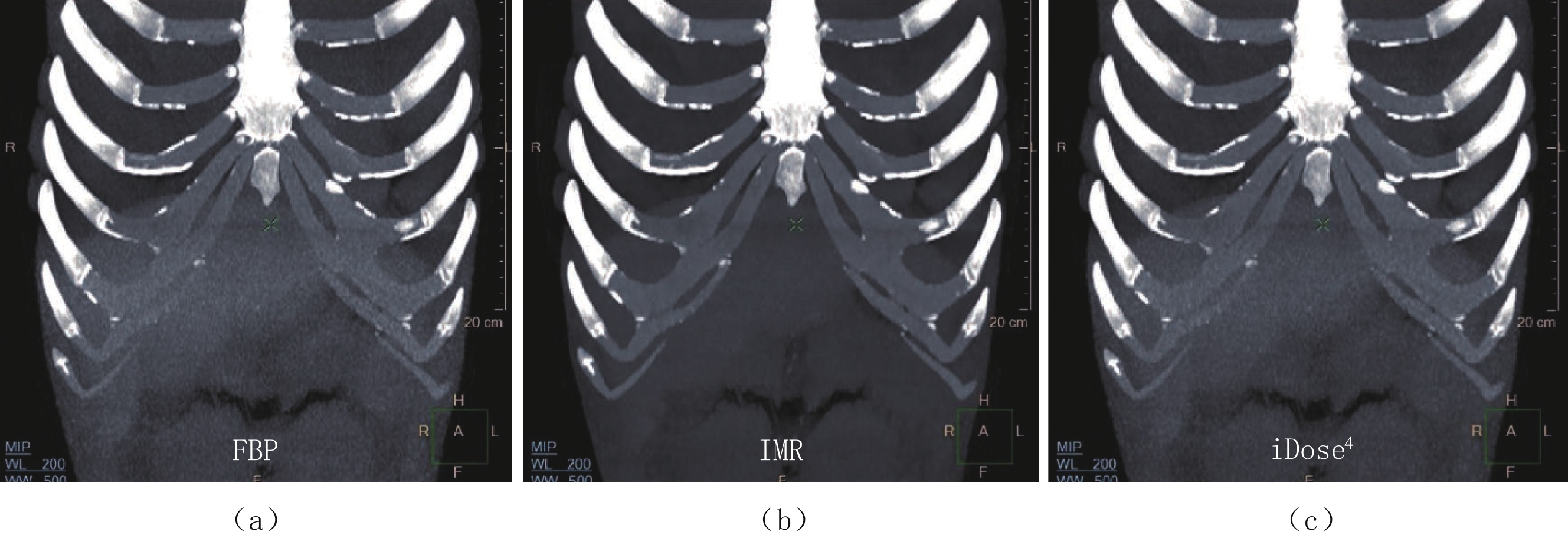
XU T J, WANG X, LI Y, et al. Application of IMR, iDose4 and FBP reconstruction algorithms in CT scanning of rhinoplasty[J]. CT Theory and Applications, 2022, 31(3): 357-364. DOI: 10.15953/j.ctta.2021.072. (in Chinese).

|
| [1] |
NUARA M J, LOCH R B, SAXON S A. Reconstructive rhinoplasty using multiplanar carved costal cartilage[J]. JAMA Facial Plastic Surgery, 2016, 18(3): 207−211. doi: 10.1001/jamafacial.2015.2251
|
| [2] |
ZELKEN J, CHANG C S, HSIAO Y C. The PI graft for correction of severe saddle nose deformity[J]. Facial Plastic Surgery, 2016, 32(4): 452−459. doi: 10.1055/s-0036-1584555
|
| [3] |
JANG Y J, YU M S. Rhinoplasty for the Asian nose[J]. Facial Plastic Surgery, 2010, 26(2): 93−101. doi: 10.1055/s-0030-1253507
|
| [4] |
殷硕, 杨英, 徐同江, 等. 胸部多排螺旋CT扫描在肋软骨隆鼻术整形中的临床应用研究[J]. CT理论与应用研究, 2021,30(3): 347−353. DOI: 10.15953/j.1004-4140.2021.30.03.08.
YIN S, YANG Y, XU T J, et al. Clinical application of multi-slice spiral CT scan in chest for costal cartilage augmentation rhinoplasty[J]. CT Theory and Applications, 2021, 30(3): 347−353. DOI: 10.15953/j.1004-4140.2021.30.03.08. (in Chinese).
|
| [5] |
LEACH L, SHAMIL E, MALATA C M. Indications and long-term outcomes of open augmentation rhinoplasty with autogenous L-shaped costal cartilage strut grafts: A single plastic Surgeon’s experience[J]. The Polish Otolaryngology, 2018, 72(3): 26−32.
|
| [6] |
李婷婷, 张永高, 高剑波, 等. FBP、iDose4和 IMR3种重建算法对低剂量胸部CT图像质量的影响[J]. 实用放射学杂志, 2016,32(5): 777−780. doi: 10.3969/j.issn.1002-1671.2016.05.030
LI T T, ZHANG Y G, GAO J B, et al. Impact of reconstruction techniques on low dose chest CT image quality: Comparison of FBP, iDose4 and IMR[J]. Journal of Practical Radiology, 2016, 32(5): 777−780. (in Chinese). doi: 10.3969/j.issn.1002-1671.2016.05.030
|
| [7] |
马继光, 蔡磊, 王克明, 等. 自体颗粒肋软骨移植隆鼻的临床应用[J]. 中华整形外科杂志, 2016,32(1): 25−28. doi: 10.3760/cma.j.issn.1009-4598.2016.01.007
MA J G, CAI L, WANG K M, et al. Costal cartilage for rhinoplasty[J]. Chinese Journal of Plastic Surgery, 2016, 32(1): 25−28. (in Chinese). doi: 10.3760/cma.j.issn.1009-4598.2016.01.007
|
| [8] |
蔡武, 胡春洪, 王希明, 等. “四低”联合自动管电流调节和全模型迭代重建技术在头颈部CT血管成像中的应用[J]. 中华医学杂志, 2018,98(1): 30−35. doi: 10.3760/cma.j.issn.0376-2491.2018.01.007
CAI W, HU C H, WANG X M, et al. Applied research of "quadri-low" combined with automatic tube current modulation and iterative model reconstruction technology in head and neck CT angiography[J]. National Medical Journal of China, 2018, 98(1): 30−35. (in Chinese). doi: 10.3760/cma.j.issn.0376-2491.2018.01.007
|
| [9] |
方林, 李亮. 自适应统计迭代重建技术在降低CT辐射剂量中的应用进展[J]. CT理论与应用研究, 2013,22(2): 207−214.
FANG L, LI L. Application progress of adaptive statistical iterative reconstruction in reducing radiation dose[J]. CT Theory and Applications, 2013, 22(2): 207−214. (in Chinese).
|
| [10] |
KIMI S. Augmentation rhinoplasty using silicone implants[J]. Facial Plastic Surgery Clinics of North America, 2018, 26(3): 285−293. doi: 10.1016/j.fsc.2018.03.003
|
| [11] |
HALPERN E J, GINGOLD E L, WHITE H, et al. Evaluation of coronary artery image quality with knowledge-based iterative model recon-struction[J]. Academic Radiology, 2014, 21(6): 805−811. doi: 10.1016/j.acra.2014.02.017
|
| [12] |
HUANG M P, LIANG C H, ZHAO Z J, et al. Evaluation of image quality and radiation dose at prospective ECG-triggered axial 256-slice multi-detector CT in infants with congenital heart disease[J]. Pediatric Radiology, 2011, 41(7): 858−866. doi: 10.1007/s00247-011-2079-2
|
| [13] |
蒋骏, 黄美萍, 雷益, 等. 全模型迭代重建技术在心脏CT成像中应用的实验研究[J]. 中华放射学杂志, 2015,(6): 473−477. doi: 10.3760/cma.j.issn.1005-1201.2015.06.017
JIANG J, HUANG M P, LEI Y, et al. Application of iterative model reconstruction iterative reconstruction in cardiac CT imaging-an animal experimental study[J]. Chinese Journal of Radiology, 2015, (6): 473−477. (in Chinese). doi: 10.3760/cma.j.issn.1005-1201.2015.06.017
|
| [14] |
SCHINDERA S T, LARS D, MÜLLER H C, et al. Iterative reconstruction algorithm for abdominal multidetector CT at different tube voltages: Assessment of diagnostic accuracy, image quality, and radiation dose in a phantom study[J]. Radiology, 2011, 260(2): 454−462. doi: 10.1148/radiol.11102217
|
| [1] | HAO Qi, ZHANG Yan, LI Xingpeng, LIU Mengke, SUN Xiaoli, WANG Rengui. The Application Value of CT Lymphangiography in Diagnosis and Grading in Patients with Primary Chyluria[J]. CT Theory and Applications, 2022, 31(4): 459-468. DOI: 10.15953/j.ctta.2022.106 |
| [2] | ZHANG Yan, HAO Qi, LIU Mengke, LI Xingpeng, SUN Xiaoli, WANG Rengui. Application of CT Lymphangiography in the Diagnosiis of Primary Pulmonary Lymphedema[J]. CT Theory and Applications, 2022, 31(4): 441-447. DOI: 10.15953/j.ctta.2022.096 |
| [3] | GUO Jia, ZHANG Yan, LIU Mengke, LI Xingpeng, HAO Qi, WANG Rengui. CT and CT Lymphangiography Findings of Congenital Pulmonary Lymphangiectasis[J]. CT Theory and Applications, 2022, 31(4): 433-440. DOI: 10.15953/j.ctta.2022.116 |
| [4] | YU Xingyao, LIU Chao, FENG Qianqian, XU Lin. The Value of MSCT Signs in Differential Diagnosis of Pathological Types of Acute Appendicitis[J]. CT Theory and Applications, 2022, 31(2): 251-258. DOI: 10.15953/j.1004-4140.2022.31.02.13 |
| [5] | LI Ya-chao, YU Bei, LI Xiao-li. The Relationship of between Subacromial Impingement Syndrome and Shoulder Typing under MSCT[J]. CT Theory and Applications, 2015, 24(5): 753-759. DOI: 10.15953/j.1004-4140.2015.24.05.14 |
| [6] | WU De-hong, YANG Song, GONG Xiao-hong, FU Chuan-ming, LIU Chao, CHEN Ping-you. The CT Diagnosis of Cystic Type Intestinal Duplications[J]. CT Theory and Applications, 2014, 23(5): 871-876. |
| [7] | WU Meng-nan, PENG Zhen-peng, HU Xiao-shu, DONG Zhi, LUO Yan-ji, FENG Shi-ting, YANG Xu-feng. The Classification Diagnosis of CT and its Imaging Features in Infantile Hepatic Hemangioendothelioma[J]. CT Theory and Applications, 2014, 23(4): 663-668. |
| [8] | SHI Ying-qin, PAN Jin-xiao. The Research of Reconstruction Speed for the Feldkamp-type-VOI Algorithm[J]. CT Theory and Applications, 2011, 20(1): 55-61. |
| [9] | WANG Ge, Seung WookLee. Grangeat-Type and Katsevich-Type Algorithms for Cone-Beam CT[J]. CT Theory and Applications, 2003, 12(2): 45-55. |
| [10] | Wang Hong-sheng, Wang Feng-yun. A Signification of Sediment Grouping after TACE Intraarterial Infusion of Iodized Oil[J]. CT Theory and Applications, 2001, 10(1): 5-7. |
| 1. |
武占鑫. 纵隔支气管源性囊肿的CT诊断特点分析. 影像研究与医学应用. 2025(01): 136-138 .

| |
| 2. |
陈君如,刀娅,杨仕武,王舒钰,代如涛,张熙晨,胡玉磊,马永钰,吴骏. 基于三维重建技术胸腔镜治疗婴儿复杂解剖支气管源性囊肿1例. 中国微创外科杂志. 2024(12): 846-848 .
|
Supported by: Beijing Renhe Information Technology Co. Ltd 

 DownLoad: