ISSN 1004-4140
CN 11-3017/P
| Citation: |
YANG Z X, ZHANG Y Q, LIU A S, et al. Diagnostic Value of Dual-source CT Dual-energy Virtual Non-contrasting Extramural Vascular Invasions of Rectal Cancer[J]. CT Theory and Applications, 2024, 33(1): 42-48. DOI: 10.15953/j.ctta.2022.231. (in Chinese).

|
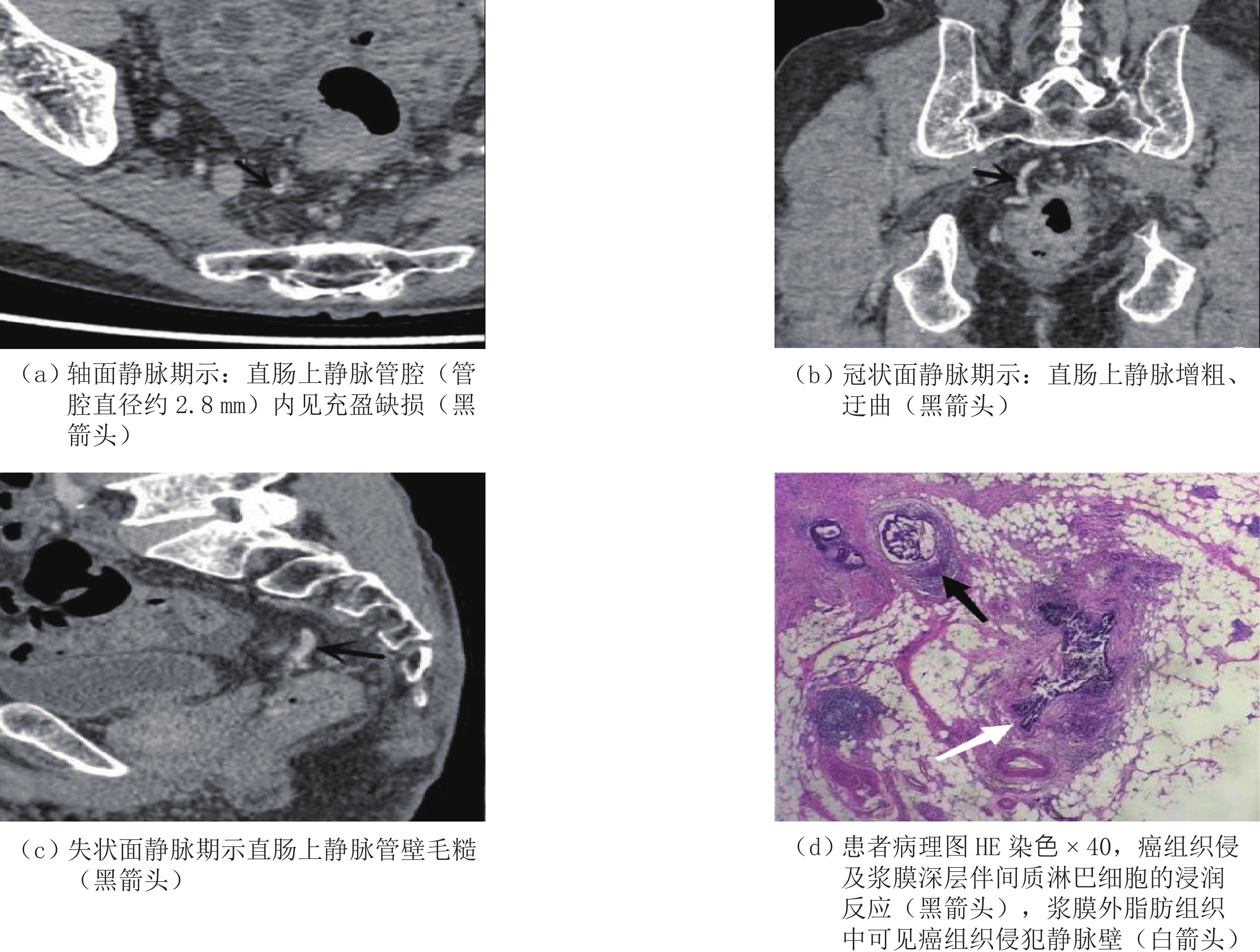
Objective: To investigate the value of dual-source CT dual-energy virtual plain scan in the preoperative diagnosis of extramural vascular invasion (EMVI) in rectal cancer. Methods: A total of 150 patients with rectal cancer (64 females (42.7%) and 86 males (57.3%), with an average age of (62.3±11.8) years) who were scheduled for surgical treatment in our hospital from November 2019 to December 2021 were selected for the preoperative dual-source CT dual-energy virtual plain scan;. Two senior radiologists independently analyzed preoperative imaging data (including ctEMVI status, local lymph node infiltration, and distant metastasis) to determine the existence of preoperative EMVI. With pathological results as the gold standard, the sensitivity, specificity, accuracy, positive predictive value, and negative predictive value of VNC in the diagnosis of EMVI were evaluated, and the area under the receiver operating characteristic curve (ROC) was used to evaluate the diagnostic efficiency. Results: Among 150 patients with rectal cancer, 56 (37.3%) were positive for EMVI and 94 (62.7%) were negative for EMVI. The accuracy, sensitivity, specificity, positive predictive value, and negative predictive value of EMVI evaluation by physician 1 were 86%, 80.36%, 89.36%, 81.82%, and 88.42%, respectively, and the AUC was 0.831 (95%CI, 0.756~0.905). The accuracy, sensitivity, specificity, positive predictive value, and negative predictive value of physician 2 were 88.67%, 80.36%, 93.62%, 88.24%, and 88.89%, respectively, and the AUC was 0.870 (95%CI, 0.802~0.938). The consistency of the evaluation results between physician 1 and physician 2 was high (k=0.943). Conclusion: Dual-source CT dual-energy virtual plain scan has a certain diagnostic value in the preoperative evaluation of EMVI of rectal cancer.
| [1] |
SUNG H, FERLAY J, SIEGEL R L, et al. Global cancer statistics 2020: GLOBOCAN estimates of incidence and mortality worldwide for 36 cancers in 185 countries[J]. CA-A Cancer Journal for Clinicians, 2021, 71(3): 209−249. DOI: 10.3322/caac.21660.
|
| [2] |
CHER H T, RAGHUNANDAN V, PIYAPORN B, et al. Extramural venous invasion by gastrointestinal malignancies: CT appearances[J]. Abdominal Imaging, 2011, 36(5): 491−502. DOI: 10.1007/s00261-010-9667-8.
|
| [3] |
CHAND M, BHODAY J, BHOME R, et al. mrEMVI status should be used in addition to pEMVI for treatment decision making in rectal cancer to prevent under-reporting of extramural venous invasion[J]. European Journal of Surgical Oncology, 2013, 39(2): 66−79.
|
| [4] |
PRAMPOLINIF, TASCHINIS, PECCHI, et al. Magnetic resonance imaging performed before and after preoperative chemoradiotherapy chemoradiotherapy in rectal cancer: Predictive factors of recurrence and prognostic significance of MR-detected extramural venous invasion[J]. Abdominal Radiology, 2020, 45(10): 2941−2949. DOI: 10.1007/s00261-018-1838-z.
|
| [5] |
MASSUCCO P, FONTANA A P, BALBO M A, et al. MRI-detected extramural vascular invasion (mrEMVI) as the best predictive factor to identify candidates to total neoadjuvant therapy in locally advanced rectal cancer[J]. Journal of Surgical Oncology, 2022, 125(6): 1024−1031. DOI: 10.1002/jso.26818.
|
| [6] |
TAE H K, SUNGMIN W, SANGWON H, et al. The diagnostic performance of MRI for detection of extramural venous invasion in colorectal cancer: A systematic review and meta-analysis of the literature[J]. American Journal of Roentgenology, 2019, 213(3): 575−585. DOI: 10.2214/AJR.19.21112.
|
| [7] |
TRIVEDI H, CHAMARTHY U, DICARLO L, et al. Prognostic factors of overall survival for patients with stage Ⅱ colon cancer[J]. Acta Medica Academiae Scientiarum Hungaricae, 2014, 43(2): 134−143. DOI: 10.5644/ama2006-124.112.
|
| [8] |
白晨. 基于高分辨率MRI影像组学方法直肠癌EMVI诊断模型的研究[D]. 长春: 吉林大学, 2022.
BAI C. Study of extramural venous invasion diagnostic model for rectal cancer based on high-resolution MRI radiomics method[D]. Changchun: Jilin University, 2022. DOI:10.27162/d.cnki.gjlin.2022.007006. (in Chinese).
|
| [9] |
中华人民共和国国家卫生健康委员会医政医管局, 中华医学会肿瘤学分会. 中国结直肠癌诊疗规范(2020年版)[J]. 中国实用外科杂志, 2020, 40(6): 601-625.
Hospital Authority of National Health Commission of the People's Republic of China, Chinese Society of Oncology, Chinese Medical Association. Chinese protocol of diagnosis and treatment of colorectal cancer (2020 edition)[J]. Chinese Journal of Practical Surgery, 2020, 40(6): 601-625. DOI:10.19538/j.cjps.issn1005-2208.2020.06.01. (in Chinese).
|
| [10] |
BENSON A B, VENOOK A P, AL-HAWARY M M, et al. Rectal cancer, version 2.2018 clinical practice guidelines in oncology[J]. Journal of the National Comprehensive Cancer Network, 2018, 16(7): 874−901. DOI: 10.6004/jnccn.2018.0061.
|
| [11] |
唐雪, 江长思, 张世派, 等. 直肠MR造影对直肠癌壁外血管侵犯的诊断价值[J]. 医学影像学杂志, 2022,32(4): 638−642. DOI: 1006-9011(2022)04-0638-05.
TANG X, JIANG C S, ZHANG S P, et al. Diagnosis value of MR rectography in assessment for extravasational vascular invasion of rectal cancers[J]. Journal of Medical Imaging, 2022, 32(4): 638−642. DOI: 1006-9011(2022)04-0638-05. (in Chinese).
|
| [12] |
路凯, 兰国宾, 戴士林. 双源CT双能量虚拟平扫对结直肠良恶性肿瘤的鉴别诊断价值研究[J]. 中国CT和MRI杂志, 2021,19(9): 131−134. DOI: 10.3969/j.issn.1672-5131.2021.09.042.
LU K, LAN G B, DAI S L. Differential diagnosis value of dual-source CT dual-energy virtual non-contrast on benign and malignant colorectal tumors[J]. Chinese Journal of CT and MRI, 2021, 19(9): 131−134. DOI: 10.3969/j.issn.1672-5131.2021.09.042. (in Chinese).
|
| [13] |
ZHEN G, ZHANG X Y, LI X T, et al. Correlation and prognostic value of CT-detected extramural venous invasion and pathological lymph-vascular invasion in colon cancer[J]. Abdominal Radiology, 2022, 47(4): 1232−1243. DOI: 10.1007/s00261-022-03414-7.
|
| [14] |
TRIPATHI P, GUO W, RAO S, et al. Additional value of MRI-detected EMVI scoring system in rectal cancer: Applicability in predicting synchronous metastasis[J]. Tumori Journal, 2020, 106(4): 286−294. DOI: 10.1177/0300891620901745.
|
| [15] |
TRIPATHI P, RAO S X, ZENG M S. Clinical value of MRI-detected extramural venous invasion in rectal cancer[J]. Journal of Digestive Diseases, 2017, 18(1): 2−12. DOI: 10.1111/1751-2980.12439.
|
| [16] |
BRUCE M, JENNIFER S. Somatostatin receptor scintigraphy of neuroendocrine tumors of the abdomen and pelvis[J]. Seminars in Roentgenology, 2016, 51(2): 112−122. DOI: 10.1053/j.ro.2016.02.008.
|
| [17] |
虞云杰, 陈孝娟, 李鹏, 等. 多层螺旋CT在直肠癌术前诊断及⾎管侵犯评估中的应⽤价值[J]. 中国CT和MRI杂志, 2020,18(2): 117−120. DOI: 10.3969/j.issn.1672-5131.2020.02.035.
YU Y J, CHEN X J, LI P, et al. Application value of multi-slice spiral CT in the preoperative diagnosis of rectal cancer and assessment of vascular invasion[J]. Chinese Journal of CT and MRI, 2020, 18(2): 117−120. DOI: 10.3969/j.issn.1672-5131.2020.02.035. (in Chinese).
|
| [18] |
SOHN B, LIM J S, KIM H, et al. MRI-detected extramural vascular invasion is an independent prognostic factor for synchronous metastasis in patients with rectal cancer[J]. European Radiology, 2015, 25(5): 1347−1355. DOI: 10.1007/s00330-014-3527-9.
|
| [19] |
CHAND M, EVANS J, SWIFT R I, et al. The prognostic significance of postchemoradiotherapy high-resolution MRI and histopathology detected extramural venous invasion in rectal cancer[J]. Annals of Surgery, 2015, 261(3): 473−479. DOI: 10.1097/SLA.0000000000000848.
|
Supported by: Beijing Renhe Information Technology Co. Ltd 


 DownLoad: